 Movie
Movie Controller


+ Open data

- Basic information
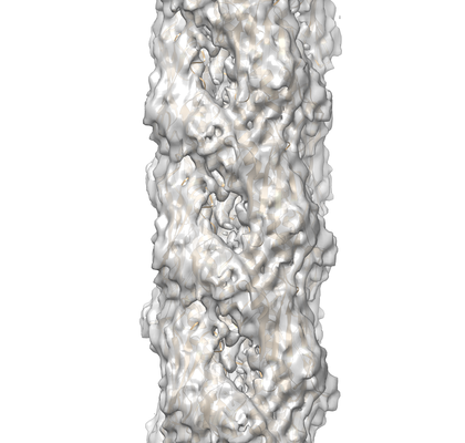
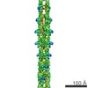
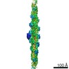
| Entry | Database: EMDB / ID: EMD-8740 | |||||||||||||||
|---|---|---|---|---|---|---|---|---|---|---|---|---|---|---|---|---|
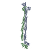
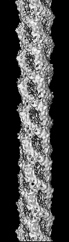
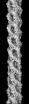
| Title | Cryo-EM reconstruction of PAK pilus from Pseudomonas aeruginosa | |||||||||||||||
 Map data Map data | PAK pilus from Pseudomonas aeruginosa | |||||||||||||||
 Sample Sample |
| |||||||||||||||
 Keywords Keywords | Melted helix / type IV pili / PROTEIN FIBRIL | |||||||||||||||
| Function / homology |  Function and homology information Function and homology informationtype IV pilus / type IV pilus-dependent motility / cell adhesion / membrane Similarity search - Function | |||||||||||||||
| Biological species |  Pseudomonas aeruginosa PAK (bacteria) Pseudomonas aeruginosa PAK (bacteria) | |||||||||||||||
| Method | helical reconstruction / cryo EM / Resolution: 8.0 Å | |||||||||||||||
 Authors Authors | Wang F / Osinksi T | |||||||||||||||
| Funding support |  United States, United States,  Canada, Canada,  France, 4 items France, 4 items
| |||||||||||||||
 Citation Citation | Journal: Structure / Year: 2017 Title: Cryoelectron Microscopy Reconstructions of the Pseudomonas aeruginosa and Neisseria gonorrhoeae Type IV Pili at Sub-nanometer Resolution. Authors: Fengbin Wang / Mathieu Coureuil / Tomasz Osinski / Albina Orlova / Tuba Altindal / Gaël Gesbert / Xavier Nassif / Edward H Egelman / Lisa Craig / Abstract: We report here cryoelectron microscopy reconstructions of type IV pili (T4P) from two important human pathogens, Pseudomonas aeruginosa and Neisseria gonorrhoeae, at ∼ 8 and 5 Å resolution, ...We report here cryoelectron microscopy reconstructions of type IV pili (T4P) from two important human pathogens, Pseudomonas aeruginosa and Neisseria gonorrhoeae, at ∼ 8 and 5 Å resolution, respectively. The two structures reveal distinct arrangements of the pilin globular domains on the pilus surfaces, which impart different helical parameters, but similar packing of the conserved N-terminal α helices, α1, in the filament core. In contrast to the continuous α helix seen in the X-ray crystal structures of the P. aeruginosa and N. gonorrhoeae pilin subunits, α1 in the pilus filaments has a melted segment located between conserved helix-breaking residues Gly14 and Pro22, as seen for the Neisseria meningitidis T4P. Using mutagenesis we show that Pro22 is critical for pilus assembly, as are Thr2 and Glu5, which are positioned to interact in the hydrophobic filament core. These structures provide a framework for understanding T4P assembly, function, and biophysical properties. | |||||||||||||||
| History |
|
- Structure visualization
| Movie |
Movie viewer |
|---|---|
| Structure viewer | EM map: SurfViewMolmilJmol/JSmol |
| Supplemental images |
- Downloads & links
-EMDB archive
| Map data | emd_8740.map.gz | 5.7 MB | EMDB map data format | |
|---|---|---|---|---|
| Header (meta data) | emd-8740-v30.xmlemd-8740.xml | 12 KB 12 KB | Display Display | EMDB header |
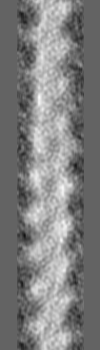
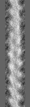
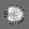
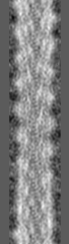
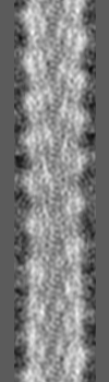
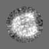
| Images |  emd_8740.png emd_8740.png | 133.2 KB | ||
| Filedesc metadata | emd-8740.cif.gz | 5.3 KB | ||
| Archive directory |  http://ftp.pdbj.org/pub/emdb/structures/EMD-8740ftp://ftp.pdbj.org/pub/emdb/structures/EMD-8740 http://ftp.pdbj.org/pub/emdb/structures/EMD-8740ftp://ftp.pdbj.org/pub/emdb/structures/EMD-8740 | HTTPS FTP |
-Related structure data
| Related structure data |  5vxyMC  8739C  5vxxC C: citing same article ( M: atomic model generated by this map |
|---|---|
| Similar structure data |
-Links
| EMDB pages | EMDB (EBI/PDBe) / EMDataResource |
|---|---|
| Related items in Molecule of the Month |

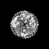
-Map
| File | Download / File: emd_8740.map.gz / Format: CCP4 / Size: 13.4 MB / Type: IMAGE STORED AS FLOATING POINT NUMBER (4 BYTES) | ||||||||||||||||||||||||||||||||||||||||||||||||||||||||||||||||||||
|---|---|---|---|---|---|---|---|---|---|---|---|---|---|---|---|---|---|---|---|---|---|---|---|---|---|---|---|---|---|---|---|---|---|---|---|---|---|---|---|---|---|---|---|---|---|---|---|---|---|---|---|---|---|---|---|---|---|---|---|---|---|---|---|---|---|---|---|---|---|
| Annotation | PAK pilus from Pseudomonas aeruginosa | ||||||||||||||||||||||||||||||||||||||||||||||||||||||||||||||||||||
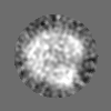
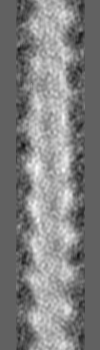
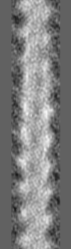
| Projections & slices | Image control
Images are generated by Spider. generated in cubic-lattice coordinate | ||||||||||||||||||||||||||||||||||||||||||||||||||||||||||||||||||||
| Voxel size | X=Y=Z: 1.05 Å | ||||||||||||||||||||||||||||||||||||||||||||||||||||||||||||||||||||
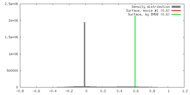
| Density |
| ||||||||||||||||||||||||||||||||||||||||||||||||||||||||||||||||||||
| Symmetry | Space group: 1 | ||||||||||||||||||||||||||||||||||||||||||||||||||||||||||||||||||||
| Details | EMDB XML:
CCP4 map header:
| ||||||||||||||||||||||||||||||||||||||||||||||||||||||||||||||||||||
 Z (Sec.)
 Y (Row.)
 X (Col.)


-Supplemental data
- Sample components
-Entire : Pseudomonas aeruginosa Type IV pilin filament
| Entire | Name: Pseudomonas aeruginosa Type IV pilin filament |
|---|---|
| Components |
|
-Supramolecule #1: Pseudomonas aeruginosa Type IV pilin filament
| Supramolecule | Name: Pseudomonas aeruginosa Type IV pilin filament / type: complex / ID: 1 / Parent: 0 / Macromolecule list: all |
|---|---|
| Source (natural) | Organism: Pseudomonas aeruginosa PAK (bacteria) / Strain: PAK/2pfs |
-Macromolecule #1: Fimbrial protein
| Macromolecule | Name: Fimbrial protein / type: protein_or_peptide / ID: 1 / Number of copies: 21 / Enantiomer: LEVO |
|---|---|
| Source (natural) | Organism: Pseudomonas aeruginosa PAK (bacteria) / Strain: PAK/2pfs |
| Molecular weight | Theoretical: 15.02118 KDa |
| Sequence | String: FTLIELMIVV AIIGILAAIA IPQYQNYVAR SEGASALASV NPLKTTVEEA LSRGWSVKSG TGTEDATKKE VPLGVAADAN KLGTIALKP DPADGTADIT LTFTMGGAGP KNKGKIITLT RTAADGLWKC TSDQDEQFIP KGCSR UniProtKB: Fimbrial protein |
-Experimental details
-Structure determination
| Method | cryo EM |
|---|---|
 Processing Processing | helical reconstruction |
| Aggregation state | filament |
-Sample preparation
| Concentration | 0.1 mg/mL |
|---|---|
| Buffer | pH: 9 / Details: 50 mM CHES buffer |
| Grid | Pretreatment - Type: PLASMA CLEANING / Details: unspecified |
| Vitrification | Cryogen name: ETHANE / Chamber humidity: 90 % / Instrument: FEI VITROBOT MARK IV |
- Electron microscopy
| Microscope | FEI TITAN KRIOS |
|---|---|
| Image recording | Film or detector model: FEI FALCON II (4k x 4k) / Detector mode: INTEGRATING / Average exposure time: 2.0 sec. / Average electron dose: 20.0 e/Å2 Details: Images were stored containing seven parts, where each part represented a set of frames corresponding to a dose of ~20 electrons per Angstrom^2. The full dose image stack was used for the ...Details: Images were stored containing seven parts, where each part represented a set of frames corresponding to a dose of ~20 electrons per Angstrom^2. The full dose image stack was used for the estimation of the CTF as well as for boxing filaments. Only the first two parts were used for the reconstruction (~5 electrons per Angstrom^2) |
| Electron beam | Acceleration voltage: 300 kV / Electron source:  FIELD EMISSION GUN FIELD EMISSION GUN |
| Electron optics | Illumination mode: FLOOD BEAM / Imaging mode: BRIGHT FIELD |
| Experimental equipment |  Model: Titan Krios / Image courtesy: FEI Company |
-Image processing
| Final reconstruction | Applied symmetry - Helical parameters - Δz: 10.5 Å Applied symmetry - Helical parameters - Δ&Phi: 87.3 ° Applied symmetry - Helical parameters - Axial symmetry: C1 (asymmetric) Resolution.type: BY AUTHOR / Resolution: 8.0 Å / Resolution method: OTHER / Software - Name: SPIDER / Details: model-map FSC 0.38 cut-off / Number images used: 31231 |
|---|---|
| Startup model | Type of model: OTHER / Details: featureless cylinder |
| Final angle assignment | Type: NOT APPLICABLE / Software - Name: SPIDER |
-Atomic model buiding 1
| Refinement | Space: REAL |
|---|---|
| Output model | PDB-5vxy: |
