 Movie
Movie Controller


+ Open data

- Basic information
| Entry | Database: PDB / ID: 5jyg | ||||||
|---|---|---|---|---|---|---|---|
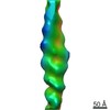
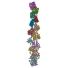
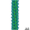
| Title | Cryo-EM structure of the MamK filament at 6.5 A | ||||||
 Components Components | Actin-like ATPase | ||||||
 Keywords Keywords | STRUCTURAL PROTEIN / Actin-like / Magnetosome / bacteria | ||||||
| Function / homology |  Function and homology information Function and homology informationmagnetosome assembly / magnetosome membrane / Hydrolases; Acting on acid anhydrides; In phosphorus-containing anhydrides / cytoskeleton / GTP binding / ATP hydrolysis activity / ATP binding / metal ion binding / cytoplasm Similarity search - Function | ||||||
| Biological species |  Magnetospirillum magneticum (bacteria) Magnetospirillum magneticum (bacteria) | ||||||
| Method | ELECTRON MICROSCOPY / helical reconstruction / cryo EM / Resolution: 6.5 Å | ||||||
 Authors Authors | Bergeron, J.R.C. / Hutto, R. / Kollman, J.M. | ||||||
 Citation Citation | Journal: Protein Sci / Year: 2017 Title: Structure of the magnetosome-associated actin-like MamK filament at subnanometer resolution. Authors: Julien R C Bergeron / Rachel Hutto / Ertan Ozyamak / Nancy Hom / Jesse Hansen / Olga Draper / Meghan E Byrne / Sepehr Keyhani / Arash Komeili / Justin M Kollman /   Abstract: Magnetotactic bacteria possess cellular compartments called magnetosomes that sense magnetic fields. Alignment of magnetosomes in the bacterial cell is necessary for their function, and this is ...Magnetotactic bacteria possess cellular compartments called magnetosomes that sense magnetic fields. Alignment of magnetosomes in the bacterial cell is necessary for their function, and this is achieved through anchoring of magnetosomes to filaments composed of the protein MamK. MamK is an actin homolog that polymerizes upon ATP binding. Here, we report the structure of the MamK filament at ∼6.5 Å, obtained by cryo-Electron Microscopy. This structure confirms our previously reported double-stranded, nonstaggered architecture, and reveals the molecular basis for filament formation. While MamK is closest in sequence to the bacterial actin MreB, the longitudinal contacts along each MamK strand most closely resemble those of eukaryotic actin. In contrast, the cross-strand interface, with a surprisingly limited set of contacts, is novel among actin homologs and gives rise to the nonstaggered architecture. | ||||||
| History |
|
- Structure visualization
| Movie |
Movie viewer |
|---|---|
| Structure viewer | Molecule: MolmilJmol/JSmol |
- Downloads & links
-Download
| PDBx/mmCIF format | 5jyg.cif.gz | 751.8 KB | Display | PDBx/mmCIF format |
|---|---|---|---|---|
| PDB format | pdb5jyg.ent.gz | 607.9 KB | Display | PDB format |
| PDBx/mmJSON format | 5jyg.json.gz | Tree view | PDBx/mmJSON format | |
| Others |  Other downloads Other downloads |
-Validation report
| Arichive directory | https://data.pdbj.org/pub/pdb/validation_reports/jy/5jygftp://data.pdbj.org/pub/pdb/validation_reports/jy/5jyg | HTTPS FTP |
|---|
-Related structure data
| Related structure data | 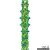 8180MC M: map data used to model this data C: citing same article ( |
|---|---|
| Similar structure data |

-Links
 PDBj
PDBj


- Assembly
| Deposited unit | 
|
|---|---|
| 1 |
|
-Components
| #1: Protein | Mass: 37656.180 Da / Num. of mol.: 14 Source method: isolated from a genetically manipulated source Source: (gene. exp.) Magnetospirillum magneticum (strain AMB-1 / ATCC 700264) (bacteria)Strain: AMB-1 / ATCC 700264 / Gene: amb0965 / Production host: #2: Chemical | ChemComp-MG /   Mass: 24.305 Da / Num. of mol.: 14 / Source method: obtained synthetically / Formula: Mg Mass: 24.305 Da / Num. of mol.: 14 / Source method: obtained synthetically / Formula: Mg#3: Chemical | ChemComp-ADP /   Mass: 427.201 Da / Num. of mol.: 14 / Source method: obtained synthetically / Formula: C10H15N5O10P2 / Comment: ADP, energy-carrying molecule*YM Mass: 427.201 Da / Num. of mol.: 14 / Source method: obtained synthetically / Formula: C10H15N5O10P2 / Comment: ADP, energy-carrying molecule*YM |
|---|
-Experimental details
-Experiment
| Experiment | Method: ELECTRON MICROSCOPY |
|---|---|
| EM experiment | Aggregation state: FILAMENT / 3D reconstruction method: helical reconstruction |
- Sample preparation
| Component | Name: Magnetosome-associated MamK filament / Type: ORGANELLE OR CELLULAR COMPONENT / Details: ADP-bound MamK / Entity ID: #1 / Source: RECOMBINANT |
|---|---|
| Source (natural) | Organism: Magnetospirillum magneticum AMB-1 (bacteria) |
| Source (recombinant) | Organism: |
| Buffer solution | pH: 7 |
| Specimen | Conc.: 0.5 mg/ml / Embedding applied: NO / Shadowing applied: NO / Staining applied: NO / Vitrification applied: YES |
| Vitrification | Cryogen name: ETHANE |
- Electron microscopy imaging
| Experimental equipment |  Model: Tecnai F20 / Image courtesy: FEI Company |
|---|---|
| Microscopy | Model: FEI TECNAI F20 |
| Electron gun | Electron source:  FIELD EMISSION GUN / Accelerating voltage: 200 kV / Illumination mode: OTHER FIELD EMISSION GUN / Accelerating voltage: 200 kV / Illumination mode: OTHER |
| Electron lens | Mode: OTHER |
| Image recording | Electron dose: 40 e/Å2 / Film or detector model: GATAN K2 SUMMIT (4k x 4k) |
- Processing
| EM software | Name: Rosetta / Version: 3.5 / Category: model refinement |
|---|---|
| CTF correction | Type: PHASE FLIPPING AND AMPLITUDE CORRECTION |
| Helical symmerty | Angular rotation/subunit: 24 ° / Axial rise/subunit: 53.8 Å / Axial symmetry: C2 |
| 3D reconstruction | Resolution: 6.5 Å / Resolution method: FSC 0.143 CUT-OFF / Num. of particles: 8129 / Symmetry type: HELICAL |
| Atomic model building | Protocol: OTHER |
