National Natural Science Foundation of China (NSFC)
12034006
中国
National Natural Science Foundation of China (NSFC)
32071209
中国
National Natural Science Foundation of China (NSFC)
31971122
中国
National Natural Science Foundation of China (NSFC)
32200994
中国
引用
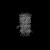
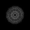
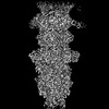
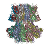
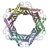
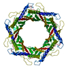
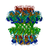
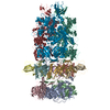
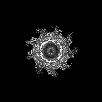
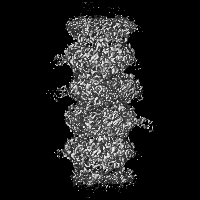
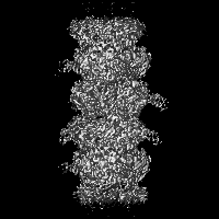
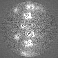
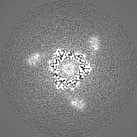
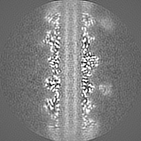
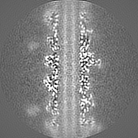
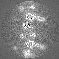
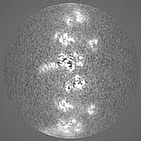
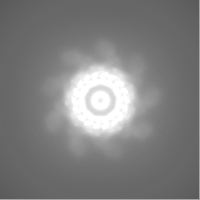
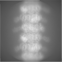
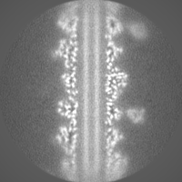
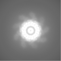
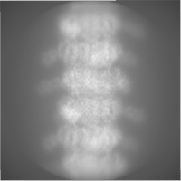
ジャーナル: Int J Mol Sci / 年: 2024 タイトル: Structures of Mature and Urea-Treated Empty Bacteriophage T5: Insights into Siphophage Infection and DNA Ejection. 著者: Yuning Peng / Huanrong Tang / Hao Xiao / Wenyuan Chen / Jingdong Song / Jing Zheng / Hongrong Liu / 要旨: T5 is a siphophage that has been extensively studied by structural and biochemical methods. However, the complete in situ structures of T5 before and after DNA ejection remain unknown. In this study, ...T5 is a siphophage that has been extensively studied by structural and biochemical methods. However, the complete in situ structures of T5 before and after DNA ejection remain unknown. In this study, we used cryo-electron microscopy (cryo-EM) to determine the structures of mature T5 (a laboratory-adapted, fiberless T5 mutant) and urea-treated empty T5 (lacking the tip complex) at near-atomic resolutions. Atomic models of the head, connector complex, tail tube, and tail tip were built for mature T5, and atomic models of the connector complex, comprising the portal protein pb7, adaptor protein p144, and tail terminator protein p142, were built for urea-treated empty T5. Our findings revealed that the aforementioned proteins did not undergo global conformational changes before and after DNA ejection, indicating that these structural features were conserved among most myophages and siphophages. The present study elucidates the underlying mechanisms of siphophage infection and DNA ejection.
 ムービー
ムービー コントローラー


 データを開く
 基本情報
基本情報
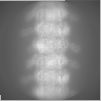
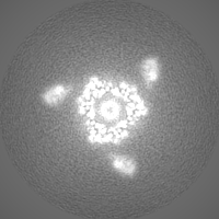
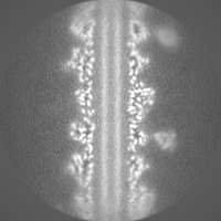
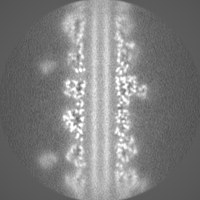
 マップデータ
 試料
試料 キーワード
キーワード 機能・相同性情報
 Escherichia phage T5 (ファージ)
 データ登録者
 中国, 4件
  引用
引用 構造の表示
構造の表示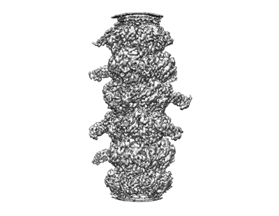
 ダウンロードとリンク
 emd_60712.png
 http://ftp.pdbj.org/pub/emdb/structures/EMD-60712

 Z (Sec.)
 Y (Row.)
 X (Col.)


 試料の構成要素
 解析
解析 電子顕微鏡法
 FIELD EMISSION GUN
