[English] 日本語
 Yorodumi
- EMDB-5789: Cryo-electron microscopy of an immature 50S ribosomal subunit (45... -
+ Open data
- Basic information
| Entry | Database: EMDB / ID: EMD-5789 | |||||||||
|---|---|---|---|---|---|---|---|---|---|---|
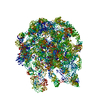
| Title | Cryo-electron microscopy of an immature 50S ribosomal subunit (45S particle) from Bacillus depleted of RbgA (YlqF), conformation 1 | |||||||||
 Map data Map data | 3D reconstruction of an immature 50S ribosomal subunit (45S particle) from Bacillus subtilis depleted of RbgA (YlqF), conformation 1 | |||||||||
 Sample Sample |
| |||||||||
 Keywords Keywords | Ribosome assembly / 50S subunit / RbgA / YlqF / GTPase / 45S subunit | |||||||||
| Biological species |  | |||||||||
| Method | single particle reconstruction / cryo EM / Resolution: 13.0 Å | |||||||||
 Authors Authors | Jomaa A / Jain N / Davis JH / Williamson JR / Britton RA / Ortega J | |||||||||
 Citation Citation |  Journal: Nucleic Acids Res / Year: 2014 Journal: Nucleic Acids Res / Year: 2014Title: Functional domains of the 50S subunit mature late in the assembly process. Authors: Ahmad Jomaa / Nikhil Jain / Joseph H Davis / James R Williamson / Robert A Britton / Joaquin Ortega /  Abstract: Despite the identification of many factors that facilitate ribosome assembly, the molecular mechanisms by which they drive ribosome biogenesis are poorly understood. Here, we analyze the late stages ...Despite the identification of many factors that facilitate ribosome assembly, the molecular mechanisms by which they drive ribosome biogenesis are poorly understood. Here, we analyze the late stages of assembly of the 50S subunit using Bacillus subtilis cells depleted of RbgA, a highly conserved GTPase. We found that RbgA-depleted cells accumulate late assembly intermediates bearing sub-stoichiometric quantities of ribosomal proteins L16, L27, L28, L33a, L35 and L36. Using a novel pulse labeling/quantitative mass spectrometry technique, we show that this particle is physiologically relevant and is capable of maturing into a complete 50S particle. Cryo-electron microscopy and chemical probing revealed that the central protuberance, the GTPase associating region and tRNA-binding sites in this intermediate are unstructured. These findings demonstrate that key functional sites of the 50S subunit remain unstructured until late stages of maturation, preventing the incomplete subunit from prematurely engaging in translation. Finally, structural and biochemical analysis of a ribosome particle depleted of L16 indicate that L16 binding is necessary for the stimulation of RbgA GTPase activity and, in turn, release of this co-factor, and for conversion of the intermediate to a complete 50S subunit. | |||||||||
| History |
|
- Structure visualization
| Movie |
 Movie viewer Movie viewer |
|---|---|
| Structure viewer | EM map:  SurfView SurfView Molmil Molmil Jmol/JSmol Jmol/JSmol |
| Supplemental images |
- Downloads & links
-EMDB archive
| Map data |  emd_5789.map.gz emd_5789.map.gz | 7.4 MB |  EMDB map data format EMDB map data format | |
|---|---|---|---|---|
| Header (meta data) |  emd-5789-v30.xml emd-5789-v30.xml emd-5789.xml emd-5789.xml | 11.2 KB 11.2 KB | Display Display |  EMDB header EMDB header |
| Images |  emd_5789.tif emd_5789.tif | 373.7 KB | ||
| Archive directory |  http://ftp.pdbj.org/pub/emdb/structures/EMD-5789 http://ftp.pdbj.org/pub/emdb/structures/EMD-5789 ftp://ftp.pdbj.org/pub/emdb/structures/EMD-5789 ftp://ftp.pdbj.org/pub/emdb/structures/EMD-5789 | HTTPS FTP |
-Validation report
| Summary document |  emd_5789_validation.pdf.gz emd_5789_validation.pdf.gz | 78.4 KB | Display |  EMDB validaton report EMDB validaton report |
|---|---|---|---|---|
| Full document |  emd_5789_full_validation.pdf.gz emd_5789_full_validation.pdf.gz | 77.5 KB | Display | |
| Data in XML |  emd_5789_validation.xml.gz emd_5789_validation.xml.gz | 494 B | Display | |
| Arichive directory |  https://ftp.pdbj.org/pub/emdb/validation_reports/EMD-5789 https://ftp.pdbj.org/pub/emdb/validation_reports/EMD-5789 ftp://ftp.pdbj.org/pub/emdb/validation_reports/EMD-5789 ftp://ftp.pdbj.org/pub/emdb/validation_reports/EMD-5789 | HTTPS FTP |
-Related structure data
- Links
Links
| EMDB pages |  EMDB (EBI/PDBe) / EMDB (EBI/PDBe) /  EMDataResource EMDataResource |
|---|---|
| Related items in Molecule of the Month |
- Map
Map
| File |  Download / File: emd_5789.map.gz / Format: CCP4 / Size: 7.8 MB / Type: IMAGE STORED AS FLOATING POINT NUMBER (4 BYTES) Download / File: emd_5789.map.gz / Format: CCP4 / Size: 7.8 MB / Type: IMAGE STORED AS FLOATING POINT NUMBER (4 BYTES) | ||||||||||||||||||||||||||||||||||||||||||||||||||||||||||||
|---|---|---|---|---|---|---|---|---|---|---|---|---|---|---|---|---|---|---|---|---|---|---|---|---|---|---|---|---|---|---|---|---|---|---|---|---|---|---|---|---|---|---|---|---|---|---|---|---|---|---|---|---|---|---|---|---|---|---|---|---|---|
| Annotation | 3D reconstruction of an immature 50S ribosomal subunit (45S particle) from Bacillus subtilis depleted of RbgA (YlqF), conformation 1 | ||||||||||||||||||||||||||||||||||||||||||||||||||||||||||||
| Projections & slices | Image control
Images are generated by Spider. | ||||||||||||||||||||||||||||||||||||||||||||||||||||||||||||
| Voxel size | X=Y=Z: 2.54 Å | ||||||||||||||||||||||||||||||||||||||||||||||||||||||||||||
| Density |
| ||||||||||||||||||||||||||||||||||||||||||||||||||||||||||||
| Symmetry | Space group: 1 | ||||||||||||||||||||||||||||||||||||||||||||||||||||||||||||
| Details | EMDB XML:
CCP4 map header:
| ||||||||||||||||||||||||||||||||||||||||||||||||||||||||||||
-Supplemental data
- Sample components
-Entire : Immature 50S ribosomal subunit (45S particle) from Bacillus subti...
| Entire | Name: Immature 50S ribosomal subunit (45S particle) from Bacillus subtilis depleted of RbgA (YlqF), conformation 1 |
|---|---|
| Components |
|
-Supramolecule #1000: Immature 50S ribosomal subunit (45S particle) from Bacillus subti...
| Supramolecule | Name: Immature 50S ribosomal subunit (45S particle) from Bacillus subtilis depleted of RbgA (YlqF), conformation 1 type: sample / ID: 1000 / Number unique components: 1 |
|---|---|
| Molecular weight | Theoretical: 1.6 MDa |
-Supramolecule #1: 45 subunit
| Supramolecule | Name: 45 subunit / type: complex / ID: 1 / Recombinant expression: No / Database: NCBI Ribosome-details: ribosome-prokaryote: LSU 50S, LSU RNA 23S, LSU RNA 5S |
|---|---|
| Source (natural) | Organism:  |
| Molecular weight | Theoretical: 1.6 MDa |
-Experimental details
-Structure determination
| Method | cryo EM |
|---|---|
 Processing Processing | single particle reconstruction |
| Aggregation state | particle |
- Sample preparation
| Buffer | pH: 7.5 Details: 10 mM Tris-HCl, pH 7.5, 10 mM magnesium acetate, 60 mM ammonium chloride, 3 mM 2-mercaptoethanol |
|---|---|
| Grid | Details: 400 mesh holey carbon grids with an additional layer (5-10 nm) of thin carbon. Grids were glow discharged at 5 mA for 15 seconds. |
| Vitrification | Cryogen name: ETHANE / Chamber humidity: 100 % / Chamber temperature: 77 K / Instrument: FEI VITROBOT MARK III Method: Grids were blotted twice, for 7 seconds each time, before being plunged into liquid ethane. |
- Electron microscopy
| Microscope | JEOL 2010F |
|---|---|
| Temperature | Min: 77 K / Max: 77 K |
| Alignment procedure | Legacy - Astigmatism: Objective lens astigmatism was corrected at 300,000 times magnification |
| Date | Feb 15, 2012 |
| Image recording | Category: FILM / Film or detector model: KODAK SO-163 FILM / Digitization - Scanner: NIKON SUPER COOLSCAN 9000 / Digitization - Sampling interval: 12.7 µm / Number real images: 300 / Average electron dose: 20 e/Å2 / Bits/pixel: 8 |
| Tilt angle min | 0 |
| Tilt angle max | 0 |
| Electron beam | Acceleration voltage: 200 kV / Electron source:  FIELD EMISSION GUN FIELD EMISSION GUN |
| Electron optics | Illumination mode: FLOOD BEAM / Imaging mode: BRIGHT FIELD / Cs: 1 mm / Nominal defocus max: 3.9 µm / Nominal defocus min: 1.5 µm / Nominal magnification: 50000 |
| Sample stage | Specimen holder: Gatan 914 cryo-holder / Specimen holder model: GATAN LIQUID NITROGEN |
- Image processing
| CTF correction | Details: ctFFIND |
|---|---|
| Final reconstruction | Algorithm: OTHER / Resolution.type: BY AUTHOR / Resolution: 13.0 Å / Resolution method: OTHER / Software - Name: Xmipp / Number images used: 20655 |
 Movie
Movie Controller


 Z (Sec.)
 Y (Row.)
 X (Col.)


