[English] 日本語
 Yorodumi
- EMDB-5754: Maximizing the potential of electron cryomicroscopy data collecte... -
+ Open data
- Basic information
| Entry | Database: EMDB / ID: EMD-5754 | |||||||||
|---|---|---|---|---|---|---|---|---|---|---|
| Title | Maximizing the potential of electron cryomicroscopy data collected using direct detectors | |||||||||
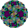
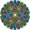
 Map data Map data | Reconstruction of mature STIV virion | |||||||||
 Sample Sample |
| |||||||||
 Keywords Keywords | Electron microscopy / Direct detectors / Near-atomic resolution / Sulfolobus turreted icosahedral virus | |||||||||
| Biological species |    Sulfolobus turreted icosahedral virus Sulfolobus turreted icosahedral virus | |||||||||
| Method | single particle reconstruction / cryo EM / Resolution: 5.8 Å | |||||||||
 Authors Authors | Veesler D / Campbell MG / Cheng A / Fu CY / Murez Z / Johnson JE / Potter CS / Carragher B | |||||||||
 Citation Citation |  Journal: J Struct Biol / Year: 2013 Journal: J Struct Biol / Year: 2013Title: Maximizing the potential of electron cryomicroscopy data collected using direct detectors. Authors: David Veesler / Melody G Campbell / Anchi Cheng / Chi-Yu Fu / Zachary Murez / John E Johnson / Clinton S Potter / Bridget Carragher /  Abstract: Single-particle electron cryomicroscopy is undergoing a technical revolution due to the recent developments of direct detectors. These new recording devices detect electrons directly (i.e. without ...Single-particle electron cryomicroscopy is undergoing a technical revolution due to the recent developments of direct detectors. These new recording devices detect electrons directly (i.e. without conversion into light) and feature significantly improved detective quantum efficiencies and readout rates as compared to photographic films or CCDs. We evaluated here the potential of one such detector (Gatan K2 Summit) to enable the achievement of near-atomic resolution reconstructions of biological specimens when coupled to a widely used, mid-range transmission electron microscope (FEI TF20 Twin). Compensating for beam-induced motion and stage drift provided a 4.4Å resolution map of Sulfolobus turreted icosahedral virus (STIV), which we used as a test particle in this study. Several motion correction and dose fractionation procedures were explored and we describe their influence on the resolution of the final reconstruction. We also compared the quality of this data to that collected with a FEI Titan Krios microscope equipped with a Falcon I direct detector, which provides a benchmark for data collected using a high-end electron microscope. | |||||||||
| History |
|
- Structure visualization
| Movie |
 Movie viewer Movie viewer |
|---|---|
| Structure viewer | EM map:  SurfView SurfView Molmil Molmil Jmol/JSmol Jmol/JSmol |
| Supplemental images |
- Downloads & links
-EMDB archive
| Map data |  emd_5754.map.gz emd_5754.map.gz | 3.3 GB |  EMDB map data format EMDB map data format | |
|---|---|---|---|---|
| Header (meta data) |  emd-5754-v30.xml emd-5754-v30.xml emd-5754.xml emd-5754.xml | 10.2 KB 10.2 KB | Display Display |  EMDB header EMDB header |
| Images |  400_5754.gif 400_5754.gif 80_5754.gif 80_5754.gif | 91.4 KB 5.5 KB | ||
| Archive directory |  http://ftp.pdbj.org/pub/emdb/structures/EMD-5754 http://ftp.pdbj.org/pub/emdb/structures/EMD-5754 ftp://ftp.pdbj.org/pub/emdb/structures/EMD-5754 ftp://ftp.pdbj.org/pub/emdb/structures/EMD-5754 | HTTPS FTP |
-Validation report
| Summary document |  emd_5754_validation.pdf.gz emd_5754_validation.pdf.gz | 78.9 KB | Display |  EMDB validaton report EMDB validaton report |
|---|---|---|---|---|
| Full document |  emd_5754_full_validation.pdf.gz emd_5754_full_validation.pdf.gz | 78 KB | Display | |
| Data in XML |  emd_5754_validation.xml.gz emd_5754_validation.xml.gz | 494 B | Display | |
| Arichive directory |  https://ftp.pdbj.org/pub/emdb/validation_reports/EMD-5754 https://ftp.pdbj.org/pub/emdb/validation_reports/EMD-5754 ftp://ftp.pdbj.org/pub/emdb/validation_reports/EMD-5754 ftp://ftp.pdbj.org/pub/emdb/validation_reports/EMD-5754 | HTTPS FTP |
-Related structure data
| Similar structure data |
|---|
- Links
Links
| EMDB pages |  EMDB (EBI/PDBe) / EMDB (EBI/PDBe) /  EMDataResource EMDataResource |
|---|
- Map
Map
| File |  Download / File: emd_5754.map.gz / Format: CCP4 / Size: 3.9 GB / Type: IMAGE STORED AS FLOATING POINT NUMBER (4 BYTES) Download / File: emd_5754.map.gz / Format: CCP4 / Size: 3.9 GB / Type: IMAGE STORED AS FLOATING POINT NUMBER (4 BYTES) | ||||||||||||||||||||||||||||||||||||||||||||||||||||||||||||||||||||
|---|---|---|---|---|---|---|---|---|---|---|---|---|---|---|---|---|---|---|---|---|---|---|---|---|---|---|---|---|---|---|---|---|---|---|---|---|---|---|---|---|---|---|---|---|---|---|---|---|---|---|---|---|---|---|---|---|---|---|---|---|---|---|---|---|---|---|---|---|---|
| Annotation | Reconstruction of mature STIV virion | ||||||||||||||||||||||||||||||||||||||||||||||||||||||||||||||||||||
| Projections & slices | Image control
Images are generated by Spider. | ||||||||||||||||||||||||||||||||||||||||||||||||||||||||||||||||||||
| Voxel size | X=Y=Z: 1.21 Å | ||||||||||||||||||||||||||||||||||||||||||||||||||||||||||||||||||||
| Density |
| ||||||||||||||||||||||||||||||||||||||||||||||||||||||||||||||||||||
| Symmetry | Space group: 1 | ||||||||||||||||||||||||||||||||||||||||||||||||||||||||||||||||||||
| Details | EMDB XML:
CCP4 map header:
| ||||||||||||||||||||||||||||||||||||||||||||||||||||||||||||||||||||
-Supplemental data
- Sample components
-Entire : Mature Sulfolobus Turreted Icosahedral Virus
| Entire | Name: Mature Sulfolobus Turreted Icosahedral Virus |
|---|---|
| Components |
|
-Supramolecule #1000: Mature Sulfolobus Turreted Icosahedral Virus
| Supramolecule | Name: Mature Sulfolobus Turreted Icosahedral Virus / type: sample / ID: 1000 / Oligomeric state: icosahedral / Number unique components: 1 |
|---|---|
| Molecular weight | Theoretical: 75 MDa |
-Supramolecule #1: Sulfolobus turreted icosahedral virus
| Supramolecule | Name: Sulfolobus turreted icosahedral virus / type: virus / ID: 1 / NCBI-ID: 269145 / Sci species name: Sulfolobus turreted icosahedral virus / Sci species strain: YNPRC179 / Database: NCBI / Virus type: VIRION / Virus isolate: STRAIN / Virus enveloped: Yes / Virus empty: No |
|---|---|
| Host (natural) | Organism:   Sulfolobus solfataricus (archaea) / Strain: 2-2-12 / synonym: ARCHAEA Sulfolobus solfataricus (archaea) / Strain: 2-2-12 / synonym: ARCHAEA |
| Molecular weight | Theoretical: 75 MDa |
| Virus shell | Shell ID: 1 / Diameter: 730 Å |
-Experimental details
-Structure determination
| Method | cryo EM |
|---|---|
 Processing Processing | single particle reconstruction |
| Aggregation state | particle |
- Sample preparation
| Buffer | pH: 3.5 Details: 23 mM KH2PO4, 19 mM (NH4)2SO4, 1 mM MgSO4, 2 mM CaCl2 |
|---|---|
| Grid | Details: plasma cleaned C-flat holey carbon grids (CF-1.2/1.3, Protochips) |
| Vitrification | Cryogen name: ETHANE / Chamber temperature: 94 K / Instrument: GATAN CRYOPLUNGE 3 / Details: Vitrification was carried out at room temperature. / Method: Blot for 3 seconds before plunging. |
- Electron microscopy
| Microscope | FEI TECNAI F20 |
|---|---|
| Temperature | Average: 90 K |
| Alignment procedure | Legacy - Astigmatism: Objective lens astigmatism was corrected at 41322 times magnification |
| Date | Nov 30, 2012 |
| Image recording | Category: CCD / Film or detector model: GATAN K2 (4k x 4k) / Number real images: 754 / Average electron dose: 22 e/Å2 Details: Every movie is composed of sixteen frames recorded by the direct electron detector. |
| Electron beam | Acceleration voltage: 200 kV / Electron source:  FIELD EMISSION GUN FIELD EMISSION GUN |
| Electron optics | Calibrated magnification: 41322 / Illumination mode: FLOOD BEAM / Imaging mode: BRIGHT FIELD / Cs: 2.0 mm / Nominal defocus max: 3.7 µm / Nominal defocus min: 0.45 µm |
| Sample stage | Specimen holder: Nitrogen cooled / Specimen holder model: GATAN LIQUID NITROGEN |
| Experimental equipment |  Model: Tecnai F20 / Image courtesy: FEI Company |
- Image processing
| Details | The final reconstruction was sharpened with a negative temperature factor of 650 A^2. |
|---|---|
| CTF correction | Details: Each particle |
| Final reconstruction | Algorithm: OTHER / Resolution.type: BY AUTHOR / Resolution: 5.8 Å / Resolution method: OTHER / Software - Name: Frealign Details: The final reconstruction was computed using the first ten frames of each movie and sharpened with a negative temperature factor of 650 A^2. Number images used: 4446 |
 Movie
Movie Controller





 Z (Sec.)
 Y (Row.)
 X (Col.)


