ムービー
ムービー コントローラー


+ データを開く

- 基本情報
基本情報
| 登録情報 |  | |||||||||
|---|---|---|---|---|---|---|---|---|---|---|
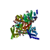
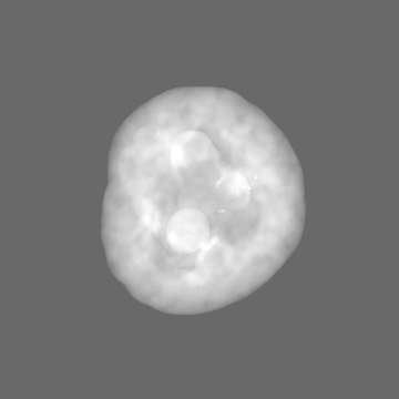
| タイトル | Full-length apo human voltage-gated sodium channel 1.8 (NaV1.8) | |||||||||
 マップデータ マップデータ | ||||||||||
 試料 試料 |
| |||||||||
 キーワード キーワード | ion channel / MEMBRANE PROTEIN | |||||||||
| 機能・相同性 |  機能・相同性情報 機能・相同性情報bundle of His cell action potential / AV node cell action potential / clathrin complex / regulation of atrial cardiac muscle cell membrane depolarization / membrane depolarization during action potential / voltage-gated monoatomic ion channel activity involved in regulation of presynaptic membrane potential / cardiac muscle cell action potential involved in contraction / voltage-gated sodium channel complex / regulation of monoatomic ion transmembrane transport / sensory perception ...bundle of His cell action potential / AV node cell action potential / clathrin complex / regulation of atrial cardiac muscle cell membrane depolarization / membrane depolarization during action potential / voltage-gated monoatomic ion channel activity involved in regulation of presynaptic membrane potential / cardiac muscle cell action potential involved in contraction / voltage-gated sodium channel complex / regulation of monoatomic ion transmembrane transport / sensory perception / Interaction between L1 and Ankyrins / voltage-gated sodium channel activity / odontogenesis of dentin-containing tooth / Phase 0 - rapid depolarisation / regulation of cardiac muscle contraction / regulation of heart rate / sodium ion transmembrane transport / presynaptic membrane / transmembrane transporter binding / axon / glutamatergic synapse / extracellular exosome / plasma membrane 類似検索 - 分子機能 | |||||||||
| 生物種 |  Homo sapiens (ヒト) Homo sapiens (ヒト) | |||||||||
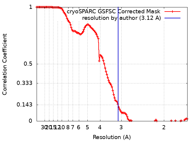
| 手法 | 単粒子再構成法 / クライオ電子顕微鏡法 / 解像度: 3.12 Å | |||||||||
 データ登録者 データ登録者 | Neumann B / McCarthy S / Gonen S | |||||||||
| 資金援助 |  米国, 2件 米国, 2件
| |||||||||
 引用 引用 | ジャーナル: Nat Commun / 年: 2025 タイトル: Structural basis of inhibition of human Na1.8 by the tarantula venom peptide Protoxin-I. 著者: Bryan Neumann / Stephen McCarthy / Shane Gonen / 要旨: Voltage-gated sodium channels (Nas) selectively permit diffusion of sodium ions across the cell membrane and, in excitable cells, are responsible for propagating action potentials. One of the nine ...Voltage-gated sodium channels (Nas) selectively permit diffusion of sodium ions across the cell membrane and, in excitable cells, are responsible for propagating action potentials. One of the nine human Na isoforms, Na1.8, is a promising target for analgesics, and selective inhibitors are of interest as therapeutics. One such inhibitor, the gating-modifier peptide Protoxin-I derived from tarantula venom, blocks channel opening by shifting the activation voltage threshold to more depolarized potentials, but the structural basis for this inhibition has not previously been determined. Using monolayer graphene grids, we report the cryogenic electron microscopy structures of full-length human apo-Na1.8 and the Protoxin-I-bound complex at 3.1 Å and 2.8 Å resolution, respectively. The apo structure shows an unexpected movement of the Domain I S4-S5 helix, and VSD was unresolvable. We find that Protoxin-I binds to and displaces the VSD S3-S4 linker, hindering translocation of the S4 helix during activation. | |||||||||
| 履歴 |
|
- 構造の表示
構造の表示
| 添付画像 |
|---|

- ダウンロードとリンク
-EMDBアーカイブ
| マップデータ | emd_46718.map.gz | 88.6 MB | EMDBマップデータ形式 | |
|---|---|---|---|---|
| ヘッダ (付随情報) | emd-46718-v30.xmlemd-46718.xml | 27 KB 27 KB | 表示 表示 | EMDBヘッダ |
| FSC (解像度算出) | emd_46718_fsc.xml | 12 KB | 表示 | FSCデータファイル |
| 画像 |  emd_46718.png emd_46718.png | 50.1 KB | ||
| マスクデータ | emd_46718_msk_1.mapemd_46718_msk_2.map | 178 MB 178 MB | マスクマップ | |
| Filedesc metadata | emd-46718.cif.gz | 8.2 KB | ||
| その他 | emd_46718_additional_1.map.gzemd_46718_additional_2.map.gzemd_46718_additional_3.map.gzemd_46718_half_map_1.map.gzemd_46718_half_map_2.map.gz | 168 MB 8.7 MB 9.3 MB 165 MB 165 MB | ||
| アーカイブディレクトリ |  http://ftp.pdbj.org/pub/emdb/structures/EMD-46718ftp://ftp.pdbj.org/pub/emdb/structures/EMD-46718 http://ftp.pdbj.org/pub/emdb/structures/EMD-46718ftp://ftp.pdbj.org/pub/emdb/structures/EMD-46718 | HTTPS FTP |
-関連構造データ


-リンク
| EMDBのページ | EMDB (EBI/PDBe) / EMDataResource |
|---|---|
| 「今月の分子」の関連する項目 |


-マップ
| ファイル | ダウンロード / ファイル: emd_46718.map.gz / 形式: CCP4 / 大きさ: 178 MB / タイプ: IMAGE STORED AS FLOATING POINT NUMBER (4 BYTES) | ||||||||||||||||||||||||||||||||||||
|---|---|---|---|---|---|---|---|---|---|---|---|---|---|---|---|---|---|---|---|---|---|---|---|---|---|---|---|---|---|---|---|---|---|---|---|---|---|
| 投影像・断面図 | 画像のコントロール
画像は Spider により作成 | ||||||||||||||||||||||||||||||||||||
| ボクセルのサイズ | X=Y=Z: 0.839 Å | ||||||||||||||||||||||||||||||||||||
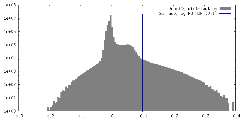
| 密度 |
| ||||||||||||||||||||||||||||||||||||
| 対称性 | 空間群: 1 | ||||||||||||||||||||||||||||||||||||
| 詳細 | EMDB XML:
|
 Z (Sec.)
 Y (Row.)
 X (Col.)


-添付データ
-マスク #1
| ファイル | emd_46718_msk_1.map | ||||||||||||
|---|---|---|---|---|---|---|---|---|---|---|---|---|---|
| 投影像・断面図 |
| ||||||||||||
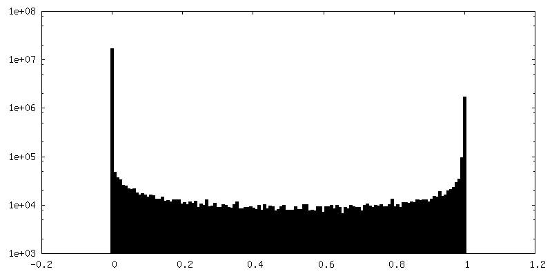
| 密度ヒストグラム |


-マスク #2
| ファイル | emd_46718_msk_2.map | ||||||||||||
|---|---|---|---|---|---|---|---|---|---|---|---|---|---|
| 投影像・断面図 |
| ||||||||||||
| 密度ヒストグラム |


-追加マップ: sharpened
| ファイル | emd_46718_additional_1.map | ||||||||||||
|---|---|---|---|---|---|---|---|---|---|---|---|---|---|
| 注釈 | sharpened | ||||||||||||
| 投影像・断面図 |
| ||||||||||||
| 密度ヒストグラム |


-追加マップ: local res
| ファイル | emd_46718_additional_2.map | ||||||||||||
|---|---|---|---|---|---|---|---|---|---|---|---|---|---|
| 注釈 | local res | ||||||||||||
| 投影像・断面図 |
| ||||||||||||
| 密度ヒストグラム |


-追加マップ: filtered
| ファイル | emd_46718_additional_3.map | ||||||||||||
|---|---|---|---|---|---|---|---|---|---|---|---|---|---|
| 注釈 | filtered | ||||||||||||
| 投影像・断面図 |
| ||||||||||||
| 密度ヒストグラム |


-ハーフマップ: #1
| ファイル | emd_46718_half_map_1.map | ||||||||||||
|---|---|---|---|---|---|---|---|---|---|---|---|---|---|
| 投影像・断面図 |
| ||||||||||||
| 密度ヒストグラム |


-ハーフマップ: #2
| ファイル | emd_46718_half_map_2.map | ||||||||||||
|---|---|---|---|---|---|---|---|---|---|---|---|---|---|
| 投影像・断面図 |
| ||||||||||||
| 密度ヒストグラム |


- 試料の構成要素
-全体 : Sodium channel protein type 10 subunit alpha
| 全体 | 名称: Sodium channel protein type 10 subunit alpha |
|---|---|
| 要素 |
|
-超分子 #1: Sodium channel protein type 10 subunit alpha
| 超分子 | 名称: Sodium channel protein type 10 subunit alpha / タイプ: complex / ID: 1 / 親要素: 0 / 含まれる分子: #1 |
|---|---|
| 由来(天然) | 生物種: Homo sapiens (ヒト) |
-分子 #1: Sodium channel protein type 10 subunit alpha
| 分子 | 名称: Sodium channel protein type 10 subunit alpha / タイプ: protein_or_peptide / ID: 1 / コピー数: 1 / 光学異性体: LEVO |
|---|---|
| 由来(天然) | 生物種: Homo sapiens (ヒト) |
| 分子量 | 理論値: 225.721281 KDa |
| 組換発現 | 生物種: Homo sapiens (ヒト) |
| 配列 | 文字列: DYKDDDDKSA WSHPQFEKGG GSGGGSGGSA WSHPQFEKEN LYFQSMEFPI GSLETNNFRR FTPESLVEIE KQIAAKQGTK KAREKHREQ KDQEEKPRPQ LDLKACNQLP KFYGELPAEL IGEPLEDLDP FYSTHRTFMV LNKGRTISRF SATRALWLFS P FNLIRRTA ...文字列: DYKDDDDKSA WSHPQFEKGG GSGGGSGGSA WSHPQFEKEN LYFQSMEFPI GSLETNNFRR FTPESLVEIE KQIAAKQGTK KAREKHREQ KDQEEKPRPQ LDLKACNQLP KFYGELPAEL IGEPLEDLDP FYSTHRTFMV LNKGRTISRF SATRALWLFS P FNLIRRTA IKVSVHSWFS LFITVTILVN CVCMTRTDLP EKIEYVFTVI YTFEALIKIL ARGFCLNEFT YLRDPWNWLD FS VITLAYV GTAIDLRGIS GLRTFRVLRA LKTVSVIPGL KVIVGALIHS VKKLADVTIL TIFCLSVFAL VGLQLFKGNL KNK CVKNDM AVNETTNYSS HRKPDIYINK RGTSDPLLCG NGSDSGHCPD GYICLKTSDN PDFNYTSFDS FAWAFLSLFR LMTQ DSWER LYQQTLRTSG KIYMIFFVLV IFLGSFYLVN LILAVVTMAY EEQNQATTDE IEAKEKKFQE ALEMLRKEQE VLAAL GIDT TSLHSHNGSP LTSKNASERR HRIKPRVSEG STEDNKSPRS DPYNQRRMSF LGLASGKRRA SHGSVFHFRS PGRDIS LPE GVTDDGVFPG DHESHRGSLL LGGGAGQQGP LPRSPLPQPS NPDSRHGEDE HQPPPTSELA PGAVDVSAFD AGQKKTF LS AEYLDEPFRA QRAMSVVSII TSVLEELEES EQKCPPCLTS LSQKYLIWDC CPMWVKLKTI LFGLVTDPFA ELTITLCI V VNTIFMAMEH HGMSPTFEAM LQIGNIVFTI FFTAEMVFKI IAFDPYYYFQ KKWNIFDCII VTVSLLELGV AKKGSLSVL RSFRLLRVFK LAKSWPTLNT LIKIIGNSVG ALGNLTIILA IIVFVFALVG KQLLGENYRN NRKNISAPHE DWPRWHMHDF FHSFLIVFR ILCGEWIENM WACMEVGQKS ICLILFLTVM VLGNLVVLNL FIALLLNSFS ADNLTAPEDD GEVNNLQVAL A RIQVFGHR TKQALCSFFS RSCPFPQPKA EPELVVKLPL SSSKAENHIA ANTARGSSGG LQAPRGPRDE HSDFIANPTV WV SVPIAEG ESDLDDLEDD GGEDAQSFQQ EVIPKGQQEQ LQQVERCGDH LTPRSPGTGT SSEDLAPSLG ETWKDESVPQ VPA EGVDDT SSSEGSTVDC LDPEEILRKI PELADDLEEP DDCFTEGCIR HCPCCKLDTT KSPWDVGWQV RKTCYRIVEH SWFE SFIIF MILLSSGSLA FEDYYLDQKP TVKALLEYTD RVFTFIFVFE MLLKWVAYGF KKYFTNAWCW LDFLIVNISL ISLTA KILE YSEVAPIKAL RTLRALRPLR ALSRFEGMRV VVDALVGAIP SIMNVLLVCL IFWLIFSIMG VNLFAGKFWR CINYTD GEF SLVPLSIVNN KSDCKIQNST GSFFWVNVKV NFDNVAMGYL ALLQVATFKG WMDIMYAAVD SREVNMQPKW EDNVYMY LY FVIFIIFGGF FTLNLFVGVI IDNFNQQKKK LGGQDIFMTE EQKKYYNAMK KLGSKKPQKP IPRPLNKFQG FVFDIVTR Q AFDITIMVLI CLNMITMMVE TDDQSEEKTK ILGKINQFFV AVFTGECVMK MFALRQYYFT NGWNVFDFIV VVLSIASLI FSAILKSLQS YFSPTLFRVI RLARIGRILR LIRAAKGIRT LLFALMMSLP ALFNIGLLLF LVMFIYSIFG MSSFPHVRWE AGIDDMFNF QTFANSMLCL FQITTSAGWD GLLSPILNTG PPYCDPNLPN SNGTRGDCGS PAVGIIFFTT YIIISFLIVV N MYIAVILE NFNVATEEST EPLSEDDFDM FYETWEKFDP EATQFITFSA LSDFADTLSG PLRIPKPNRN ILIQMDLPLV PG DKIHCLD ILFAFTKNVL GESGELDSLK ANMEEKFMAT NLSKSSYEPI ATTLRWKQED ISATVIQKAY RSYVLHRSMA LSN TPCVPR AEEEAASLPD EGFVAFTANE NCVLPDKSET ASATSFPPSY ESVTRGLSDR VNMRTSSSIQ NEDEATSMEL IAPG P UniProtKB: Sodium channel protein type 10 subunit alpha |
-分子 #4: CHOLESTEROL
| 分子 | 名称: CHOLESTEROL / タイプ: ligand / ID: 4 / コピー数: 5 / 式: CLR |
|---|---|
| 分子量 | 理論値: 386.654 Da |
| Chemical component information |  ChemComp-CLR: |
-分子 #5: 1,2-DIOLEOYL-SN-GLYCERO-3-PHOSPHOCHOLINE
| 分子 | 名称: 1,2-DIOLEOYL-SN-GLYCERO-3-PHOSPHOCHOLINE / タイプ: ligand / ID: 5 / コピー数: 3 / 式: PCW |
|---|---|
| 分子量 | 理論値: 787.121 Da |
| Chemical component information |  ChemComp-PCW: |
-分子 #6: 1-O-OCTADECYL-SN-GLYCERO-3-PHOSPHOCHOLINE
| 分子 | 名称: 1-O-OCTADECYL-SN-GLYCERO-3-PHOSPHOCHOLINE / タイプ: ligand / ID: 6 / コピー数: 7 / 式: LPE |
|---|---|
| 分子量 | 理論値: 510.708 Da |
| Chemical component information |  ChemComp-LPE: |
-分子 #7: O-[(R)-{[(2R)-2,3-bis(octadecanoyloxy)propyl]oxy}(hydroxy)phospho...
| 分子 | 名称: O-[(R)-{[(2R)-2,3-bis(octadecanoyloxy)propyl]oxy}(hydroxy)phosphoryl]-L-serine タイプ: ligand / ID: 7 / コピー数: 2 / 式: P5S |
|---|---|
| 分子量 | 理論値: 792.075 Da |
| Chemical component information |  ChemComp-P5S: |
-実験情報
-構造解析
| 手法 | クライオ電子顕微鏡法 |
|---|---|
 解析 解析 | 単粒子再構成法 |
| 試料の集合状態 | particle |
-試料調製
| 濃度 | 0.25 mg/mL | ||||||||||||
|---|---|---|---|---|---|---|---|---|---|---|---|---|---|
| 緩衝液 | pH: 7.5 構成要素:
| ||||||||||||
| グリッド | モデル: Quantifoil R2/4 / 材質: GOLD / メッシュ: 300 / 支持フィルム - #0 - Film type ID: 1 / 支持フィルム - #0 - 材質: CARBON / 支持フィルム - #0 - トポロジー: HOLEY / 支持フィルム - #1 - Film type ID: 2 / 支持フィルム - #1 - 材質: GRAPHENE / 支持フィルム - #1 - トポロジー: CONTINUOUS / 支持フィルム - #1 - Film thickness: 0.4 / 前処理 - タイプ: GLOW DISCHARGE / 前処理 - 時間: 30 sec. / 前処理 - 雰囲気: AIR 詳細: Negatively glow discharged with the graphene facing up | ||||||||||||
| 凍結 | 凍結剤: ETHANE / チャンバー内湿度: 96 % / チャンバー内温度: 283 K / 装置: LEICA EM GP |
- 電子顕微鏡法
| 顕微鏡 | FEI TITAN KRIOS |
|---|---|
| 特殊光学系 | エネルギーフィルター - 名称: GIF Bioquantum / エネルギーフィルター - スリット幅: 20 eV |
| 撮影 | フィルム・検出器のモデル: GATAN K3 (6k x 4k) / 実像数: 13124 / 平均電子線量: 60.0 e/Å2 |
| 電子線 | 加速電圧: 300 kV / 電子線源:  FIELD EMISSION GUN FIELD EMISSION GUN |
| 電子光学系 | 照射モード: FLOOD BEAM / 撮影モード: BRIGHT FIELD / Cs: 2.7 mm / 最大 デフォーカス(公称値): 2.5 µm / 最小 デフォーカス(公称値): 1.0 µm / 倍率(公称値): 105000 |
| 試料ステージ | 試料ホルダーモデル: FEI TITAN KRIOS AUTOGRID HOLDER ホルダー冷却材: NITROGEN |
| 実験機器 |  モデル: Titan Krios / 画像提供: FEI Company |