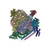
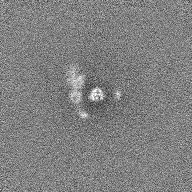
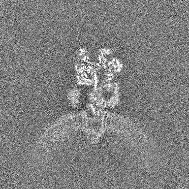
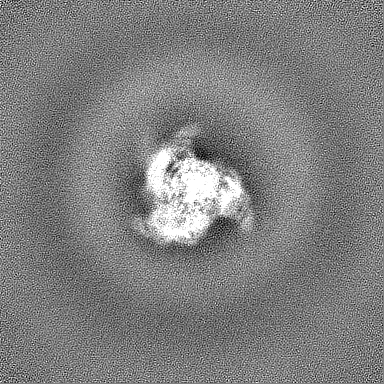
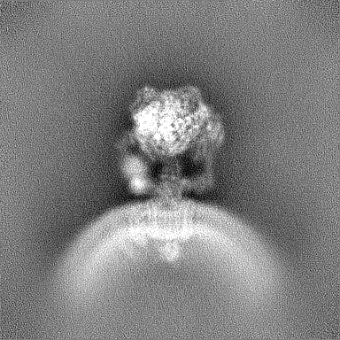
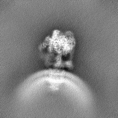
登録情報 データベース : EMDB / ID : EMD-44355タイトル Synaptic Vesicle V-ATPase with synaptophysin and SidK, State 1 複合体 : Synaptic Vesicle V-ATPase with synaptophysin and SidK, State 3 / / / / 機能・相同性 分子機能 ドメイン・相同性 構成要素
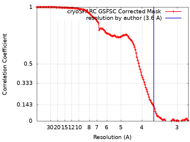
/ / / / / / / / / / / / / / / / / / / / / / / / / / / / / / / / / / / / / / / / / / / / / / / / / / / / / / / / / / / / / / / / / / / / / / / / / / / / / / / / / / / / / / / / / / / / / / / / / / / / / / / / / / / / / / / / / / / / / / / / / / / / / / / / / / / / / / / / / / / / / / / / / / / / / / / / / / / / / / / / / / / / / / / / / / / / / / / / / / / / / / / / / / / / / / / / / / / / / / / / / / / / / / / / / / / / / / / / 生物種 Rattus norvegicus (ドブネズミ)手法 / / 解像度 : 3.6 Å Coupland CE / Rubinstein JL 資金援助 Organization Grant number 国 Canadian Institutes of Health Research (CIHR) PJT166152
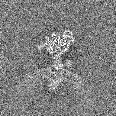
ジャーナル : Science / 年 : 2024タイトル : High-resolution electron cryomicroscopy of V-ATPase in native synaptic vesicles.著者 : Claire E Coupland / Ryan Karimi / Stephanie A Bueler / Yingke Liang / Gautier M Courbon / Justin M Di Trani / Cassandra J Wong / Rayan Saghian / Ji-Young Youn / Lu-Yang Wang / John L Rubinstein / 要旨 : Intercellular communication in the nervous system occurs through the release of neurotransmitters into the synaptic cleft between neurons. In the presynaptic neuron, the proton pumping vesicular- or ... Intercellular communication in the nervous system occurs through the release of neurotransmitters into the synaptic cleft between neurons. In the presynaptic neuron, the proton pumping vesicular- or vacuolar-type ATPase (V-ATPase) powers neurotransmitter loading into synaptic vesicles (SVs), with the V complex dissociating from the membrane region of the enzyme before exocytosis. We isolated SVs from rat brain using SidK, a V-ATPase-binding bacterial effector protein. Single-particle electron cryomicroscopy allowed high-resolution structure determination of V-ATPase within the native SV membrane. In the structure, regularly spaced cholesterol molecules decorate the enzyme's rotor and the abundant SV protein synaptophysin binds the complex stoichiometrically. ATP hydrolysis during vesicle loading results in a loss of the V region of V-ATPase from the SV membrane, suggesting that loading is sufficient to induce dissociation of the enzyme. 履歴 登録 2024年3月31日 - ヘッダ(付随情報) 公開 2024年6月26日 - マップ公開 2024年6月26日 - 更新 2024年7月24日 - 現状 2024年7月24日 処理サイト : RCSB / 状態 : 公開
すべて表示 表示を減らす
 ムービー
ムービー コントローラー


 データを開く
 基本情報
基本情報
 マップデータ
 試料
試料 キーワード
キーワード 機能・相同性情報
 データ登録者
 カナダ, 1件
  引用
引用 構造の表示
構造の表示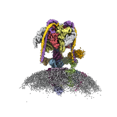
 ダウンロードとリンク
 emd_44355.png
 http://ftp.pdbj.org/pub/emdb/structures/EMD-44355


 Z (Sec.)
 Y (Row.)
 X (Col.)


 試料の構成要素
 解析
解析 電子顕微鏡法
 FIELD EMISSION GUN