 Movie
Movie Controller

[English] 日本語
 Yorodumi
- EMDB-44350: Synaptic Vesicle V-ATPase with synaptophysin and SidK, State 3, Vo -
+ Open data

- Basic information
| Entry |  | |||||||||
|---|---|---|---|---|---|---|---|---|---|---|
| Title | Synaptic Vesicle V-ATPase with synaptophysin and SidK, State 3, Vo | |||||||||
 Map data Map data | ||||||||||
 Sample Sample |
| |||||||||
 Keywords Keywords | Membrane / Synaptic / Complex / PROTON TRANSPORT | |||||||||
| Function / homology |  Function and homology information Function and homology informationvestibular calyx terminal / regulation of opioid receptor signaling pathway / Metabolism of Angiotensinogen to Angiotensins / Ion channel transport / Transferrin endocytosis and recycling / Amino acids regulate mTORC1 / Insulin receptor recycling / RHOA GTPase cycle / negative regulation of autophagic cell death / eye pigmentation ...vestibular calyx terminal / regulation of opioid receptor signaling pathway / Metabolism of Angiotensinogen to Angiotensins / Ion channel transport / Transferrin endocytosis and recycling / Amino acids regulate mTORC1 / Insulin receptor recycling / RHOA GTPase cycle / negative regulation of autophagic cell death / eye pigmentation / central nervous system maturation / positive regulation of transforming growth factor beta1 production / rostrocaudal neural tube patterning / synaptic vesicle lumen acidification / regulation of synaptic vesicle priming / proton-transporting V-type ATPase, V0 domain / cellular response to increased oxygen levels / extrinsic component of synaptic vesicle membrane / vacuolar proton-transporting V-type ATPase, V1 domain / endosome to plasma membrane protein transport / vacuolar proton-transporting V-type ATPase, V0 domain / lysosomal lumen acidification / clathrin-coated vesicle membrane / endosomal lumen acidification / proton-transporting V-type ATPase complex / basal part of cell / head morphogenesis / neuron spine / protein localization to cilium / vacuolar proton-transporting V-type ATPase complex / osteoclast development / vacuolar acidification / regulation of short-term neuronal synaptic plasticity / : / ROS and RNS production in phagocytes / dendritic spine membrane / syntaxin-1 binding / Neutrophil degranulation / presynaptic active zone / cholesterol binding / neuron projection terminus / response to amyloid-beta / homeostatic process / ATPase activator activity / regulation of MAPK cascade / autophagosome membrane / proton-transporting ATPase activity, rotational mechanism / cilium assembly / regulation of macroautophagy / regulation of neuronal synaptic plasticity / transporter activator activity / positive regulation of Wnt signaling pathway / angiotensin maturation / receptor-mediated endocytosis of virus by host cell / RNA endonuclease activity / endoplasmic reticulum-Golgi intermediate compartment membrane / excitatory synapse / SH2 domain binding / receptor-mediated endocytosis / proton transmembrane transport / SNARE binding / neuromuscular junction / protein homooligomerization / regulation of long-term neuronal synaptic plasticity / modulation of chemical synaptic transmission / Schaffer collateral - CA1 synapse / transmembrane transport / small GTPase binding / endocytosis / terminal bouton / synaptic vesicle / melanosome / positive regulation of canonical Wnt signaling pathway / synaptic vesicle membrane / ATPase binding / presynapse / signaling receptor activity / cell body / presynaptic membrane / intracellular iron ion homeostasis / positive regulation of ERK1 and ERK2 cascade / lysosome / endosome membrane / apical plasma membrane / nuclear speck / neuron projection / postsynaptic density / cilium / external side of plasma membrane / protein domain specific binding / axon / lysosomal membrane / ubiquitin protein ligase binding / centrosome / synapse / endoplasmic reticulum membrane / protein-containing complex binding / perinuclear region of cytoplasm / Golgi apparatus / protein-containing complex Similarity search - Function | |||||||||
| Biological species |  | |||||||||
| Method | single particle reconstruction / cryo EM / Resolution: 3.2 Å | |||||||||
 Authors Authors | Coupland CE / Rubinstein JL | |||||||||
| Funding support |  Canada, 1 items Canada, 1 items
| |||||||||
 Citation Citation | Journal: Science / Year: 2024 Title: High-resolution electron cryomicroscopy of V-ATPase in native synaptic vesicles. Authors: Claire E Coupland / Ryan Karimi / Stephanie A Bueler / Yingke Liang / Gautier M Courbon / Justin M Di Trani / Cassandra J Wong / Rayan Saghian / Ji-Young Youn / Lu-Yang Wang / John L Rubinstein / Abstract: Intercellular communication in the nervous system occurs through the release of neurotransmitters into the synaptic cleft between neurons. In the presynaptic neuron, the proton pumping vesicular- or ...Intercellular communication in the nervous system occurs through the release of neurotransmitters into the synaptic cleft between neurons. In the presynaptic neuron, the proton pumping vesicular- or vacuolar-type ATPase (V-ATPase) powers neurotransmitter loading into synaptic vesicles (SVs), with the V complex dissociating from the membrane region of the enzyme before exocytosis. We isolated SVs from rat brain using SidK, a V-ATPase-binding bacterial effector protein. Single-particle electron cryomicroscopy allowed high-resolution structure determination of V-ATPase within the native SV membrane. In the structure, regularly spaced cholesterol molecules decorate the enzyme's rotor and the abundant SV protein synaptophysin binds the complex stoichiometrically. ATP hydrolysis during vesicle loading results in a loss of the V region of V-ATPase from the SV membrane, suggesting that loading is sufficient to induce dissociation of the enzyme. | |||||||||
| History |
|
- Structure visualization
| Supplemental images |
|---|

- Downloads & links
-EMDB archive
| Map data | emd_44350.map.gz | 97.3 MB | EMDB map data format | |
|---|---|---|---|---|
| Header (meta data) | emd-44350-v30.xmlemd-44350.xml | 28.9 KB 28.9 KB | Display Display | EMDB header |
| FSC (resolution estimation) | emd_44350_fsc.xml | 9.9 KB | Display | FSC data file |
| Images |  emd_44350.png emd_44350.png | 46.3 KB | ||
| Filedesc metadata | emd-44350.cif.gz | 8.7 KB | ||
| Others | emd_44350_half_map_1.map.gzemd_44350_half_map_2.map.gz | 95.5 MB 95.5 MB | ||
| Archive directory |  http://ftp.pdbj.org/pub/emdb/structures/EMD-44350ftp://ftp.pdbj.org/pub/emdb/structures/EMD-44350 http://ftp.pdbj.org/pub/emdb/structures/EMD-44350ftp://ftp.pdbj.org/pub/emdb/structures/EMD-44350 | HTTPS FTP |
-Related structure data
| Related structure data |  9b8oMC 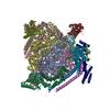 9b8pC  9b8qC  9brbC  9brcC  9brdC C: citing same article ( M: atomic model generated by this map |
|---|---|
| Similar structure data |

-Links
| EMDB pages | EMDB (EBI/PDBe) / EMDataResource |
|---|---|
| Related items in Molecule of the Month |


-Map
| File | Download / File: emd_44350.map.gz / Format: CCP4 / Size: 103 MB / Type: IMAGE STORED AS FLOATING POINT NUMBER (4 BYTES) | ||||||||||||||||||||||||||||||||||||
|---|---|---|---|---|---|---|---|---|---|---|---|---|---|---|---|---|---|---|---|---|---|---|---|---|---|---|---|---|---|---|---|---|---|---|---|---|---|
| Projections & slices | Image control
Images are generated by Spider. | ||||||||||||||||||||||||||||||||||||
| Voxel size | X=Y=Z: 1.03 Å | ||||||||||||||||||||||||||||||||||||
| Density |
| ||||||||||||||||||||||||||||||||||||
| Symmetry | Space group: 1 | ||||||||||||||||||||||||||||||||||||
| Details | EMDB XML:
|
 Z (Sec.)
 Y (Row.)
 X (Col.)


-Supplemental data
-Half map: #2
| File | emd_44350_half_map_1.map | ||||||||||||
|---|---|---|---|---|---|---|---|---|---|---|---|---|---|
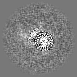
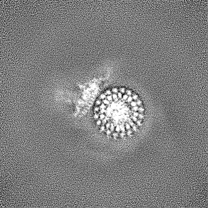
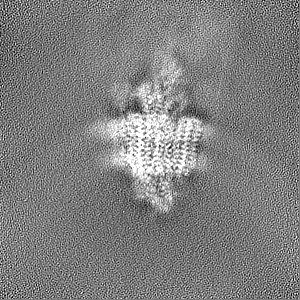
| Projections & Slices |
| ||||||||||||
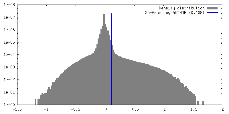
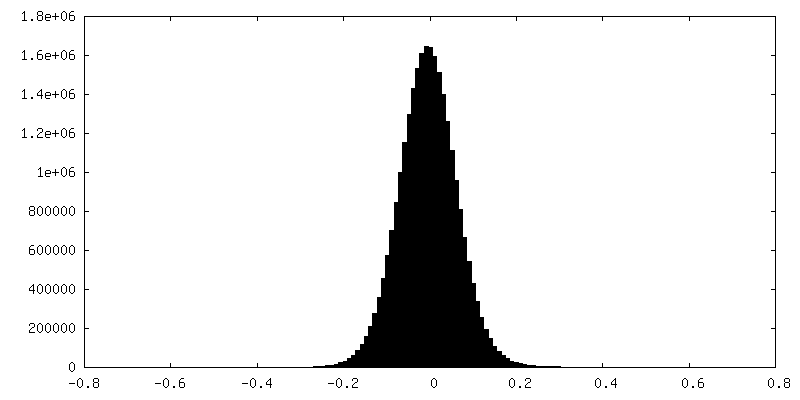
| Density Histograms |


-Half map: #1
| File | emd_44350_half_map_2.map | ||||||||||||
|---|---|---|---|---|---|---|---|---|---|---|---|---|---|
| Projections & Slices |
| ||||||||||||
| Density Histograms |


- Sample components
+Entire : Synaptic vesicle V-ATPase with synaptophysin and SidK, state 3, Vo
+Supramolecule #1: Synaptic vesicle V-ATPase with synaptophysin and SidK, state 3, Vo
+Macromolecule #1: ATPase H+-transporting V1 subunit D
+Macromolecule #2: V-type proton ATPase subunit S1
+Macromolecule #3: Synaptophysin
+Macromolecule #4: V-type proton ATPase 116 kDa subunit a 1
+Macromolecule #5: ATPase, H+ transporting, V0 subunit B (Predicted), isoform CRA_a
+Macromolecule #6: V-type proton ATPase subunit
+Macromolecule #7: V-type proton ATPase subunit e 2
+Macromolecule #8: Rnasek protein
+Macromolecule #9: V-type proton ATPase 16 kDa proteolipid subunit c
+Macromolecule #10: Renin receptor
+Macromolecule #11: V-type proton ATPase subunit F
+Macromolecule #14: 1,2-DIACYL-SN-GLYCERO-3-PHOSPHOCHOLINE

+Macromolecule #15: methyl (3R,6Z,10E,14E)-3,7,11,15,19-pentamethylicosa-6,10,14,18-t...
+Macromolecule #16: 2-acetamido-2-deoxy-beta-D-glucopyranose

+Macromolecule #17: (7R)-4,7-DIHYDROXY-N,N,N-TRIMETHYL-10-OXO-3,5,9-TRIOXA-4-PHOSPHAH...
+Macromolecule #18: PHOSPHATIDYLETHANOLAMINE

+Macromolecule #19: CHOLESTEROL

-Experimental details
-Structure determination
| Method | cryo EM |
|---|---|
 Processing Processing | single particle reconstruction |
| Aggregation state | particle |
-Sample preparation
| Buffer | pH: 7.4 |
|---|---|
| Grid | Model: Homemade / Material: GRAPHENE OXIDE / Support film - Material: GRAPHENE OXIDE / Support film - topology: CONTINUOUS / Pretreatment - Type: GLOW DISCHARGE / Pretreatment - Time: 15 sec. |
| Vitrification | Cryogen name: ETHANE / Chamber humidity: 100 % / Chamber temperature: 277 K / Instrument: FEI VITROBOT MARK IV |
- Electron microscopy
| Microscope | TFS KRIOS |
|---|---|
| Image recording | Film or detector model: TFS FALCON 4i (4k x 4k) / Average electron dose: 37.5 e/Å2 |
| Electron beam | Acceleration voltage: 300 kV / Electron source:  FIELD EMISSION GUN FIELD EMISSION GUN |
| Electron optics | Illumination mode: FLOOD BEAM / Imaging mode: BRIGHT FIELD / Nominal defocus max: 1.9000000000000001 µm / Nominal defocus min: 1.0 µm |
| Sample stage | Specimen holder model: FEI TITAN KRIOS AUTOGRID HOLDER / Cooling holder cryogen: NITROGEN |
| Experimental equipment |  Model: Titan Krios / Image courtesy: FEI Company |

