 Movie
Movie Controller


+ Open data

- Basic information
| Entry |  | |||||||||
|---|---|---|---|---|---|---|---|---|---|---|
| Title | Structure of Apo CXCR4/Gi complex | |||||||||
 Map data Map data | ||||||||||
 Sample Sample |
| |||||||||
 Keywords Keywords | GPCR / chemokine receptor / SIGNALING PROTEIN | |||||||||
| Function / homology |  Function and homology information Function and homology informationC-X-C motif chemokine 12 receptor activity / positive regulation of macrophage migration inhibitory factor signaling pathway / myosin light chain binding / CXCL12-activated CXCR4 signaling pathway / Specification of primordial germ cells / myelin maintenance / Developmental Lineage of Multipotent Pancreatic Progenitor Cells / C-X-C chemokine receptor activity / positive regulation of vasculature development / Signaling by ROBO receptors ...C-X-C motif chemokine 12 receptor activity / positive regulation of macrophage migration inhibitory factor signaling pathway / myosin light chain binding / CXCL12-activated CXCR4 signaling pathway / Specification of primordial germ cells / myelin maintenance / Developmental Lineage of Multipotent Pancreatic Progenitor Cells / C-X-C chemokine receptor activity / positive regulation of vasculature development / Signaling by ROBO receptors / Formation of definitive endoderm / C-C chemokine receptor activity / C-C chemokine binding / anchoring junction / Chemokine receptors bind chemokines / dendritic cell chemotaxis / cellular response to cytokine stimulus / cell leading edge / positive regulation of oligodendrocyte differentiation / Binding and entry of HIV virion / regulation of cell adhesion / coreceptor activity / adenylate cyclase inhibitor activity / positive regulation of protein localization to cell cortex / T cell migration / positive regulation of relaxation of smooth muscle / Adenylate cyclase inhibitory pathway / D2 dopamine receptor binding / adenylate cyclase-inhibiting serotonin receptor signaling pathway / G protein-coupled serotonin receptor binding / neurogenesis / cellular response to forskolin / regulation of mitotic spindle organization / chemokine-mediated signaling pathway / cell chemotaxis / ubiquitin binding / Regulation of insulin secretion / neuropeptide signaling pathway / calcium-mediated signaling / response to prostaglandin E / positive regulation of cholesterol biosynthetic process / brain development / negative regulation of insulin secretion / G protein-coupled receptor binding / response to peptide hormone / G protein-coupled receptor activity / adenylate cyclase-modulating G protein-coupled receptor signaling pathway / G-protein beta/gamma-subunit complex binding / centriolar satellite / response to virus / adenylate cyclase-inhibiting G protein-coupled receptor signaling pathway / Olfactory Signaling Pathway / Activation of the phototransduction cascade / G protein-coupled acetylcholine receptor signaling pathway / G beta:gamma signalling through PLC beta / Presynaptic function of Kainate receptors / Thromboxane signalling through TP receptor / Activation of G protein gated Potassium channels / Inhibition of voltage gated Ca2+ channels via Gbeta/gamma subunits / G-protein activation / Glucagon signaling in metabolic regulation / G beta:gamma signalling through CDC42 / Prostacyclin signalling through prostacyclin receptor / G beta:gamma signalling through BTK / Synthesis, secretion, and inactivation of Glucagon-like Peptide-1 (GLP-1) / photoreceptor disc membrane / ADP signalling through P2Y purinoceptor 12 / Sensory perception of sweet, bitter, and umami (glutamate) taste / GDP binding / Glucagon-type ligand receptors / Adrenaline,noradrenaline inhibits insulin secretion / late endosome / Vasopressin regulates renal water homeostasis via Aquaporins / Glucagon-like Peptide-1 (GLP1) regulates insulin secretion / G alpha (z) signalling events / cellular response to catecholamine stimulus / ADP signalling through P2Y purinoceptor 1 / ADORA2B mediated anti-inflammatory cytokines production / G beta:gamma signalling through PI3Kgamma / adenylate cyclase-activating dopamine receptor signaling pathway / Cooperation of PDCL (PhLP1) and TRiC/CCT in G-protein beta folding / GPER1 signaling / cellular response to prostaglandin E stimulus / heterotrimeric G-protein complex / G alpha (12/13) signalling events / positive regulation of cold-induced thermogenesis / Inactivation, recovery and regulation of the phototransduction cascade / G-protein beta-subunit binding / extracellular vesicle / sensory perception of taste / adenylate cyclase-activating G protein-coupled receptor signaling pathway / sperm principal piece / Thrombin signalling through proteinase activated receptors (PARs) / signaling receptor complex adaptor activity / virus receptor activity / positive regulation of cytosolic calcium ion concentration / retina development in camera-type eye / actin binding / GTPase binding / G protein activity Similarity search - Function | |||||||||
| Biological species |  Homo sapiens (human) Homo sapiens (human) | |||||||||
| Method | single particle reconstruction / cryo EM / Resolution: 2.72 Å | |||||||||
 Authors Authors | Saotome K / McGoldrick LL / Franklin MC | |||||||||
| Funding support |  United States, 1 items United States, 1 items
| |||||||||
 Citation Citation | Journal: Nat Struct Mol Biol / Year: 2025 Title: Structural insights into CXCR4 modulation and oligomerization. Authors: Kei Saotome / Luke L McGoldrick / Jo-Hao Ho / Trudy F Ramlall / Sweta Shah / Michael J Moore / Jee Hae Kim / Raymond Leidich / William C Olson / Matthew C Franklin / Abstract: Activation of the chemokine receptor CXCR4 by its chemokine ligand CXCL12 regulates diverse cellular processes. Previously reported crystal structures of CXCR4 revealed the architecture of an ...Activation of the chemokine receptor CXCR4 by its chemokine ligand CXCL12 regulates diverse cellular processes. Previously reported crystal structures of CXCR4 revealed the architecture of an inactive, homodimeric receptor. However, many structural aspects of CXCR4 remain poorly understood. Here, we use cryo-electron microscopy to investigate various modes of human CXCR4 regulation. CXCL12 activates CXCR4 by inserting its N terminus deep into the CXCR4 orthosteric pocket. The binding of US Food and Drug Administration-approved antagonist AMD3100 is stabilized by electrostatic interactions with acidic residues in the seven-transmembrane-helix bundle. A potent antibody blocker, REGN7663, binds across the extracellular face of CXCR4 and inserts its complementarity-determining region H3 loop into the orthosteric pocket. Trimeric and tetrameric structures of CXCR4 reveal modes of G-protein-coupled receptor oligomerization. We show that CXCR4 adopts distinct subunit conformations in trimeric and tetrameric assemblies, highlighting how oligomerization could allosterically regulate chemokine receptor function. #1: Journal: Biorxiv / Year: 2024Title: Structural insights into CXCR4 modulation and oligomerization Authors: Saotome K / McGoldrick LL / Ho J / Ramlall T / Shah S / Moore MJ / Kim JH / Leidich R / Olson WC / Franklin MC | |||||||||
| History |
|
- Structure visualization
| Supplemental images |
|---|

- Downloads & links
-EMDB archive
| Map data | emd_41888.map.gz | 97.2 MB | EMDB map data format | |
|---|---|---|---|---|
| Header (meta data) | emd-41888-v30.xmlemd-41888.xml | 23.5 KB 23.5 KB | Display Display | EMDB header |
| Images | 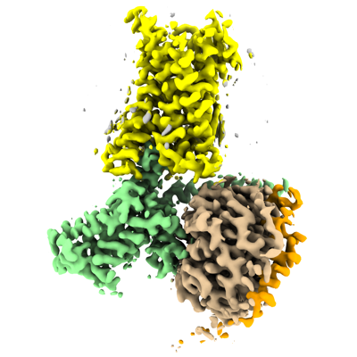 emd_41888.png emd_41888.png | 113.5 KB | ||
| Filedesc metadata | emd-41888.cif.gz | 7.1 KB | ||
| Others | emd_41888_half_map_1.map.gzemd_41888_half_map_2.map.gz | 95.6 MB 95.6 MB | ||
| Archive directory |  http://ftp.pdbj.org/pub/emdb/structures/EMD-41888ftp://ftp.pdbj.org/pub/emdb/structures/EMD-41888 http://ftp.pdbj.org/pub/emdb/structures/EMD-41888ftp://ftp.pdbj.org/pub/emdb/structures/EMD-41888 | HTTPS FTP |
-Related structure data
| Related structure data |  8u4nMC  8u4oC  8u4pC  8u4qC  8u4rC  8u4sC  8u4tC C: citing same article ( M: atomic model generated by this map |
|---|---|
| Similar structure data |


-Links
| EMDB pages | EMDB (EBI/PDBe) / EMDataResource |
|---|---|
| Related items in Molecule of the Month |


-Map
| File | Download / File: emd_41888.map.gz / Format: CCP4 / Size: 103 MB / Type: IMAGE STORED AS FLOATING POINT NUMBER (4 BYTES) | ||||||||||||||||||||||||||||||||||||
|---|---|---|---|---|---|---|---|---|---|---|---|---|---|---|---|---|---|---|---|---|---|---|---|---|---|---|---|---|---|---|---|---|---|---|---|---|---|
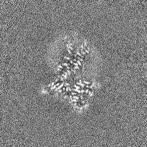
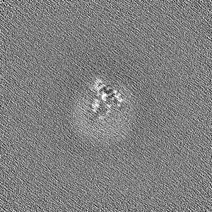
| Projections & slices | Image control
Images are generated by Spider. | ||||||||||||||||||||||||||||||||||||
| Voxel size | X=Y=Z: 0.696 Å | ||||||||||||||||||||||||||||||||||||
| Density |
| ||||||||||||||||||||||||||||||||||||
| Symmetry | Space group: 1 | ||||||||||||||||||||||||||||||||||||
| Details | EMDB XML:
|
 Z (Sec.)
 Y (Row.)
 X (Col.)


-Supplemental data
-Half map: #1
| File | emd_41888_half_map_1.map | ||||||||||||
|---|---|---|---|---|---|---|---|---|---|---|---|---|---|
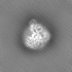
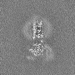
| Projections & Slices |
| ||||||||||||
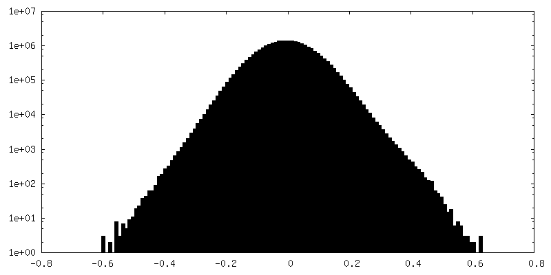
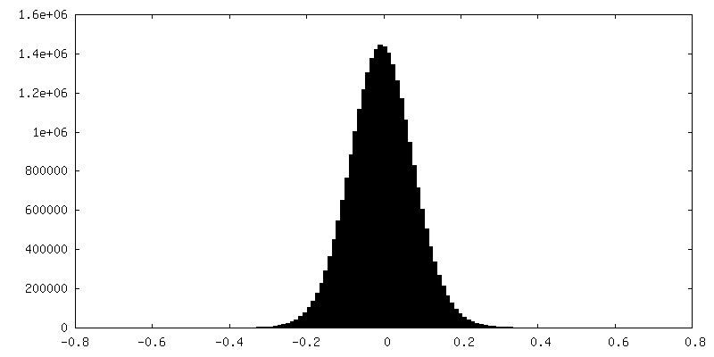
| Density Histograms |


-Half map: #2
| File | emd_41888_half_map_2.map | ||||||||||||
|---|---|---|---|---|---|---|---|---|---|---|---|---|---|
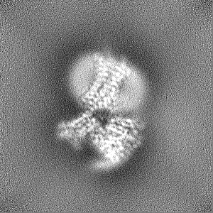
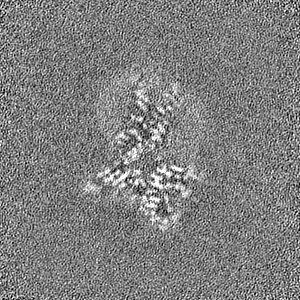
| Projections & Slices |
| ||||||||||||
| Density Histograms |

- Sample components
-Entire : Apo CXCR4/Gi complex
| Entire | Name: Apo CXCR4/Gi complex |
|---|---|
| Components |
|
-Supramolecule #1: Apo CXCR4/Gi complex
| Supramolecule | Name: Apo CXCR4/Gi complex / type: complex / ID: 1 / Parent: 0 / Macromolecule list: #1-#4 |
|---|---|
| Source (natural) | Organism: Homo sapiens (human) |
-Macromolecule #1: Guanine nucleotide-binding protein G(i) subunit alpha-1
| Macromolecule | Name: Guanine nucleotide-binding protein G(i) subunit alpha-1 type: protein_or_peptide / ID: 1 / Number of copies: 1 / Enantiomer: LEVO |
|---|---|
| Source (natural) | Organism: Homo sapiens (human) |
| Molecular weight | Theoretical: 41.591312 KDa |
| Recombinant expression | Organism:   Spodoptera frugiperda (fall armyworm) Spodoptera frugiperda (fall armyworm) |
| Sequence | String: MHHHHHHGGG GSGCTLSAED KAAVERSKMI DRNLREDGEK AAREVKLLLL GAGESGKCTI VKQMKIIHEA GYSEEECKQY KAVVYSNTI QSIIAIIRAM GRLKIDFGDS ARADDARQLF VLAGAAEEGF MTAELAGVIK RLWKDSGVQA CFNRSREYQL N DSAAYYLN ...String: MHHHHHHGGG GSGCTLSAED KAAVERSKMI DRNLREDGEK AAREVKLLLL GAGESGKCTI VKQMKIIHEA GYSEEECKQY KAVVYSNTI QSIIAIIRAM GRLKIDFGDS ARADDARQLF VLAGAAEEGF MTAELAGVIK RLWKDSGVQA CFNRSREYQL N DSAAYYLN DLDRIAQPNY IPTQQDVLRT RVKTTGIVET HFTFKDLHFK MFDVTAQRSE RKKWIHCFEG VTAIIFCVAL SD YDLVLAE DEEMNRMHAS MKLFDSICNN KWFTDTSIIL FLNKKDLFEE KIKKSPLTIC YPEYAGSNTY EEAAAYIQCQ FED LNKRKD TKEIYTHFTC STDTKNVQFV FDAVTDVIIK NNLKDCGLF UniProtKB: Guanine nucleotide-binding protein G(i) subunit alpha-1 |
-Macromolecule #2: Guanine nucleotide-binding protein G(I)/G(S)/G(T) subunit beta-1
| Macromolecule | Name: Guanine nucleotide-binding protein G(I)/G(S)/G(T) subunit beta-1 type: protein_or_peptide / ID: 2 / Number of copies: 1 / Enantiomer: LEVO |
|---|---|
| Source (natural) | Organism: Homo sapiens (human) |
| Molecular weight | Theoretical: 38.534062 KDa |
| Recombinant expression | Organism: Spodoptera frugiperda (fall armyworm) |
| Sequence | String: MHHHHHHGSS GSELDQLRQE AEQLKNQIRD ARKACADATL SQITNNIDPV GRIQMRTRRT LRGHLAKIYA MHWGTDSRLL VSASQDGKL IIWDSYTTNK VHAIPLRSSW VMTCAYAPSG NYVACGGLDN ICSIYNLKTR EGNVRVSREL AGHTGYLSCC R FLDDNQIV ...String: MHHHHHHGSS GSELDQLRQE AEQLKNQIRD ARKACADATL SQITNNIDPV GRIQMRTRRT LRGHLAKIYA MHWGTDSRLL VSASQDGKL IIWDSYTTNK VHAIPLRSSW VMTCAYAPSG NYVACGGLDN ICSIYNLKTR EGNVRVSREL AGHTGYLSCC R FLDDNQIV TSSGDTTCAL WDIETGQQTT TFTGHTGDVM SLSLAPDTRL FVSGACDASA KLWDVREGMC RQTFTGHESD IN AICFFPN GNAFATGSDD ATCRLFDLRA DQELMTYSHD NIICGITSVS FSKSGRLLLA GYDDFNCNVW DALKADRAGV LAG HDNRVS CLGVTDDGMA VATGSWDSFL KIWN UniProtKB: Guanine nucleotide-binding protein G(I)/G(S)/G(T) subunit beta-1 |
-Macromolecule #3: Guanine nucleotide-binding protein G(I)/G(S)/G(O) subunit gamma-2
| Macromolecule | Name: Guanine nucleotide-binding protein G(I)/G(S)/G(O) subunit gamma-2 type: protein_or_peptide / ID: 3 / Number of copies: 1 / Enantiomer: LEVO |
|---|---|
| Source (natural) | Organism: Homo sapiens (human) |
| Molecular weight | Theoretical: 7.861143 KDa |
| Recombinant expression | Organism: Spodoptera frugiperda (fall armyworm) |
| Sequence | String: MASNNTASIA QARKLVEQLK MEANIDRIKV SKAAADLMAY CEAHAKEDPL LTPVPASENP FREKKFFCAI L UniProtKB: Guanine nucleotide-binding protein G(I)/G(S)/G(O) subunit gamma-2 |
-Macromolecule #4: C-X-C chemokine receptor type 4
| Macromolecule | Name: C-X-C chemokine receptor type 4 / type: protein_or_peptide / ID: 4 / Number of copies: 1 / Enantiomer: LEVO |
|---|---|
| Source (natural) | Organism: Homo sapiens (human) |
| Molecular weight | Theoretical: 71.063609 KDa |
| Recombinant expression | Organism: Spodoptera frugiperda (fall armyworm) |
| Sequence | String: MKTIIALSYI FCLVFAGAPE GISIYTSDNY TEEMGSGDYD SMKEPCFREE NANFNKIFLP TIYSIIFLTG IVGNGLVILV MGYQKKLRS MTDKYRLHLS VADLLFVITL PFWAVDAVAN WYFGNFLCKA VHVIYTVSLY SSVLILAFIS LDRYLAIVHA T NSQRPRKL ...String: MKTIIALSYI FCLVFAGAPE GISIYTSDNY TEEMGSGDYD SMKEPCFREE NANFNKIFLP TIYSIIFLTG IVGNGLVILV MGYQKKLRS MTDKYRLHLS VADLLFVITL PFWAVDAVAN WYFGNFLCKA VHVIYTVSLY SSVLILAFIS LDRYLAIVHA T NSQRPRKL LAEKVVYVGV WIPALLLTIP DFIFANVSEA DDRYICDRFY PNDLWVVVFQ FQHIMVGLIL PGIVILSCYC II ISKLSHS KGHQKRKALK TTVILILAFF ACWLPYYIGI SIDSFILLEI IKQGCEFENT VHKWISITEA LAFFHCCLNP ILY AFLGAK FKTSAQHALT SVSRGSSLKI LSKGKRGGHS SVSTESESSS FHSSGRPLEV LFQGPGGGGS VSKGEELFTG VVPI LVELD GDVNGHKFSV SGEGEGDATY GKLTLKFICT TGKLPVPWPT LVTTLTYGVQ CFSRYPDHMK QHDFFKSAMP EGYVQ ERTI FFKDDGNYKT RAEVKFEGDT LVNRIELKGI DFKEDGNILG HKLEYNYNSH NVYIMADKQK NGIKVNFKIR HNIEDG SVQ LADHYQQNTP IGDGPVLLPD NHYLSTQSKL SKDPNEKRDH MVLLEFVTAA GITLGMDELY KDYKDDDDK UniProtKB: C-X-C chemokine receptor type 4 |
-Macromolecule #5: CHOLESTEROL
| Macromolecule | Name: CHOLESTEROL / type: ligand / ID: 5 / Number of copies: 1 / Formula: CLR |
|---|---|
| Molecular weight | Theoretical: 386.654 Da |
| Chemical component information |  ChemComp-CLR: |
-Experimental details
-Structure determination
| Method | cryo EM |
|---|---|
 Processing Processing | single particle reconstruction |
| Aggregation state | particle |
-Sample preparation
| Buffer | pH: 7.5 |
|---|---|
| Vitrification | Cryogen name: ETHANE |
- Electron microscopy
| Microscope | TFS GLACIOS |
|---|---|
| Image recording | Film or detector model: TFS FALCON 4i (4k x 4k) / Average electron dose: 50.0 e/Å2 |
| Electron beam | Acceleration voltage: 200 kV / Electron source:  FIELD EMISSION GUN FIELD EMISSION GUN |
| Electron optics | Illumination mode: FLOOD BEAM / Imaging mode: BRIGHT FIELD / Nominal defocus max: 2.0 µm / Nominal defocus min: 1.0 µm |
-Image processing
| Startup model | Type of model: NONE |
|---|---|
| Final reconstruction | Resolution.type: BY AUTHOR / Resolution: 2.72 Å / Resolution method: FSC 0.143 CUT-OFF / Number images used: 183399 |
| Initial angle assignment | Type: MAXIMUM LIKELIHOOD |
| Final angle assignment | Type: MAXIMUM LIKELIHOOD |
