Journal: Nat Commun / Year: 2016 Title: Structure of the ribosome post-recycling complex probed by chemical cross-linking and mass spectrometry. Authors: Kristin Kiosze-Becker / Alessandro Ori / Milan Gerovac / André Heuer / Elina Nürenberg-Goloub / Umar Jan Rashid / Thomas Becker / Roland Beckmann / Martin Beck / Robert Tampé / Abstract: Ribosome recycling orchestrated by the ATP binding cassette (ABC) protein ABCE1 can be considered as the final-or the first-step within the cyclic process of protein synthesis, connecting translation ...Ribosome recycling orchestrated by the ATP binding cassette (ABC) protein ABCE1 can be considered as the final-or the first-step within the cyclic process of protein synthesis, connecting translation termination and mRNA surveillance with re-initiation. An ATP-dependent tweezer-like motion of the nucleotide-binding domains in ABCE1 transfers mechanical energy to the ribosome and tears the ribosome subunits apart. The post-recycling complex (PRC) then re-initiates mRNA translation. Here, we probed the so far unknown architecture of the 1-MDa PRC (40S/30S·ABCE1) by chemical cross-linking and mass spectrometry (XL-MS). Our study reveals ABCE1 bound to the translational factor-binding (GTPase) site with multiple cross-link contacts of the helix-loop-helix motif to the S24e ribosomal protein. Cross-linking of the FeS cluster domain to the ribosomal protein S12 substantiates an extreme lever-arm movement of the FeS cluster domain during ribosome recycling. We were thus able to reconstitute and structurally analyse a key complex in the translational cycle, resembling the link between translation initiation and ribosome recycling.
History
Deposition
Sep 15, 2016
-
Header (metadata) release
Oct 19, 2016
-
Map release
Nov 23, 2016
-
Update
Jul 26, 2017
-
Current status
Jul 26, 2017
Processing site: PDBe / Status: Released
-
Structure visualization
Movie
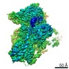
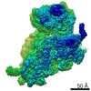
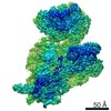
Surface view with section colored by density value
Model: Quantifoil R3/3 / Material: COPPER/PALLADIUM / Support film - Material: CARBON / Support film - topology: CONTINUOUS / Support film - Film thickness: 2.0 nm / Pretreatment - Type: GLOW DISCHARGE
Vitrification
Cryogen name: ETHANE / Chamber humidity: 100 % / Chamber temperature: 278 K / Instrument: FEI VITROBOT MARK IV
Details
Purified 30S ribosomal subunits were reconstituted with purified, recombinantly expressed ABCE1
-
Electron microscopy
Microscope
FEI TECNAI SPIRIT
Image recording
Film or detector model: TVIPS TEMCAM-F816 (8k x 8k) / Average electron dose: 20.0 e/Å2
Electron beam
Acceleration voltage: 120 kV / Electron source: OTHER
In the structure databanks used in Yorodumi, some data are registered as the other names, "COVID-19 virus" and "2019-nCoV". Here are the details of the virus and the list of structure data.
Jan 31, 2019. EMDB accession codes are about to change! (news from PDBe EMDB page)
EMDB accession codes are about to change! (news from PDBe EMDB page)
The allocation of 4 digits for EMDB accession codes will soon come to an end. Whilst these codes will remain in use, new EMDB accession codes will include an additional digit and will expand incrementally as the available range of codes is exhausted. The current 4-digit format prefixed with “EMD-” (i.e. EMD-XXXX) will advance to a 5-digit format (i.e. EMD-XXXXX), and so on. It is currently estimated that the 4-digit codes will be depleted around Spring 2019, at which point the 5-digit format will come into force.
The EM Navigator/Yorodumi systems omit the EMD- prefix.
Related info.:Q: What is EMD? / ID/Accession-code notation in Yorodumi/EM Navigator
Yorodumi is a browser for structure data from EMDB, PDB, SASBDB, etc.
This page is also the successor to EM Navigator detail page, and also detail information page/front-end page for Omokage search.
The word "yorodu" (or yorozu) is an old Japanese word meaning "ten thousand". "mi" (miru) is to see.
Related info.:EMDB / PDB / SASBDB / Comparison of 3 databanks / Yorodumi Search / Aug 31, 2016. New EM Navigator & Yorodumi / Yorodumi Papers / Jmol/JSmol / Function and homology information / Changes in new EM Navigator and Yorodumi
 Movie
Movie Controller


 Open data
 Basic information
 Map data
 Sample
 Function and homology information
 Sulfolobus solfataricus (archaea)
 Authors
 Citation
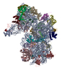
 Structure visualization
 UCSF Chimera
 Downloads & links
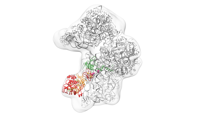 emd_4113.png
 http://ftp.pdbj.org/pub/emdb/structures/EMD-4113


 Z (Sec.)
 Y (Row.)
 X (Col.)


 Sample components
 Processing
 Electron microscopy
