 Movie
Movie Controller

[English] 日本語
 Yorodumi
- PDB-6tmf: Structure of an archaeal ABCE1-bound ribosomal post-splitting complex -
+ Open data

- Basic information
| Entry | Database: PDB / ID: 6tmf | ||||||
|---|---|---|---|---|---|---|---|
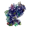
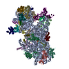
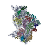
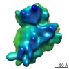
| Title | Structure of an archaeal ABCE1-bound ribosomal post-splitting complex | ||||||
 Components Components |
| ||||||
 Keywords Keywords | RIBOSOME / ABC Proteins / Ribosome Recycling / Translation | ||||||
| Function / homology |  Function and homology information Function and homology informationribonuclease P activity / tRNA 5'-leader removal / ribosomal small subunit binding / translational termination / maturation of SSU-rRNA from tricistronic rRNA transcript (SSU-rRNA, 5.8S rRNA, LSU-rRNA) / maturation of SSU-rRNA / translational initiation / ribosomal small subunit assembly / 4 iron, 4 sulfur cluster binding / ribosome biogenesis ...ribonuclease P activity / tRNA 5'-leader removal / ribosomal small subunit binding / translational termination / maturation of SSU-rRNA from tricistronic rRNA transcript (SSU-rRNA, 5.8S rRNA, LSU-rRNA) / maturation of SSU-rRNA / translational initiation / ribosomal small subunit assembly / 4 iron, 4 sulfur cluster binding / ribosome biogenesis / ribosomal small subunit biogenesis / small ribosomal subunit / small ribosomal subunit rRNA binding / cytosolic small ribosomal subunit / cytoplasmic translation / tRNA binding / oxidoreductase activity / rRNA binding / structural constituent of ribosome / ribosome / translation / iron ion binding / ribonucleoprotein complex / ATP hydrolysis activity / RNA binding / zinc ion binding / ATP binding / metal ion binding / cytoplasm / cytosol Similarity search - Function | ||||||
| Biological species |   Saccharolobus solfataricus (archaea)Thermococcus celer Vu 13 = JCM 8558 (archaea) Saccharolobus solfataricus (archaea)Thermococcus celer Vu 13 = JCM 8558 (archaea) | ||||||
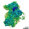
| Method | ELECTRON MICROSCOPY / single particle reconstruction / cryo EM / Resolution: 2.8 Å | ||||||
 Authors Authors | Kratzat, H. / Becker, T. / Tampe, R. / Beckmann, R. | ||||||
| Funding support |  Germany, 1items Germany, 1items
| ||||||
 Citation Citation | Journal: EMBO J / Year: 2020 Title: Molecular analysis of the ribosome recycling factor ABCE1 bound to the 30S post-splitting complex. Authors: Elina Nürenberg-Goloub / Hanna Kratzat / Holger Heinemann / André Heuer / Peter Kötter / Otto Berninghausen / Thomas Becker / Robert Tampé / Roland Beckmann / Abstract: Ribosome recycling by the twin-ATPase ABCE1 is a key regulatory process in mRNA translation and surveillance and in ribosome-associated protein quality control in Eukarya and Archaea. Here, we ...Ribosome recycling by the twin-ATPase ABCE1 is a key regulatory process in mRNA translation and surveillance and in ribosome-associated protein quality control in Eukarya and Archaea. Here, we captured the archaeal 30S ribosome post-splitting complex at 2.8 Å resolution by cryo-electron microscopy. The structure reveals the dynamic behavior of structural motifs unique to ABCE1, which ultimately leads to ribosome splitting. More specifically, we provide molecular details on how conformational rearrangements of the iron-sulfur cluster domain and hinge regions of ABCE1 are linked to closure of its nucleotide-binding sites. The combination of mutational and functional analyses uncovers an intricate allosteric network between the ribosome, regulatory domains of ABCE1, and its two structurally and functionally asymmetric ATP-binding sites. Based on these data, we propose a refined model of how signals from the ribosome are integrated into the ATPase cycle of ABCE1 to orchestrate ribosome recycling. | ||||||
| History |
|
- Structure visualization
| Movie |
Movie viewer |
|---|---|
| Structure viewer | Molecule: MolmilJmol/JSmol |
- Downloads & links
-Download
| PDBx/mmCIF format | 6tmf.cif.gz | 1.4 MB | Display | PDBx/mmCIF format |
|---|---|---|---|---|
| PDB format | pdb6tmf.ent.gz | 1.1 MB | Display | PDB format |
| PDBx/mmJSON format | 6tmf.json.gz | Tree view | PDBx/mmJSON format | |
| Others |  Other downloads Other downloads |
-Validation report
| Arichive directory | https://data.pdbj.org/pub/pdb/validation_reports/tm/6tmfftp://data.pdbj.org/pub/pdb/validation_reports/tm/6tmf | HTTPS FTP |
|---|
-Related structure data
| Related structure data |  10519MC M: map data used to model this data C: citing same article ( |
|---|---|
| Similar structure data |


-Links
 PDBj
PDBj


- Assembly
| Deposited unit | 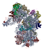
|
|---|---|
| 1 |
|
-Components
+30S ribosomal protein ... , 25 types, 25 molecules BCDEFGHIJKLMNOPQRSTUVXYZa
-Protein , 3 types, 3 molecules Wbd
| #23: Protein | Mass: 6292.359 Da / Num. of mol.: 1 / Source method: isolated from a natural source Source: (natural) Thermococcus celer Vu 13 = JCM 8558 (archaea)References: UniProt: A0A218P055 |
|---|---|
| #28: Protein | Mass: 13351.441 Da / Num. of mol.: 1 / Source method: isolated from a natural source Source: (natural) Thermococcus celer Vu 13 = JCM 8558 (archaea)References: UniProt: A0A218P167 |
| #30: Protein | Mass: 67201.469 Da / Num. of mol.: 1 / Mutation: E238A, E485A Source method: isolated from a genetically manipulated source Source: (gene. exp.) Saccharolobus solfataricus (archaea)Gene: SSOP1_0270, SULA_1305, SULB_1306, SULC_1304, SULG_06465, SULH_06465, SULI_06465, SULM_06465, SULN_06465, SULO_06475, SULZ_06710 Production host:  |
-RNA chain / Protein/peptide , 2 types, 2 molecules Ac
| #1: RNA chain | Mass: 482205.531 Da / Num. of mol.: 1 / Source method: isolated from a natural source Source: (natural) Thermococcus celer Vu 13 = JCM 8558 (archaea)References: GenBank: 1214542111 |
|---|---|
| #29: Protein/peptide | Mass: 5023.395 Da / Num. of mol.: 1 / Source method: isolated from a natural source Source: (natural) Thermococcus celer Vu 13 = JCM 8558 (archaea)References: UniProt: A0A218P2S9 |
-Non-polymers , 4 types, 37 molecules 


| #31: Chemical | ChemComp-MG /  Mass: 24.305 Da / Num. of mol.: 29 / Source method: obtained synthetically / Formula: Mg Mass: 24.305 Da / Num. of mol.: 29 / Source method: obtained synthetically / Formula: Mg#32: Chemical | ChemComp-ZN /  Mass: 65.409 Da / Num. of mol.: 4 / Source method: obtained synthetically / Formula: Zn Mass: 65.409 Da / Num. of mol.: 4 / Source method: obtained synthetically / Formula: Zn#33: Chemical |  Mass: 351.640 Da / Num. of mol.: 2 / Source method: obtained synthetically / Formula: Fe4S4 Mass: 351.640 Da / Num. of mol.: 2 / Source method: obtained synthetically / Formula: Fe4S4#34: Chemical |  Mass: 506.196 Da / Num. of mol.: 2 / Source method: obtained synthetically / Formula: C10H17N6O12P3 / Feature type: SUBJECT OF INVESTIGATION / Comment: AMP-PNP, energy-carrying molecule analogue*YM Mass: 506.196 Da / Num. of mol.: 2 / Source method: obtained synthetically / Formula: C10H17N6O12P3 / Feature type: SUBJECT OF INVESTIGATION / Comment: AMP-PNP, energy-carrying molecule analogue*YM |
|---|
-Details
| Has ligand of interest | Y |
|---|
-Experimental details
-Experiment
| Experiment | Method: ELECTRON MICROSCOPY |
|---|---|
| EM experiment | Aggregation state: PARTICLE / 3D reconstruction method: single particle reconstruction |
- Sample preparation
| Component |
| ||||||||||||||||||||||||
|---|---|---|---|---|---|---|---|---|---|---|---|---|---|---|---|---|---|---|---|---|---|---|---|---|---|
| Source (natural) |
| ||||||||||||||||||||||||
| Source (recombinant) | Organism: | ||||||||||||||||||||||||
| Buffer solution | pH: 7.5 | ||||||||||||||||||||||||
| Specimen | Embedding applied: NO / Shadowing applied: NO / Staining applied: NO / Vitrification applied: YES | ||||||||||||||||||||||||
| Vitrification | Cryogen name: ETHANE |
- Electron microscopy imaging
| Experimental equipment |  Model: Titan Krios / Image courtesy: FEI Company |
|---|---|
| Microscopy | Model: FEI TITAN KRIOS |
| Electron gun | Electron source:  FIELD EMISSION GUN / Accelerating voltage: 300 kV / Illumination mode: FLOOD BEAM FIELD EMISSION GUN / Accelerating voltage: 300 kV / Illumination mode: FLOOD BEAM |
| Electron lens | Mode: BRIGHT FIELD |
| Image recording | Electron dose: 25 e/Å2 / Film or detector model: FEI FALCON II (4k x 4k) |
| Image scans | Movie frames/image: 10 |
- Processing
| EM software | Name: RELION / Version: 3 / Category: 3D reconstruction | ||||||||||||||||||||||||
|---|---|---|---|---|---|---|---|---|---|---|---|---|---|---|---|---|---|---|---|---|---|---|---|---|---|
| CTF correction | Type: PHASE FLIPPING AND AMPLITUDE CORRECTION | ||||||||||||||||||||||||
| Symmetry | Point symmetry: C1 (asymmetric) | ||||||||||||||||||||||||
| 3D reconstruction | Resolution: 2.8 Å / Resolution method: FSC 0.143 CUT-OFF / Num. of particles: 293010 / Symmetry type: POINT | ||||||||||||||||||||||||
| Refinement | Stereochemistry target values: GeoStd + Monomer Library + CDL v1.2 | ||||||||||||||||||||||||
| Displacement parameters | Biso mean: 21.65 Å2 | ||||||||||||||||||||||||
| Refine LS restraints |
|
