 Movie
Movie Controller


+ Open data

- Basic information
| Entry | 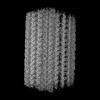 | |||||||||
|---|---|---|---|---|---|---|---|---|---|---|
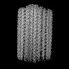
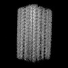
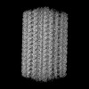
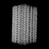
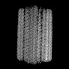
| Title | Local map of the mouse sperm doublet microtubule (Middle03) | |||||||||
 Map data Map data | Local map of the mouse sperm doublet microtubule (Middle03) | |||||||||
 Sample Sample |
| |||||||||
 Keywords Keywords | doublet microtubule / STRUCTURAL PROTEIN | |||||||||
| Biological species |  | |||||||||
| Method | single particle reconstruction / cryo EM / Resolution: 3.35 Å | |||||||||
 Authors Authors | Zhou LN / Gui M / Wu JP | |||||||||
| Funding support |  China, 1 items China, 1 items
| |||||||||
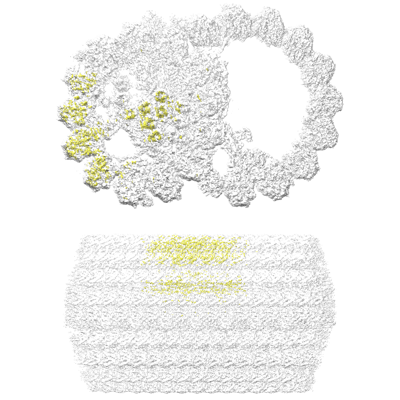
 Citation Citation | Journal: Cell / Year: 2023 Title: Structures of sperm flagellar doublet microtubules expand the genetic spectrum of male infertility. Authors: Lunni Zhou / Haobin Liu / Siyu Liu / Xiaoyu Yang / Yue Dong / Yun Pan / Zhuang Xiao / Beihong Zheng / Yan Sun / Pengyu Huang / Xixi Zhang / Jin Hu / Rui Sun / Shan Feng / Yi Zhu / Mingxi Liu ...Authors: Lunni Zhou / Haobin Liu / Siyu Liu / Xiaoyu Yang / Yue Dong / Yun Pan / Zhuang Xiao / Beihong Zheng / Yan Sun / Pengyu Huang / Xixi Zhang / Jin Hu / Rui Sun / Shan Feng / Yi Zhu / Mingxi Liu / Miao Gui / Jianping Wu / Abstract: Sperm motility is crucial for successful fertilization. Highly decorated doublet microtubules (DMTs) form the sperm tail skeleton, which propels the movement of spermatozoa. Using cryo-electron ...Sperm motility is crucial for successful fertilization. Highly decorated doublet microtubules (DMTs) form the sperm tail skeleton, which propels the movement of spermatozoa. Using cryo-electron microscopy (cryo-EM) and artificial intelligence (AI)-based modeling, we determined the structures of mouse and human sperm DMTs and built an atomic model of the 48-nm repeat of the mouse sperm DMT. Our analysis revealed 47 DMT-associated proteins, including 45 microtubule inner proteins (MIPs). We identified 10 sperm-specific MIPs, including seven classes of Tektin5 in the lumen of the A tubule and FAM166 family members that bind the intra-tubulin interfaces. Interestingly, the human sperm DMT lacks some MIPs compared with the mouse sperm DMT. We also discovered variants in 10 distinct MIPs associated with a subtype of asthenozoospermia characterized by impaired sperm motility without evident morphological abnormalities. Our study highlights the conservation and tissue/species specificity of DMTs and expands the genetic spectrum of male infertility. | |||||||||
| History |
|
- Structure visualization
| Supplemental images |
|---|
- Downloads & links
-EMDB archive
| Map data | emd_36504.map.gz | 1.8 GB |  EMDB map data format EMDB map data format | |
|---|---|---|---|---|
| Header (meta data) | emd-36504-v30.xmlemd-36504.xml | 19.3 KB 19.3 KB | Display Display | EMDB header |
| Images |  emd_36504.png emd_36504.png | 131.9 KB | ||
| Others | emd_36504_half_map_1.map.gzemd_36504_half_map_2.map.gz | 1.8 GB 1.8 GB | ||
| Archive directory |  http://ftp.pdbj.org/pub/emdb/structures/EMD-36504ftp://ftp.pdbj.org/pub/emdb/structures/EMD-36504 http://ftp.pdbj.org/pub/emdb/structures/EMD-36504ftp://ftp.pdbj.org/pub/emdb/structures/EMD-36504 | HTTPS FTP |
-Validation report
| Summary document | emd_36504_validation.pdf.gz | 1.3 MB | Display | EMDB validaton report |
|---|---|---|---|---|
| Full document | emd_36504_full_validation.pdf.gz | 1.2 MB | Display | |
| Data in XML | emd_36504_validation.xml.gz | 26.1 KB | Display | |
| Data in CIF | emd_36504_validation.cif.gz | 30.8 KB | Display | |
| Arichive directory | https://ftp.pdbj.org/pub/emdb/validation_reports/EMD-36504ftp://ftp.pdbj.org/pub/emdb/validation_reports/EMD-36504 | HTTPS FTP |
-Related structure data


-Links
| EMDB pages | EMDB (EBI/PDBe) / EMDataResource |
|---|
-Map
| File | Download / File: emd_36504.map.gz / Format: CCP4 / Size: 1.9 GB / Type: IMAGE STORED AS FLOATING POINT NUMBER (4 BYTES) | ||||||||||||||||||||||||||||||||||||
|---|---|---|---|---|---|---|---|---|---|---|---|---|---|---|---|---|---|---|---|---|---|---|---|---|---|---|---|---|---|---|---|---|---|---|---|---|---|
| Annotation | Local map of the mouse sperm doublet microtubule (Middle03) | ||||||||||||||||||||||||||||||||||||
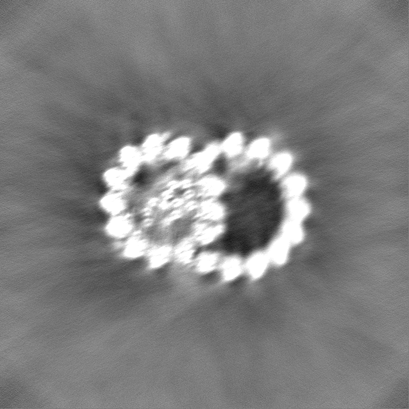
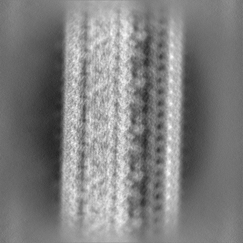
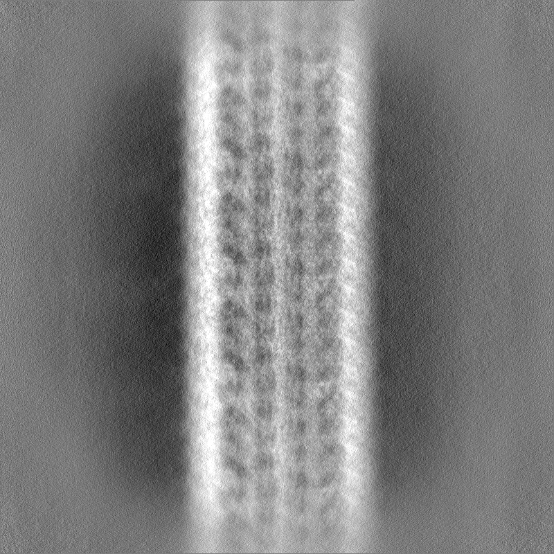
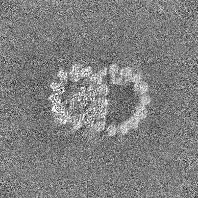
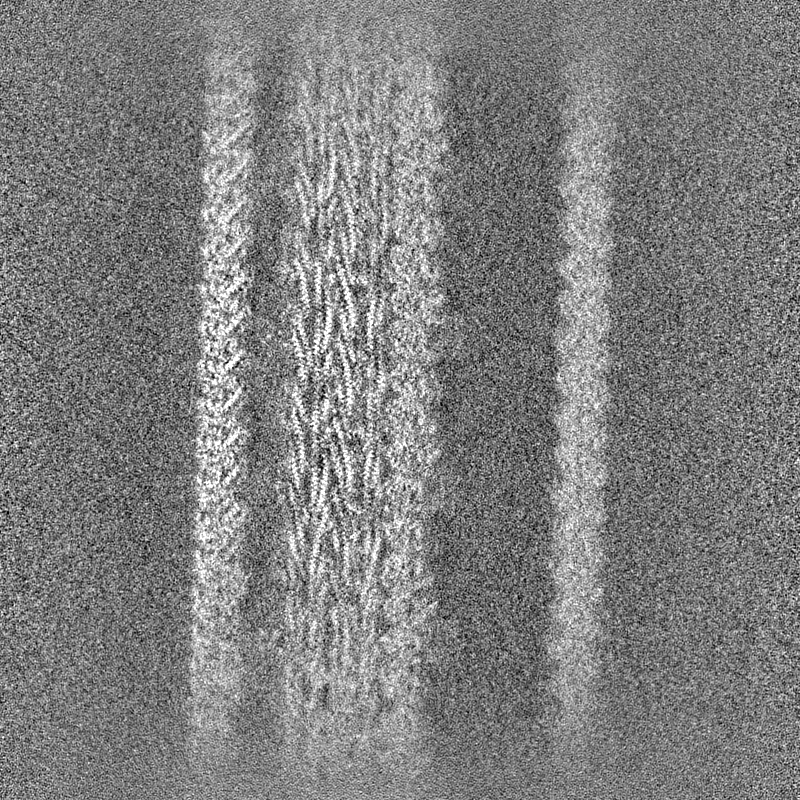
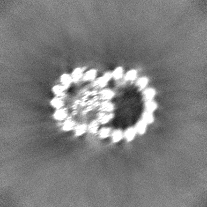
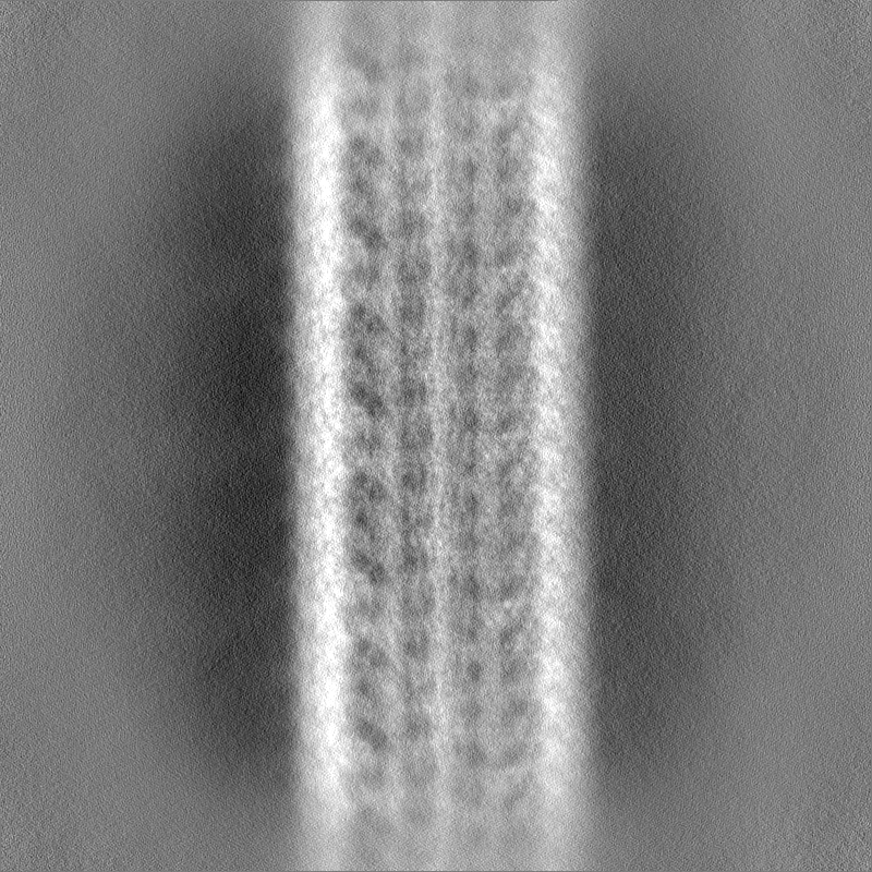
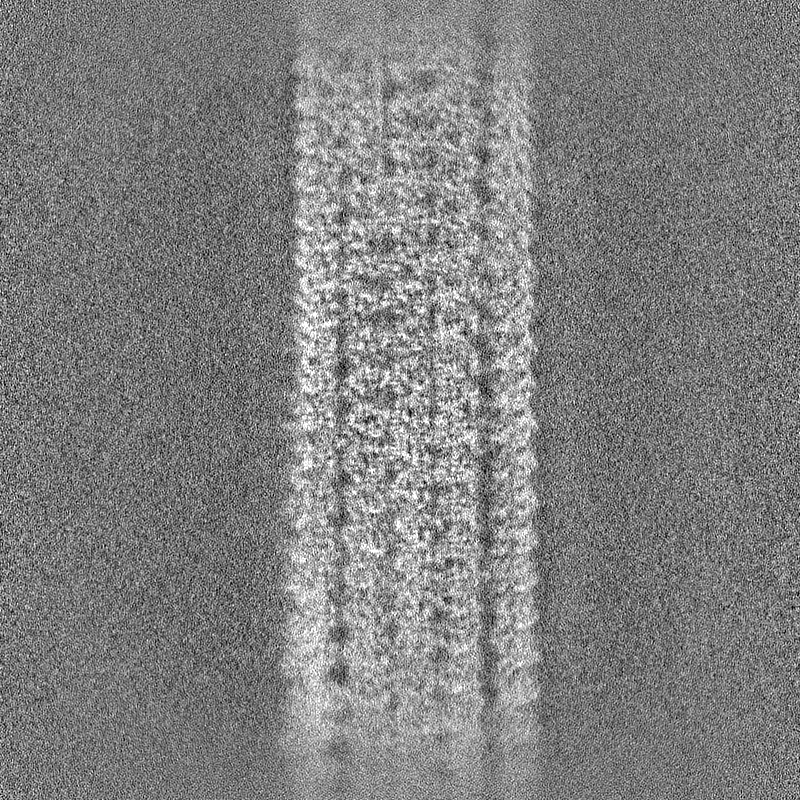
| Projections & slices | Image control
Images are generated by Spider. | ||||||||||||||||||||||||||||||||||||
| Voxel size | X=Y=Z: 1.087 Å | ||||||||||||||||||||||||||||||||||||
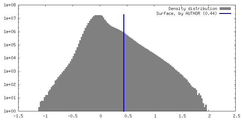
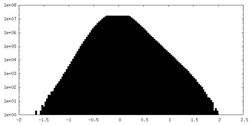
| Density |
| ||||||||||||||||||||||||||||||||||||
| Symmetry | Space group: 1 | ||||||||||||||||||||||||||||||||||||
| Details | EMDB XML:
|
 Z (Sec.)
 Y (Row.)
 X (Col.)


-Supplemental data
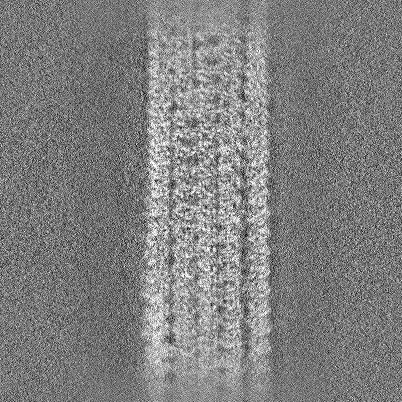
-Half map: #1
| File | emd_36504_half_map_1.map | ||||||||||||
|---|---|---|---|---|---|---|---|---|---|---|---|---|---|
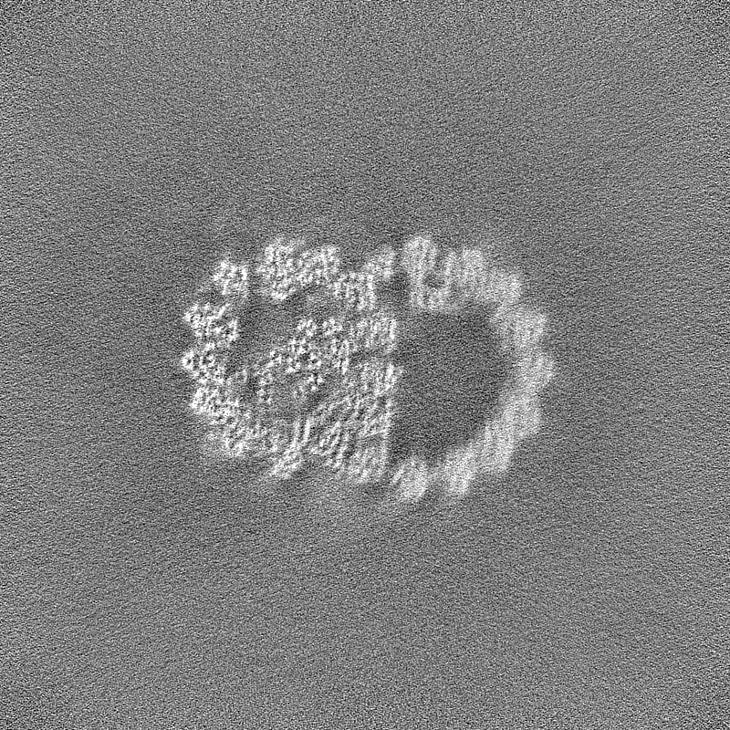
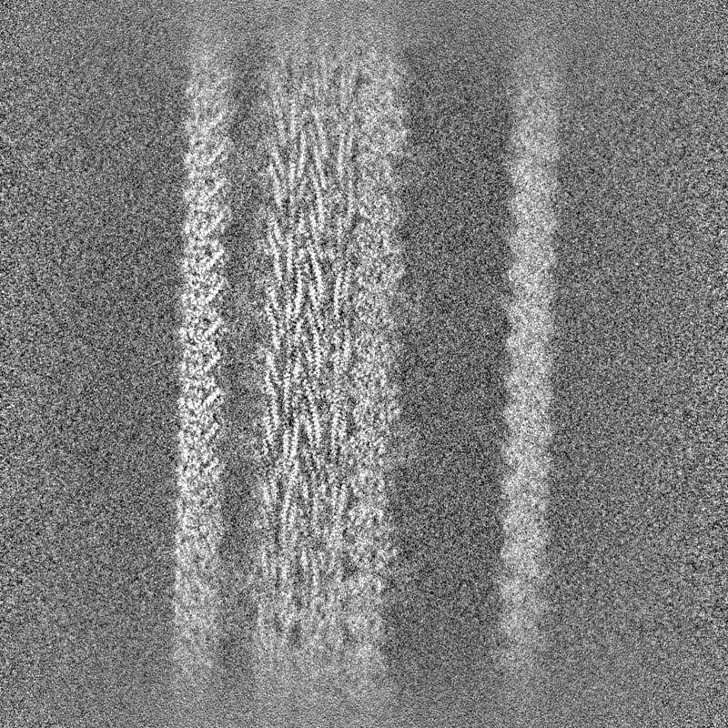
| Projections & Slices |
| ||||||||||||
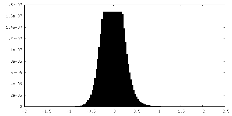
| Density Histograms |

-Half map: #2
| File | emd_36504_half_map_2.map | ||||||||||||
|---|---|---|---|---|---|---|---|---|---|---|---|---|---|
| Projections & Slices |
| ||||||||||||
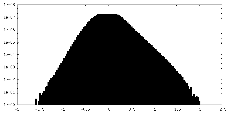
| Density Histograms |

- Sample components
-Entire : Mouse sperm DMT
| Entire | Name: Mouse sperm DMT |
|---|---|
| Components |
|
-Supramolecule #1: Mouse sperm DMT
| Supramolecule | Name: Mouse sperm DMT / type: complex / ID: 1 / Parent: 0 / Macromolecule list: #1-#49 |
|---|---|
| Source (natural) | Organism: |
-Experimental details
-Structure determination
| Method | cryo EM |
|---|---|
 Processing Processing | single particle reconstruction |
| Aggregation state | filament |
-Sample preparation
| Buffer | pH: 7.5 |
|---|---|
| Vitrification | Cryogen name: ETHANE / Instrument: FEI VITROBOT MARK IV |
- Electron microscopy
| Microscope | FEI TITAN KRIOS |
|---|---|
| Image recording | Film or detector model: GATAN K3 (6k x 4k) / Number real images: 25135 / Average exposure time: 2.56 sec. / Average electron dose: 50.0 e/Å2 |
| Electron beam | Acceleration voltage: 300 kV / Electron source:  FIELD EMISSION GUN FIELD EMISSION GUN |
| Electron optics | Illumination mode: FLOOD BEAM / Imaging mode: BRIGHT FIELD / Nominal defocus max: 2.6 µm / Nominal defocus min: 1.5 µm / Nominal magnification: 81000 |
| Sample stage | Specimen holder model: FEI TITAN KRIOS AUTOGRID HOLDER |
| Experimental equipment |  Model: Titan Krios / Image courtesy: FEI Company |
-Image processing
| Startup model | Type of model: EMDB MAP EMDB ID: |
|---|---|
| Final reconstruction | Resolution.type: BY AUTHOR / Resolution: 3.35 Å / Resolution method: FSC 0.143 CUT-OFF / Number images used: 95290 |
| Initial angle assignment | Type: NOT APPLICABLE |
| Final angle assignment | Type: NOT APPLICABLE |
