 Movie
Movie Controller


+ Open data

- Basic information
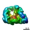
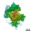
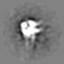
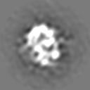
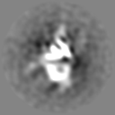
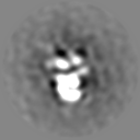
| Entry | Database: EMDB / ID: EMD-3642 | |||||||||
|---|---|---|---|---|---|---|---|---|---|---|
| Title | Full-length complex of CMG helicase with polymerase epsilon | |||||||||
 Map data Map data | ||||||||||
 Sample Sample |
| |||||||||
| Biological species |  | |||||||||
| Method | single particle reconstruction / negative staining / Resolution: 24.4 Å | |||||||||
 Authors Authors | Zhou JC / Janska A / Goswami P / Renault L / Abid Ali F / Kotecha A / Diffley JFX / Costa A | |||||||||
 Citation Citation | Journal: Proc Natl Acad Sci U S A / Year: 2017 Title: CMG-Pol epsilon dynamics suggests a mechanism for the establishment of leading-strand synthesis in the eukaryotic replisome. Authors: Jin Chuan Zhou / Agnieszka Janska / Panchali Goswami / Ludovic Renault / Ferdos Abid Ali / Abhay Kotecha / John F X Diffley / Alessandro Costa /  Abstract: The replisome unwinds and synthesizes DNA for genome duplication. In eukaryotes, the Cdc45-MCM-GINS (CMG) helicase and the leading-strand polymerase, Pol epsilon, form a stable assembly. The ...The replisome unwinds and synthesizes DNA for genome duplication. In eukaryotes, the Cdc45-MCM-GINS (CMG) helicase and the leading-strand polymerase, Pol epsilon, form a stable assembly. The mechanism for coupling DNA unwinding with synthesis is starting to be elucidated, however the architecture and dynamics of the replication fork remain only partially understood, preventing a molecular understanding of chromosome replication. To address this issue, we conducted a systematic single-particle EM study on multiple permutations of the reconstituted CMG-Pol epsilon assembly. Pol epsilon contains two flexibly tethered lobes. The noncatalytic lobe is anchored to the motor of the helicase, whereas the polymerization domain extends toward the side of the helicase. We observe two alternate configurations of the DNA synthesis domain in the CMG-bound Pol epsilon. We propose that this conformational switch might control DNA template engagement and release, modulating replisome progression. | |||||||||
| History |
|
- Structure visualization
| Movie |
 Movie viewer Movie viewer |
|---|---|
| Structure viewer | EM map: SurfViewMolmilJmol/JSmol |
| Supplemental images |
 UCSF Chimera
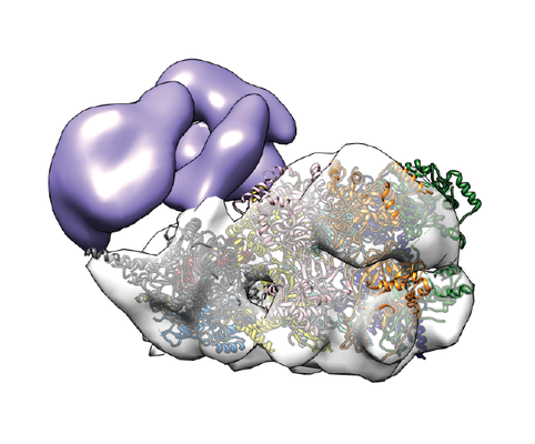
- Downloads & links
-EMDB archive
| Map data | emd_3642.map.gz | 6 MB | EMDB map data format | |
|---|---|---|---|---|
| Header (meta data) | emd-3642-v30.xmlemd-3642.xml | 7.8 KB 7.8 KB | Display Display | EMDB header |
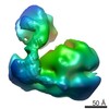
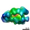
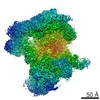
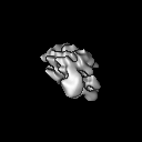
| Images |  emd_3642.png emd_3642.png | 160.9 KB | ||
| Archive directory |  http://ftp.pdbj.org/pub/emdb/structures/EMD-3642ftp://ftp.pdbj.org/pub/emdb/structures/EMD-3642 http://ftp.pdbj.org/pub/emdb/structures/EMD-3642ftp://ftp.pdbj.org/pub/emdb/structures/EMD-3642 | HTTPS FTP |
-Validation report
| Summary document | emd_3642_validation.pdf.gz | 214.1 KB | Display | EMDB validaton report |
|---|---|---|---|---|
| Full document | emd_3642_full_validation.pdf.gz | 213.2 KB | Display | |
| Data in XML | emd_3642_validation.xml.gz | 5.4 KB | Display | |
| Arichive directory | https://ftp.pdbj.org/pub/emdb/validation_reports/EMD-3642ftp://ftp.pdbj.org/pub/emdb/validation_reports/EMD-3642 | HTTPS FTP |
-Related structure data


-Links
| EMDB pages | EMDB (EBI/PDBe) / EMDataResource |
|---|
-Map
| File | Download / File: emd_3642.map.gz / Format: CCP4 / Size: 8 MB / Type: IMAGE STORED AS FLOATING POINT NUMBER (4 BYTES) | ||||||||||||||||||||||||||||||||||||||||||||||||||||||||||||
|---|---|---|---|---|---|---|---|---|---|---|---|---|---|---|---|---|---|---|---|---|---|---|---|---|---|---|---|---|---|---|---|---|---|---|---|---|---|---|---|---|---|---|---|---|---|---|---|---|---|---|---|---|---|---|---|---|---|---|---|---|---|
| Projections & slices | Image control
Images are generated by Spider. | ||||||||||||||||||||||||||||||||||||||||||||||||||||||||||||
| Voxel size | X=Y=Z: 3.65 Å | ||||||||||||||||||||||||||||||||||||||||||||||||||||||||||||
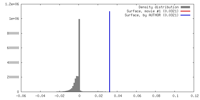
| Density |
| ||||||||||||||||||||||||||||||||||||||||||||||||||||||||||||
| Symmetry | Space group: 1 | ||||||||||||||||||||||||||||||||||||||||||||||||||||||||||||
| Details | EMDB XML:
CCP4 map header:
| ||||||||||||||||||||||||||||||||||||||||||||||||||||||||||||
 Z (Sec.)
 Y (Row.)
 X (Col.)


-Supplemental data
- Sample components
-Entire : Complex of CMG helicase with full length polymerase epsilon
| Entire | Name: Complex of CMG helicase with full length polymerase epsilon |
|---|---|
| Components |
|
-Supramolecule #1: Complex of CMG helicase with full length polymerase epsilon
| Supramolecule | Name: Complex of CMG helicase with full length polymerase epsilon type: complex / ID: 1 / Parent: 0 |
|---|---|
| Source (natural) | Organism: |
| Recombinant expression | Organism: |
-Experimental details
-Structure determination
| Method | negative staining |
|---|---|
 Processing Processing | single particle reconstruction |
| Aggregation state | particle |
-Sample preparation
| Buffer | pH: 7.6 |
|---|---|
| Staining | Type: NEGATIVE / Material: uranyl formate |
- Electron microscopy
| Microscope | JEOL 2100 |
|---|---|
| Image recording | Film or detector model: GATAN ULTRASCAN 4000 (4k x 4k) / Average electron dose: 35.0 e/Å2 |
| Electron beam | Acceleration voltage: 120 kV / Electron source: LAB6 |
| Electron optics | Illumination mode: SPOT SCAN / Imaging mode: OTHER |
-Image processing
| Final reconstruction | Resolution.type: BY AUTHOR / Resolution: 24.4 Å / Resolution method: FSC 0.143 CUT-OFF / Number images used: 24697 |
|---|---|
| Initial angle assignment | Type: PROJECTION MATCHING |
| Final angle assignment | Type: PROJECTION MATCHING |
