Movie
Movie Controller

[English] 日本語
 Yorodumi
- EMDB-34988: The structure of 30S subunit at 1 mM Magnesium concentration focu... -
+ Open data

- Basic information
| Entry |  | ||||||||||||
|---|---|---|---|---|---|---|---|---|---|---|---|---|---|
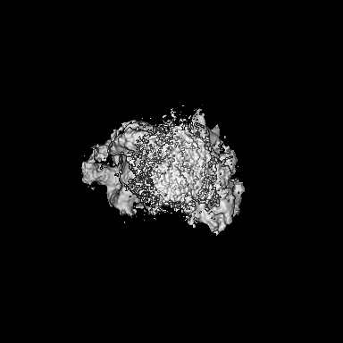
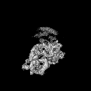
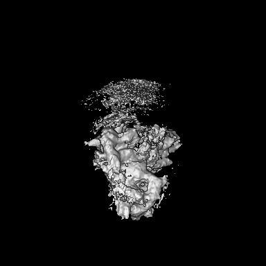
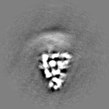
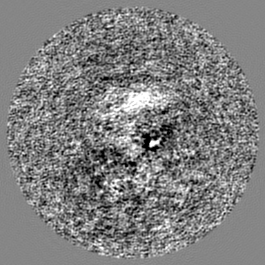
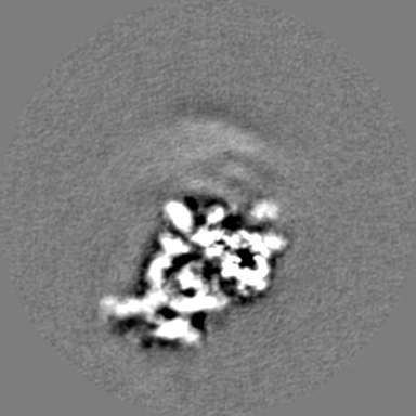
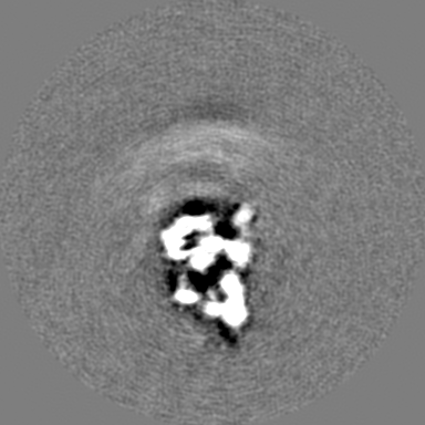
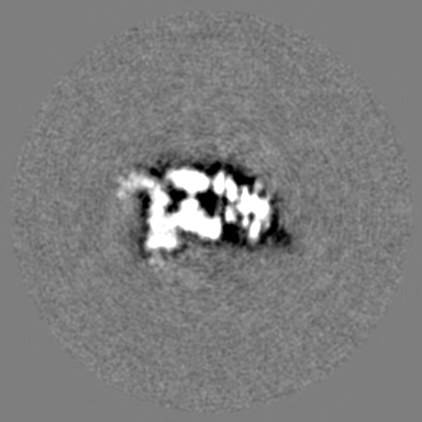
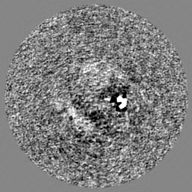
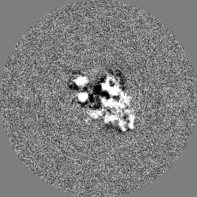
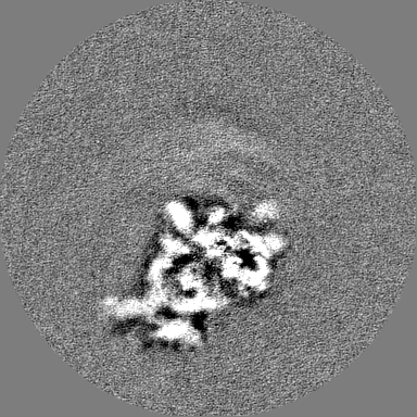
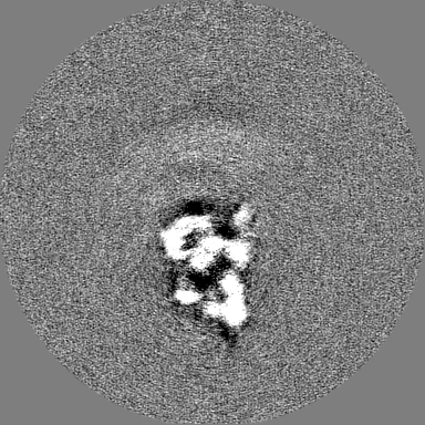
| Title | The structure of 30S subunit at 1 mM Magnesium concentration focused on h17 flexible | ||||||||||||
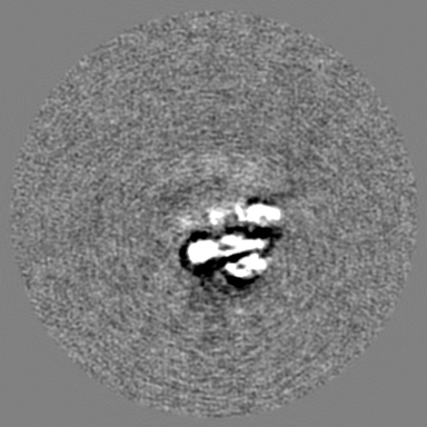
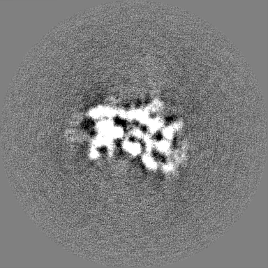
 Map data Map data | |||||||||||||
 Sample Sample |
| ||||||||||||
| Biological species |  | ||||||||||||
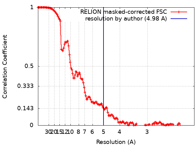
| Method | single particle reconstruction / cryo EM / Resolution: 4.98 Å | ||||||||||||
 Authors Authors | Yu T / Zeng F | ||||||||||||
| Funding support |  China, 3 items China, 3 items
| ||||||||||||
 Citation Citation | Journal: Biomolecules / Year: 2023 Title: Structural Insights into the Distortion of the Ribosomal Small Subunit at Different Magnesium Concentrations. Authors: Ting Yu / Junyi Jiang / Qianxi Yu / Xin Li / Fuxing Zeng / Abstract: Magnesium ions are abundant and play indispensable functions in the ribosome. A decrease in Mg concentration causes 70S ribosome dissociation and subsequent unfolding. Structural distortion at low Mg ...Magnesium ions are abundant and play indispensable functions in the ribosome. A decrease in Mg concentration causes 70S ribosome dissociation and subsequent unfolding. Structural distortion at low Mg concentrations has been observed in an immature pre50S, while the structural changes in mature subunits have not yet been studied. Here, we purified the 30S subunits of cells under various Mg concentrations and analyzed their structural distortion by cryo-electron microscopy. Upon systematically interrogating the structural heterogeneity within the 1 mM Mg dataset, we observed 30S particles with different levels of structural distortion in the decoding center, h17, and the 30S head. Our model showed that, when the Mg concentration decreases, the decoding center distorts, starting from h44 and followed by the shifting of h18 and h27, as well as the dissociation of ribosomal protein S12. Mg deficiency also eliminates the interactions between h17, h10, h15, and S16, resulting in the movement of h17 towards the tip of h6. More flexible structures were observed in the 30S head and platform, showing high variability in these regions. In summary, the structures resolved here showed several prominent distortion events in the decoding center and h17. The requirement for Mg in ribosomes suggests that the conformational changes reported here are likely shared due to a lack of cellular Mg in all domains of life. | ||||||||||||
| History |
|
- Structure visualization
| Supplemental images |
|---|

- Downloads & links
-EMDB archive
| Map data | emd_34988.map.gz | 201.9 MB |  EMDB map data format EMDB map data format | |
|---|---|---|---|---|
| Header (meta data) | emd-34988-v30.xmlemd-34988.xml | 14 KB 14 KB | Display Display | EMDB header |
| FSC (resolution estimation) | emd_34988_fsc.xml | 13.7 KB | Display | FSC data file |
| Images | 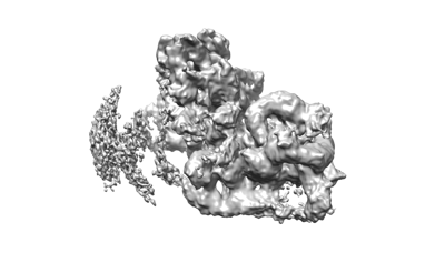 emd_34988.png emd_34988.png | 46.9 KB | ||
| Others | emd_34988_half_map_1.map.gzemd_34988_half_map_2.map.gz | 174.7 MB 174.9 MB | ||
| Archive directory |  http://ftp.pdbj.org/pub/emdb/structures/EMD-34988ftp://ftp.pdbj.org/pub/emdb/structures/EMD-34988 http://ftp.pdbj.org/pub/emdb/structures/EMD-34988ftp://ftp.pdbj.org/pub/emdb/structures/EMD-34988 | HTTPS FTP |
-Validation report
| Summary document | emd_34988_validation.pdf.gz | 846.8 KB | Display | EMDB validaton report |
|---|---|---|---|---|
| Full document | emd_34988_full_validation.pdf.gz | 846.3 KB | Display | |
| Data in XML | emd_34988_validation.xml.gz | 20.8 KB | Display | |
| Data in CIF | emd_34988_validation.cif.gz | 27.3 KB | Display | |
| Arichive directory | https://ftp.pdbj.org/pub/emdb/validation_reports/EMD-34988ftp://ftp.pdbj.org/pub/emdb/validation_reports/EMD-34988 | HTTPS FTP |
-Related structure data

-Links
| EMDB pages | EMDB (EBI/PDBe) / EMDataResource |
|---|
-Map
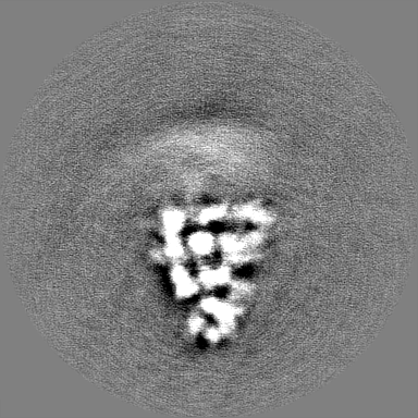
| File | Download / File: emd_34988.map.gz / Format: CCP4 / Size: 216 MB / Type: IMAGE STORED AS FLOATING POINT NUMBER (4 BYTES) | ||||||||||||||||||||||||||||||||||||
|---|---|---|---|---|---|---|---|---|---|---|---|---|---|---|---|---|---|---|---|---|---|---|---|---|---|---|---|---|---|---|---|---|---|---|---|---|---|
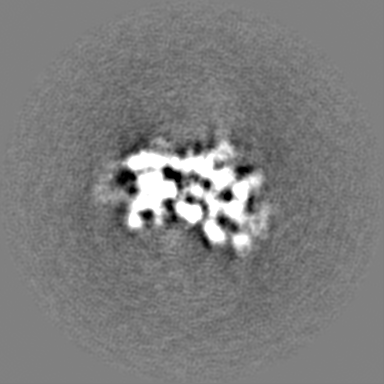
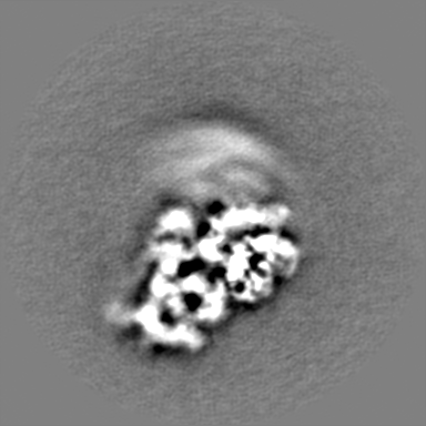
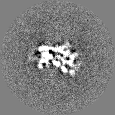
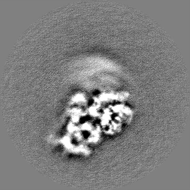
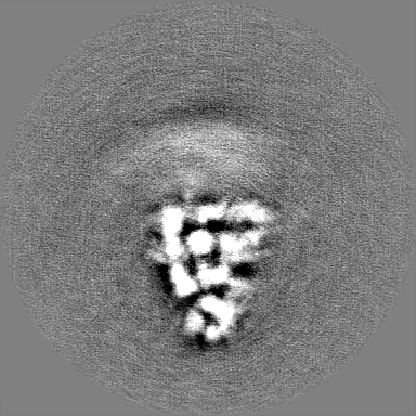
| Projections & slices | Image control
Images are generated by Spider. | ||||||||||||||||||||||||||||||||||||
| Voxel size | X=Y=Z: 1.05 Å | ||||||||||||||||||||||||||||||||||||
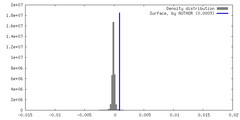
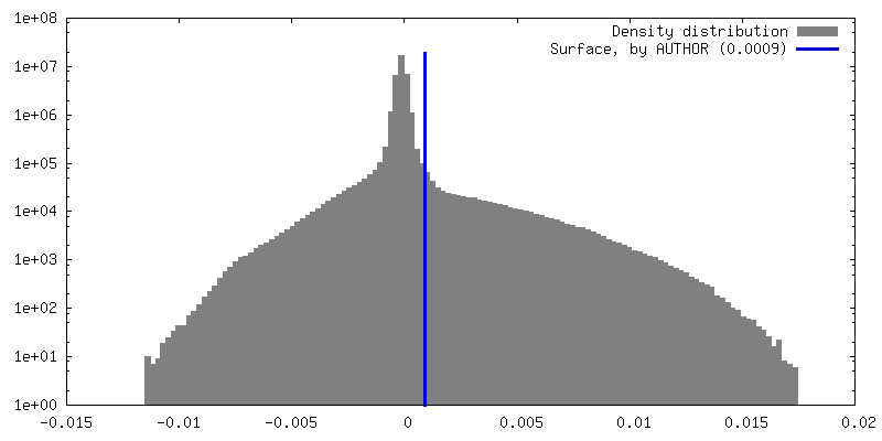
| Density |
| ||||||||||||||||||||||||||||||||||||
| Symmetry | Space group: 1 | ||||||||||||||||||||||||||||||||||||
| Details | EMDB XML:
|
 Z (Sec.)
 Y (Row.)
 X (Col.)


-Supplemental data
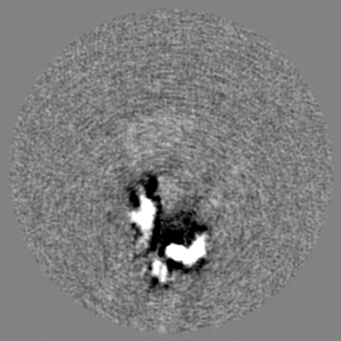
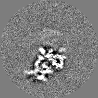
-Half map: #1
| File | emd_34988_half_map_1.map | ||||||||||||
|---|---|---|---|---|---|---|---|---|---|---|---|---|---|
| Projections & Slices |
| ||||||||||||
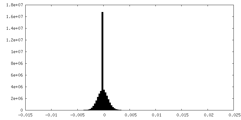
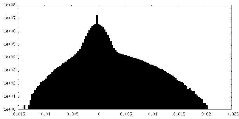
| Density Histograms |


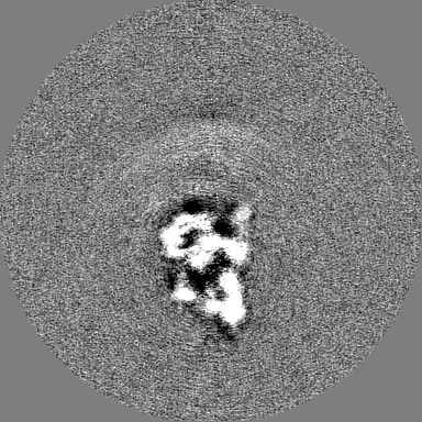
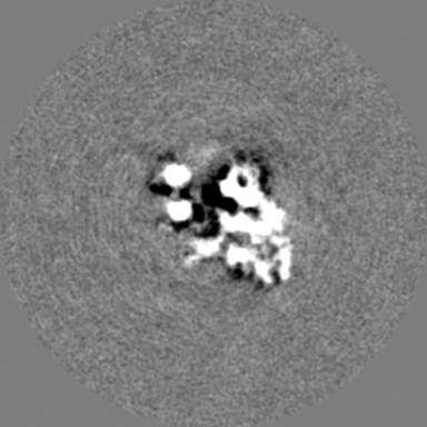
-Half map: #2
| File | emd_34988_half_map_2.map | ||||||||||||
|---|---|---|---|---|---|---|---|---|---|---|---|---|---|
| Projections & Slices |
| ||||||||||||
| Density Histograms |


- Sample components
-Entire : Ribosomal 30S subunit
| Entire | Name: Ribosomal 30S subunit |
|---|---|
| Components |
|
-Supramolecule #1: Ribosomal 30S subunit
| Supramolecule | Name: Ribosomal 30S subunit / type: complex / ID: 1 / Chimera: Yes / Parent: 0 Details: Ribosomal small subunit purified at 1 mM Magnesium concentration |
|---|---|
| Source (natural) | Organism: |
-Experimental details
-Structure determination
| Method | cryo EM |
|---|---|
 Processing Processing | single particle reconstruction |
| Aggregation state | particle |
-Sample preparation
| Buffer | pH: 7.5 Details: 20 mM HEPES-KOH pH7.5, 150 mM NH4Cl, 4 mM beta-mercaptoethanol |
|---|---|
| Grid | Model: Quantifoil R1.2/1.3 / Material: COPPER / Mesh: 300 / Support film - Material: CARBON / Support film - topology: CONTINUOUS / Support film - Film thickness: 4.0 nm / Pretreatment - Type: GLOW DISCHARGE / Pretreatment - Time: 30 sec. / Pretreatment - Atmosphere: AIR |
| Vitrification | Cryogen name: ETHANE / Chamber humidity: 100 % / Chamber temperature: 277 K / Instrument: FEI VITROBOT MARK III |
- Electron microscopy
| Microscope | FEI TITAN KRIOS |
|---|---|
| Image recording | Film or detector model: GATAN K3 (6k x 4k) / Average electron dose: 30.0 e/Å2 |
| Electron beam | Acceleration voltage: 300 kV / Electron source:  FIELD EMISSION GUN FIELD EMISSION GUN |
| Electron optics | Illumination mode: FLOOD BEAM / Imaging mode: BRIGHT FIELD / Nominal defocus max: 2.5 µm / Nominal defocus min: 1.5 µm |
| Experimental equipment |  Model: Titan Krios / Image courtesy: FEI Company |