[English] 日本語
 Yorodumi
- EMDB-34116: Spiral pentamer of the substrate-free Lon protease with an M217A ... -
+ Open data
- Basic information
| Entry |  | |||||||||
|---|---|---|---|---|---|---|---|---|---|---|
| Title | Spiral pentamer of the substrate-free Lon protease with an M217A mutation | |||||||||
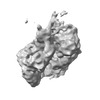
 Map data Map data | Substrate-free pentamer of MtaLon-M217A | |||||||||
 Sample Sample |
| |||||||||
 Keywords Keywords | hydrolysis / AAA proteins / HYDROLASE | |||||||||
| Biological species |  Meiothermus taiwanensis (bacteria) Meiothermus taiwanensis (bacteria) | |||||||||
| Method | single particle reconstruction / cryo EM / Resolution: 4.3 Å | |||||||||
 Authors Authors | Li S / Hsieh KY / Kuo CI / Lee SH / Ho MR / Wang CH / Zhang K / Chang CI | |||||||||
| Funding support |  Taiwan, 1 items Taiwan, 1 items
| |||||||||
 Citation Citation |  Journal: Nat Commun / Year: 2023 Journal: Nat Commun / Year: 2023Title: A 5+1 assemble-to-activate mechanism of the Lon proteolytic machine. Authors: Shanshan Li / Kan-Yen Hsieh / Chiao-I Kuo / Tzu-Chi Lin / Szu-Hui Lee / Yi-Ru Chen / Chun-Hsiung Wang / Meng-Ru Ho / See-Yeun Ting / Kaiming Zhang / Chung-I Chang /   Abstract: Many AAA+ (ATPases associated with diverse cellular activities) proteins function as protein or DNA remodelers by threading the substrate through the central pore of their hexameric assemblies. In ...Many AAA+ (ATPases associated with diverse cellular activities) proteins function as protein or DNA remodelers by threading the substrate through the central pore of their hexameric assemblies. In this ATP-dependent translocating state, the substrate is gripped by the pore loops of the ATPase domains arranged in a universal right-handed spiral staircase organization. However, the process by which a AAA+ protein is activated to adopt this substrate-pore-loop arrangement remains unknown. We show here, using cryo-electron microscopy (cryo-EM), that the activation process of the Lon AAA+ protease may involve a pentameric assembly and a substrate-dependent incorporation of the sixth protomer to form the substrate-pore-loop contacts seen in the translocating state. Based on the structural results, we design truncated monomeric mutants that inhibit Lon activity by binding to the native pentamer and demonstrated that expressing these monomeric mutants in Escherichia coli cells containing functional Lon elicits specific phenotypes associated with lon deficiency, including the inhibition of persister cell formation. These findings uncover a substrate-dependent assembly process for the activation of a AAA+ protein and demonstrate a targeted approach to selectively inhibit its function within cells. | |||||||||
| History |
|
- Structure visualization
| Supplemental images |
|---|
- Downloads & links
-EMDB archive
| Map data |  emd_34116.map.gz emd_34116.map.gz | 71 MB |  EMDB map data format EMDB map data format | |
|---|---|---|---|---|
| Header (meta data) |  emd-34116-v30.xml emd-34116-v30.xml emd-34116.xml emd-34116.xml | 17.8 KB 17.8 KB | Display Display |  EMDB header EMDB header |
| Images |  emd_34116.png emd_34116.png | 49.3 KB | ||
| Filedesc metadata |  emd-34116.cif.gz emd-34116.cif.gz | 5.6 KB | ||
| Others |  emd_34116_half_map_1.map.gz emd_34116_half_map_1.map.gz emd_34116_half_map_2.map.gz emd_34116_half_map_2.map.gz | 134.1 MB 134.1 MB | ||
| Archive directory |  http://ftp.pdbj.org/pub/emdb/structures/EMD-34116 http://ftp.pdbj.org/pub/emdb/structures/EMD-34116 ftp://ftp.pdbj.org/pub/emdb/structures/EMD-34116 ftp://ftp.pdbj.org/pub/emdb/structures/EMD-34116 | HTTPS FTP |
-Validation report
| Summary document |  emd_34116_validation.pdf.gz emd_34116_validation.pdf.gz | 888.9 KB | Display |  EMDB validaton report EMDB validaton report |
|---|---|---|---|---|
| Full document |  emd_34116_full_validation.pdf.gz emd_34116_full_validation.pdf.gz | 888.5 KB | Display | |
| Data in XML |  emd_34116_validation.xml.gz emd_34116_validation.xml.gz | 14.5 KB | Display | |
| Data in CIF |  emd_34116_validation.cif.gz emd_34116_validation.cif.gz | 17.1 KB | Display | |
| Arichive directory |  https://ftp.pdbj.org/pub/emdb/validation_reports/EMD-34116 https://ftp.pdbj.org/pub/emdb/validation_reports/EMD-34116 ftp://ftp.pdbj.org/pub/emdb/validation_reports/EMD-34116 ftp://ftp.pdbj.org/pub/emdb/validation_reports/EMD-34116 | HTTPS FTP |
-Related structure data
| Related structure data | 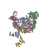 7yphC 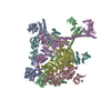 7ypiC 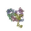 7ypjC 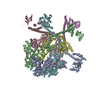 7ypkC 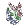 7yuhC 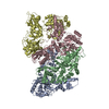 7yumC 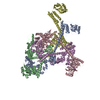 7yupC 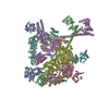 7yutC  7yuuC 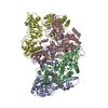 7yuvC  7yuwC  7yuxC  8k3yC C: citing same article ( |
|---|
- Links
Links
| EMDB pages |  EMDB (EBI/PDBe) / EMDB (EBI/PDBe) /  EMDataResource EMDataResource |
|---|
- Map
Map
| File |  Download / File: emd_34116.map.gz / Format: CCP4 / Size: 144.7 MB / Type: IMAGE STORED AS FLOATING POINT NUMBER (4 BYTES) Download / File: emd_34116.map.gz / Format: CCP4 / Size: 144.7 MB / Type: IMAGE STORED AS FLOATING POINT NUMBER (4 BYTES) | ||||||||||||||||||||||||||||||||||||
|---|---|---|---|---|---|---|---|---|---|---|---|---|---|---|---|---|---|---|---|---|---|---|---|---|---|---|---|---|---|---|---|---|---|---|---|---|---|
| Annotation | Substrate-free pentamer of MtaLon-M217A | ||||||||||||||||||||||||||||||||||||
| Projections & slices | Image control
Images are generated by Spider. | ||||||||||||||||||||||||||||||||||||
| Voxel size | X=Y=Z: 1.061 Å | ||||||||||||||||||||||||||||||||||||
| Density |
| ||||||||||||||||||||||||||||||||||||
| Symmetry | Space group: 1 | ||||||||||||||||||||||||||||||||||||
| Details | EMDB XML:
|
-Supplemental data
-Half map: Half map A
| File | emd_34116_half_map_1.map | ||||||||||||
|---|---|---|---|---|---|---|---|---|---|---|---|---|---|
| Annotation | Half map A | ||||||||||||
| Projections & Slices |
| ||||||||||||
| Density Histograms |
-Half map: Half map B
| File | emd_34116_half_map_2.map | ||||||||||||
|---|---|---|---|---|---|---|---|---|---|---|---|---|---|
| Annotation | Half map B | ||||||||||||
| Projections & Slices |
| ||||||||||||
| Density Histograms |
- Sample components
-Entire : Substrate-free pentamer of Lon protease with M217A mutation
| Entire | Name: Substrate-free pentamer of Lon protease with M217A mutation |
|---|---|
| Components |
|
-Supramolecule #1: Substrate-free pentamer of Lon protease with M217A mutation
| Supramolecule | Name: Substrate-free pentamer of Lon protease with M217A mutation type: complex / ID: 1 / Parent: 0 / Macromolecule list: all |
|---|---|
| Source (natural) | Organism:  Meiothermus taiwanensis (bacteria) Meiothermus taiwanensis (bacteria) |
| Molecular weight | Theoretical: 440 KDa |
-Macromolecule #1: Substrate-free pentamer of Lon protease with M217A mutation
| Macromolecule | Name: Substrate-free pentamer of Lon protease with M217A mutation type: protein_or_peptide / ID: 1 / Enantiomer: LEVO |
|---|---|
| Source (natural) | Organism:  Meiothermus taiwanensis (bacteria) Meiothermus taiwanensis (bacteria) |
| Recombinant expression | Organism:  |
| Sequence | String: MRLELPVIPL RNTVILPHTT TPVDVGRAKS KRAVEEAMGA DRLIFLVAQR DPEVDDPAPD DLYTWGVQA VVKQAMRLPD GTLQVMVEAR ARAQVTDYIP GPYLRARGEV FSEIFPIDEA V VRVLVEEL KEAFEKYVAN HKSLRLDRYQ LEAVKGTSDP AMLADTIAYH ...String: MRLELPVIPL RNTVILPHTT TPVDVGRAKS KRAVEEAMGA DRLIFLVAQR DPEVDDPAPD DLYTWGVQA VVKQAMRLPD GTLQVMVEAR ARAQVTDYIP GPYLRARGEV FSEIFPIDEA V VRVLVEEL KEAFEKYVAN HKSLRLDRYQ LEAVKGTSDP AMLADTIAYH ATWTVAEKQE IL ELTDLEA RLKKVLGLLS RDLERFELDK RVAQRVKEQa DTNQREYYLR EQMKAIQKEL GGE DGLSDL EALRKKIEEV GMPEAVKTKA LKELDRLERM QQGSPEATVA RTYLDWLTEV PWSK ADPEV LDINHTRQVL DEDHYGLKDV KERILEYLAV RQLTQGLDVR NKAPILVLVG PPGVG KTSL GRSIARSMNR KFHRISLGGV RDEAEIRGHR RTYIGAMPGK LIHAMKQVGV INPVIL LDE IDKMSSDWRG DPASAMLEVL DPEQNNTFTD HYLDVPYDLS KVFFITTANT LQTIPRP LL DRMEVIEIPG YTNMEKQAIA RQYLWPKQVR ESGMEGRIEV TDAAILRVIS EYTREAGV R GLERELGKIA RKGAKFWLEG AWEGLRTIDA SDIPTYLGIP RYRPDKAETE PQVGTAQGL AWTPVGGTLL TIEVAAVPGS GKLSLTGQLG EVMKESAQAA LTYLRAHTQD YGLPEDFYNK VDLHVHVPD GATPKDGPSA GITMATAIAS ALSRRPARMD IAMTGEVSLR GKVMPIGGVK E KLLAAHQA GIHKIVLPKD NEAQLEELPK EVLEGLEIKL VEDVGEVLEY LLLPEPTMPP VV QPSDNRQ QPGAGA |
-Experimental details
-Structure determination
| Method | cryo EM |
|---|---|
 Processing Processing | single particle reconstruction |
| Aggregation state | particle |
- Sample preparation
| Concentration | 1 mg/mL | ||||||||||||||||||
|---|---|---|---|---|---|---|---|---|---|---|---|---|---|---|---|---|---|---|---|
| Buffer | pH: 8 Component:
| ||||||||||||||||||
| Grid | Model: Quantifoil R1.2/1.3 / Material: COPPER / Mesh: 200 / Support film - Material: CARBON / Pretreatment - Type: GLOW DISCHARGE / Pretreatment - Time: 60 sec. | ||||||||||||||||||
| Vitrification | Cryogen name: ETHANE / Chamber humidity: 100 % / Chamber temperature: 277 K | ||||||||||||||||||
| Details | Lon protease with M217A mutation was incubated with alpha-S1-casein and ADP |
- Electron microscopy
| Microscope | FEI TITAN KRIOS |
|---|---|
| Image recording | Film or detector model: GATAN K3 BIOQUANTUM (6k x 4k) / Number grids imaged: 1 / Number real images: 10600 / Average exposure time: 2.0 sec. / Average electron dose: 49.0 e/Å2 |
| Electron beam | Acceleration voltage: 300 kV / Electron source:  FIELD EMISSION GUN FIELD EMISSION GUN |
| Electron optics | Illumination mode: FLOOD BEAM / Imaging mode: BRIGHT FIELD / Cs: 2.7 mm / Nominal defocus max: 2.2 µm / Nominal defocus min: 1.4000000000000001 µm / Nominal magnification: 81000 |
| Sample stage | Cooling holder cryogen: NITROGEN |
| Experimental equipment |  Model: Titan Krios / Image courtesy: FEI Company |
 Movie
Movie Controller




 Z (Sec.)
 Y (Row.)
 X (Col.)


