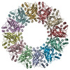
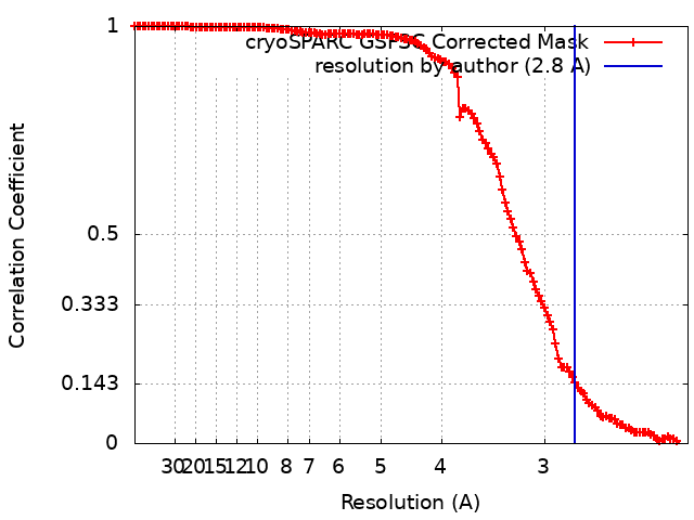
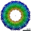
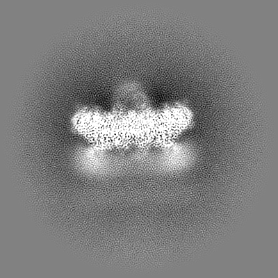
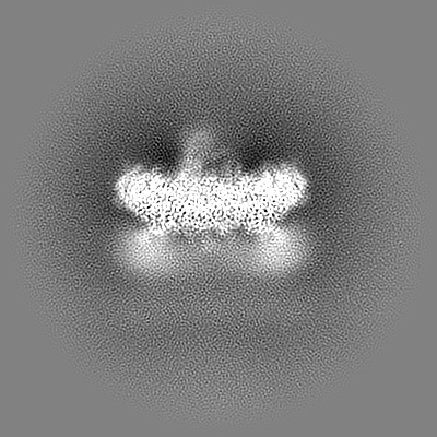
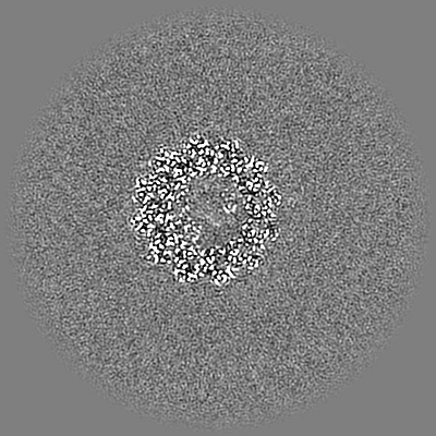
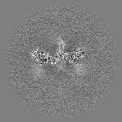
Journal: Sci Adv / Year: 2022 Title: Molecular architecture of the Chikungunya virus replication complex. Authors: Yaw Bia Tan / David Chmielewski / Michelle Cheok Yien Law / Kuo Zhang / Yu He / Muyuan Chen / Jing Jin / Dahai Luo / Abstract: To better understand how positive-strand (+) RNA viruses assemble membrane-associated replication complexes (RCs) to synthesize, process, and transport viral RNA in virus-infected cells, we ...To better understand how positive-strand (+) RNA viruses assemble membrane-associated replication complexes (RCs) to synthesize, process, and transport viral RNA in virus-infected cells, we determined both the high-resolution structure of the core RNA replicase of chikungunya virus and the native RC architecture in its cellular context at subnanometer resolution, using in vitro reconstitution and in situ electron cryotomography, respectively. Within the core RNA replicase, the viral polymerase nsP4, which is in complex with nsP2 helicase-protease, sits in the central pore of the membrane-anchored nsP1 RNA-capping ring. The addition of a large cytoplasmic ring next to the C terminus of nsP1 forms the holo-RNA-RC as observed at the neck of spherules formed in virus-infected cells. These results represent a major conceptual advance in elucidating the molecular mechanisms of RNA virus replication and the principles underlying the molecular architecture of RCs, likely to be shared with many pathogenic (+) RNA viruses.
Supramolecule #3: nsP4 RdRp; docked within nsP1 central cavity
Supramolecule
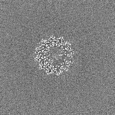
Name: nsP4 RdRp; docked within nsP1 central cavity / type: complex / ID: 3 / Parent: 1 / Macromolecule list: #2 Details: viral RdRp protein, single copy nsP4 slotted in the central pore of nsP1
Source (natural)
Organism: Onyong-nyong virus
+
Supramolecule #4: nsP2 helicase-protease; docked on top of viral complex with synth...
Supramolecule
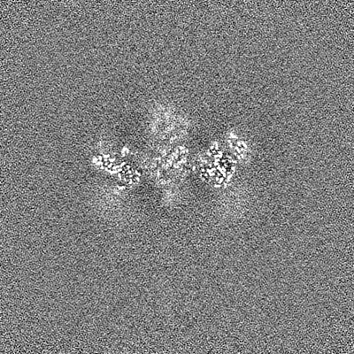
Name: nsP2 helicase-protease; docked on top of viral complex with synthetic RNA bound type: complex / ID: 4 / Parent: 1 / Macromolecule list: #3-#4 / Details: only helicase density was observed
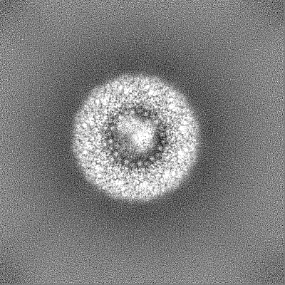
Name: mRNA-capping enzyme nsP1,affinity-tag (strepII-3XFLAG) type: protein_or_peptide / ID: 1 Details: 1. affinity-tag (strepII-3XFLAG) insertion in between residue number 517-552; 2. replaced the last residue A535 with short peptide-tag NGL (corresponding to position 571-573) Number of copies: 12 / Enantiomer: LEVO EC number: Transferases; Transferring one-carbon groups; Methyltransferases
Name: RNA-directed RNA polymerase nsP4 / type: protein_or_peptide / ID: 2 Details: Sourced from virus isolate cDNA, not available in uniprot database at the time of biocuration Number of copies: 1 / Enantiomer: LEVO / EC number: polynucleotide adenylyltransferase
Name: Protease nsP2 / type: protein_or_peptide / ID: 3 Details: helicase region is built on visible 3D electron density map but not protease region Number of copies: 1 / Enantiomer: LEVO / EC number: polynucleotide 5'-phosphatase
Model: Quantifoil R1.2/1.3 / Material: COPPER / Mesh: 300 / Support film - Material: GRAPHENE / Support film - topology: CONTINUOUS / Support film - Film thickness: 3.45 / Pretreatment - Type: GLOW DISCHARGE / Pretreatment - Time: 10 sec. / Pretreatment - Atmosphere: OTHER
Vitrification
Cryogen name: ETHANE / Chamber humidity: 100 % / Chamber temperature: 273.15 K / Instrument: FEI VITROBOT MARK III / Details: wait 45s, blot 2s at force -2.
Details
The sample was in vitro reconstituted and purified via affinity pulldown and ion-exchanged.
-
Electron microscopy
Microscope
FEI TITAN KRIOS
Temperature
Min: 70.0 K / Max: 80.0 K
Image recording
Film or detector model: GATAN K2 SUMMIT (4k x 4k) / Detector mode: COUNTING / Digitization - Dimensions - Width: 3710 pixel / Digitization - Dimensions - Height: 3838 pixel / Number grids imaged: 1 / Number real images: 7865 / Average exposure time: 5.0 sec. / Average electron dose: 34.0 e/Å2 / Details: movie-mode at 40 FPS
Electron beam
Acceleration voltage: 300 kV / Electron source: FIELD EMISSION GUN
Number classes: 6 / Avg.num./class: 95000 / Software - Name: cryoSPARC (ver. 3.3.1) / Software - details: 3D Classification / Details: the 6th class has the highest number of particles
In the structure databanks used in Yorodumi, some data are registered as the other names, "COVID-19 virus" and "2019-nCoV". Here are the details of the virus and the list of structure data.
Jan 31, 2019. EMDB accession codes are about to change! (news from PDBe EMDB page)
EMDB accession codes are about to change! (news from PDBe EMDB page)
The allocation of 4 digits for EMDB accession codes will soon come to an end. Whilst these codes will remain in use, new EMDB accession codes will include an additional digit and will expand incrementally as the available range of codes is exhausted. The current 4-digit format prefixed with “EMD-” (i.e. EMD-XXXX) will advance to a 5-digit format (i.e. EMD-XXXXX), and so on. It is currently estimated that the 4-digit codes will be depleted around Spring 2019, at which point the 5-digit format will come into force.
The EM Navigator/Yorodumi systems omit the EMD- prefix.
Related info.:Q: What is EMD? / ID/Accession-code notation in Yorodumi/EM Navigator
Yorodumi is a browser for structure data from EMDB, PDB, SASBDB, etc.
This page is also the successor to EM Navigator detail page, and also detail information page/front-end page for Omokage search.
The word "yorodu" (or yorozu) is an old Japanese word meaning "ten thousand". "mi" (miru) is to see.
Related info.:EMDB / PDB / SASBDB / Comparison of 3 databanks / Yorodumi Search / Aug 31, 2016. New EM Navigator & Yorodumi / Yorodumi Papers / Jmol/JSmol / Function and homology information / Changes in new EM Navigator and Yorodumi
 Movie
Movie Controller

 Yorodumi
 Open data
 Basic information
 Map data
 Sample
 Keywords
 Function and homology information
 Chikungunya virus strain S27-African prototype /
  Authors
 Singapore, 1 items
  Citation
 Structure visualization
 Downloads & links
 emd_33591.png
 http://ftp.pdbj.org/pub/emdb/structures/EMD-33591


 Z (Sec.)
 Y (Row.)
 X (Col.)


 Sample components
 Homo sapiens (human)


 Processing
 Electron microscopy
 FIELD EMISSION GUN