 Movie
Movie Controller

[English] 日本語
 Yorodumi
- EMDB-27268: In situ structure of the non-replicative Chikungunya virus nonstr... -
+ Open data

- Basic information
| Entry |  | |||||||||
|---|---|---|---|---|---|---|---|---|---|---|
| Title | In situ structure of the non-replicative Chikungunya virus nonstructural protein complex | |||||||||
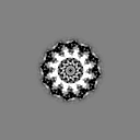
 Map data Map data | Subtomogram average of nsp1-2-4 ring | |||||||||
 Sample Sample |
| |||||||||
| Biological species |   Chikungunya virus Chikungunya virus | |||||||||
| Method | subtomogram averaging / cryo EM / Resolution: 9.9 Å | |||||||||
 Authors Authors | Yaw Bia T / Chmielewski D / Yien Law MC / Zhang K / He Y / Chen M / Jin J / Luo D | |||||||||
| Funding support |  United States, 2 items United States, 2 items
| |||||||||
 Citation Citation | Journal: Sci Adv / Year: 2022 Title: Molecular architecture of the Chikungunya virus replication complex. Authors: Yaw Bia Tan / David Chmielewski / Michelle Cheok Yien Law / Kuo Zhang / Yu He / Muyuan Chen / Jing Jin / Dahai Luo /  Abstract: To better understand how positive-strand (+) RNA viruses assemble membrane-associated replication complexes (RCs) to synthesize, process, and transport viral RNA in virus-infected cells, we ...To better understand how positive-strand (+) RNA viruses assemble membrane-associated replication complexes (RCs) to synthesize, process, and transport viral RNA in virus-infected cells, we determined both the high-resolution structure of the core RNA replicase of chikungunya virus and the native RC architecture in its cellular context at subnanometer resolution, using in vitro reconstitution and in situ electron cryotomography, respectively. Within the core RNA replicase, the viral polymerase nsP4, which is in complex with nsP2 helicase-protease, sits in the central pore of the membrane-anchored nsP1 RNA-capping ring. The addition of a large cytoplasmic ring next to the C terminus of nsP1 forms the holo-RNA-RC as observed at the neck of spherules formed in virus-infected cells. These results represent a major conceptual advance in elucidating the molecular mechanisms of RNA virus replication and the principles underlying the molecular architecture of RCs, likely to be shared with many pathogenic (+) RNA viruses. | |||||||||
| History |
|
- Structure visualization
| Supplemental images |
|---|

- Downloads & links
-EMDB archive
| Map data | emd_27268.map.gz | 274.1 KB |  EMDB map data format EMDB map data format | |
|---|---|---|---|---|
| Header (meta data) | emd-27268-v30.xmlemd-27268.xml | 13.2 KB 13.2 KB | Display Display | EMDB header |
| Images | 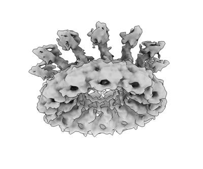 emd_27268.png emd_27268.png | 64.6 KB | ||
| Others | emd_27268_half_map_1.map.gzemd_27268_half_map_2.map.gz | 284.1 KB 275.3 KB | ||
| Archive directory |  http://ftp.pdbj.org/pub/emdb/structures/EMD-27268ftp://ftp.pdbj.org/pub/emdb/structures/EMD-27268 http://ftp.pdbj.org/pub/emdb/structures/EMD-27268ftp://ftp.pdbj.org/pub/emdb/structures/EMD-27268 | HTTPS FTP |
-Related structure data


-Links
| EMDB pages | EMDB (EBI/PDBe) / EMDataResource |
|---|
-Map
| File | Download / File: emd_27268.map.gz / Format: CCP4 / Size: 8 MB / Type: IMAGE STORED AS FLOATING POINT NUMBER (4 BYTES) | ||||||||||||||||||||||||||||||||||||
|---|---|---|---|---|---|---|---|---|---|---|---|---|---|---|---|---|---|---|---|---|---|---|---|---|---|---|---|---|---|---|---|---|---|---|---|---|---|
| Annotation | Subtomogram average of nsp1-2-4 ring | ||||||||||||||||||||||||||||||||||||
| Projections & slices | Image control
Images are generated by Spider. | ||||||||||||||||||||||||||||||||||||
| Voxel size | X=Y=Z: 3.54 Å | ||||||||||||||||||||||||||||||||||||
| Density |
| ||||||||||||||||||||||||||||||||||||
| Symmetry | Space group: 1 | ||||||||||||||||||||||||||||||||||||
| Details | EMDB XML:
|
 Z (Sec.)
 Y (Row.)
 X (Col.)


-Supplemental data
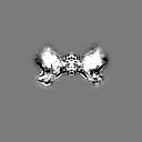
-Half map: even half map
| File | emd_27268_half_map_1.map | ||||||||||||
|---|---|---|---|---|---|---|---|---|---|---|---|---|---|
| Annotation | even half map | ||||||||||||
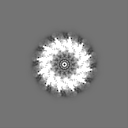
| Projections & Slices |
| ||||||||||||
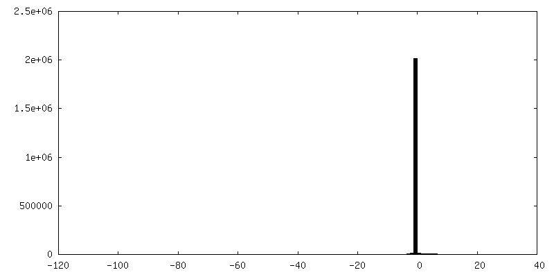
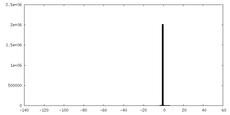
| Density Histograms |


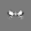
-Half map: odd half map
| File | emd_27268_half_map_2.map | ||||||||||||
|---|---|---|---|---|---|---|---|---|---|---|---|---|---|
| Annotation | odd half map | ||||||||||||
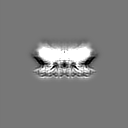
| Projections & Slices |
| ||||||||||||
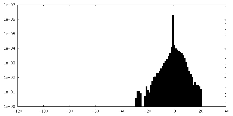
| Density Histograms |

- Sample components
-Entire : Chikungunya virus replicase complex
| Entire | Name: Chikungunya virus replicase complex |
|---|---|
| Components |
|
-Supramolecule #1: Chikungunya virus replicase complex
| Supramolecule | Name: Chikungunya virus replicase complex / type: complex / Chimera: Yes / ID: 1 / Parent: 0 |
|---|---|
| Source (natural) | Organism: Chikungunya virus |
-Experimental details
-Structure determination
| Method | cryo EM |
|---|---|
 Processing Processing | subtomogram averaging |
| Aggregation state | particle |
-Sample preparation
| Buffer | pH: 7 |
|---|---|
| Vitrification | Cryogen name: ETHANE |
- Electron microscopy
| Microscope | FEI TALOS ARCTICA |
|---|---|
| Image recording | Film or detector model: GATAN K2 SUMMIT (4k x 4k) / Detector mode: COUNTING / Average electron dose: 1.76 e/Å2 |
| Electron beam | Acceleration voltage: 200 kV / Electron source:  FIELD EMISSION GUN FIELD EMISSION GUN |
| Electron optics | Illumination mode: FLOOD BEAM / Imaging mode: BRIGHT FIELD / Nominal defocus max: 5.0 µm / Nominal defocus min: 2.0 µm |
| Experimental equipment |  Model: Talos Arctica / Image courtesy: FEI Company |
-Image processing
| Final reconstruction | Applied symmetry - Point group: C12 (12 fold cyclic) / Resolution.type: BY AUTHOR / Resolution: 9.9 Å / Resolution method: FSC 0.143 CUT-OFF / Number subtomograms used: 5707 |
|---|---|
| Extraction | Number tomograms: 30 / Number images used: 5707 |
| Final angle assignment | Type: PROJECTION MATCHING |
