[English] 日本語
 Yorodumi
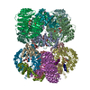
- EMDB-3243: Dodecameric Chaetomium thermophilum Rvb1/Rvb2 complex in stretche... -
+ Open data
- Basic information
| Entry | Database: EMDB / ID: EMD-3243 | |||||||||
|---|---|---|---|---|---|---|---|---|---|---|
| Title | Dodecameric Chaetomium thermophilum Rvb1/Rvb2 complex in stretched conformation | |||||||||
 Map data Map data | Cryo-EM reconstruction of Chaetomium thermophilum dodecameric Rvb1/Rvb2 complex in stretched conformation | |||||||||
 Sample Sample |
| |||||||||
 Keywords Keywords | AAA+ proteins / cryo-electron microscopy / Rvb1 / Rvb2 / Chaetomium thermophilum | |||||||||
| Function / homology |  Function and homology information Function and homology informationR2TP complex / Swr1 complex / Ino80 complex / box C/D snoRNP assembly / NuA4 histone acetyltransferase complex / DNA helicase activity / DNA helicase / chromatin remodeling / DNA repair / regulation of transcription by RNA polymerase II ...R2TP complex / Swr1 complex / Ino80 complex / box C/D snoRNP assembly / NuA4 histone acetyltransferase complex / DNA helicase activity / DNA helicase / chromatin remodeling / DNA repair / regulation of transcription by RNA polymerase II / ATP hydrolysis activity / ATP binding Similarity search - Function | |||||||||
| Biological species |  Chaetomium thermophilum (fungus) Chaetomium thermophilum (fungus) | |||||||||
| Method | single particle reconstruction / cryo EM / Resolution: 21.4 Å | |||||||||
 Authors Authors | Silva-Martin N / Dauden MI / Glatt S / Hoffmann NA / Kastritis P / Bork P / Beck M / Mueller CW | |||||||||
 Citation Citation |  Journal: PLoS One / Year: 2016 Journal: PLoS One / Year: 2016Title: The Combination of X-Ray Crystallography and Cryo-Electron Microscopy Provides Insight into the Overall Architecture of the Dodecameric Rvb1/Rvb2 Complex. Authors: Noella Silva-Martin / María I Daudén / Sebastian Glatt / Niklas A Hoffmann / Panagiotis Kastritis / Peer Bork / Martin Beck / Christoph W Müller /  Abstract: The Rvb1/Rvb2 complex is an essential component of many cellular pathways. The Rvb1/Rvb2 complex forms a dodecameric assembly where six copies of each subunit form two heterohexameric rings. However, ...The Rvb1/Rvb2 complex is an essential component of many cellular pathways. The Rvb1/Rvb2 complex forms a dodecameric assembly where six copies of each subunit form two heterohexameric rings. However, due to conformational variability, the way the two rings pack together is still not fully understood. Here, we present the crystal structure and two cryo-electron microscopy reconstructions of the dodecameric, full-length Rvb1/Rvb2 complex, all showing that the interaction between the two heterohexameric rings is mediated through the Rvb1/Rvb2-specific domain II. Two conformations of the Rvb1/Rvb2 dodecamer are present in solution: a stretched conformation also present in the crystal, and a compact conformation. Novel asymmetric features observed in the reconstruction of the compact conformation provide additional insight into the plasticity of the Rvb1/Rvb2 complex. | |||||||||
| History |
|
- Structure visualization
| Movie |
 Movie viewer Movie viewer |
|---|---|
| Structure viewer | EM map:  SurfView SurfView Molmil Molmil Jmol/JSmol Jmol/JSmol |
| Supplemental images |
- Downloads & links
-EMDB archive
| Map data |  emd_3243.map.gz emd_3243.map.gz | 1.1 MB |  EMDB map data format EMDB map data format | |
|---|---|---|---|---|
| Header (meta data) |  emd-3243-v30.xml emd-3243-v30.xml emd-3243.xml emd-3243.xml | 11.2 KB 11.2 KB | Display Display |  EMDB header EMDB header |
| FSC (resolution estimation) |  emd_3243_fsc.xml emd_3243_fsc.xml | 2.6 KB | Display |  FSC data file FSC data file |
| Images |  emd_3243.tif emd_3243.tif | 45.7 KB | ||
| Archive directory |  http://ftp.pdbj.org/pub/emdb/structures/EMD-3243 http://ftp.pdbj.org/pub/emdb/structures/EMD-3243 ftp://ftp.pdbj.org/pub/emdb/structures/EMD-3243 ftp://ftp.pdbj.org/pub/emdb/structures/EMD-3243 | HTTPS FTP |
-Validation report
| Summary document |  emd_3243_validation.pdf.gz emd_3243_validation.pdf.gz | 248.3 KB | Display |  EMDB validaton report EMDB validaton report |
|---|---|---|---|---|
| Full document |  emd_3243_full_validation.pdf.gz emd_3243_full_validation.pdf.gz | 247.4 KB | Display | |
| Data in XML |  emd_3243_validation.xml.gz emd_3243_validation.xml.gz | 6.6 KB | Display | |
| Arichive directory |  https://ftp.pdbj.org/pub/emdb/validation_reports/EMD-3243 https://ftp.pdbj.org/pub/emdb/validation_reports/EMD-3243 ftp://ftp.pdbj.org/pub/emdb/validation_reports/EMD-3243 ftp://ftp.pdbj.org/pub/emdb/validation_reports/EMD-3243 | HTTPS FTP |
-Related structure data
- Links
Links
| EMDB pages |  EMDB (EBI/PDBe) / EMDB (EBI/PDBe) /  EMDataResource EMDataResource |
|---|
- Map
Map
| File |  Download / File: emd_3243.map.gz / Format: CCP4 / Size: 1.5 MB / Type: IMAGE STORED AS FLOATING POINT NUMBER (4 BYTES) Download / File: emd_3243.map.gz / Format: CCP4 / Size: 1.5 MB / Type: IMAGE STORED AS FLOATING POINT NUMBER (4 BYTES) | ||||||||||||||||||||||||||||||||||||||||||||||||||||||||||||
|---|---|---|---|---|---|---|---|---|---|---|---|---|---|---|---|---|---|---|---|---|---|---|---|---|---|---|---|---|---|---|---|---|---|---|---|---|---|---|---|---|---|---|---|---|---|---|---|---|---|---|---|---|---|---|---|---|---|---|---|---|---|
| Annotation | Cryo-EM reconstruction of Chaetomium thermophilum dodecameric Rvb1/Rvb2 complex in stretched conformation | ||||||||||||||||||||||||||||||||||||||||||||||||||||||||||||
| Projections & slices | Image control
Images are generated by Spider. | ||||||||||||||||||||||||||||||||||||||||||||||||||||||||||||
| Voxel size | X=Y=Z: 4.34 Å | ||||||||||||||||||||||||||||||||||||||||||||||||||||||||||||
| Density |
| ||||||||||||||||||||||||||||||||||||||||||||||||||||||||||||
| Symmetry | Space group: 1 | ||||||||||||||||||||||||||||||||||||||||||||||||||||||||||||
| Details | EMDB XML:
CCP4 map header:
| ||||||||||||||||||||||||||||||||||||||||||||||||||||||||||||
-Supplemental data
- Sample components
-Entire : Full-length Chaetomium thermophilum Rvb1/Rvb2 dodecameric complex...
| Entire | Name: Full-length Chaetomium thermophilum Rvb1/Rvb2 dodecameric complex in stretched conformation |
|---|---|
| Components |
|
-Supramolecule #1000: Full-length Chaetomium thermophilum Rvb1/Rvb2 dodecameric complex...
| Supramolecule | Name: Full-length Chaetomium thermophilum Rvb1/Rvb2 dodecameric complex in stretched conformation type: sample / ID: 1000 Oligomeric state: Dodecamer formed by two hetero-hexamers of Rvb1/Rvb2 Number unique components: 2 |
|---|---|
| Molecular weight | Theoretical: 621 KDa |
-Macromolecule #1: Rvb1
| Macromolecule | Name: Rvb1 / type: protein_or_peptide / ID: 1 / Name.synonym: RuvBL1, Pontin, TIP49a / Number of copies: 6 / Oligomeric state: Double hetero-hexamer with Rvb2 / Recombinant expression: Yes |
|---|---|
| Source (natural) | Organism:  Chaetomium thermophilum (fungus) / synonym: thermophilic filamentous fungus Chaetomium thermophilum (fungus) / synonym: thermophilic filamentous fungus |
| Molecular weight | Theoretical: 50.384 KDa |
| Recombinant expression | Organism:  |
| Sequence | UniProtKB: RuvB-like helicase InterPro: AAA+ ATPase domain, P-loop containing nucleoside triphosphate hydrolase, RuvB-like, TIP49, P-loop domain |
-Macromolecule #2: Rvb2
| Macromolecule | Name: Rvb2 / type: protein_or_peptide / ID: 2 / Name.synonym: RuvBL2, Reptin, TIP49b / Number of copies: 6 / Oligomeric state: Double hetero-hexamer with Rvb2 / Recombinant expression: Yes |
|---|---|
| Source (natural) | Organism:  Chaetomium thermophilum (fungus) / synonym: thermophilic filamentous fungus Chaetomium thermophilum (fungus) / synonym: thermophilic filamentous fungus |
| Molecular weight | Theoretical: 53.1458 KDa |
| Recombinant expression | Organism:  |
| Sequence | UniProtKB: RuvB-like helicase InterPro: AAA+ ATPase domain, P-loop containing nucleoside triphosphate hydrolase, RuvB-like, TIP49, P-loop domain |
-Experimental details
-Structure determination
| Method | cryo EM |
|---|---|
 Processing Processing | single particle reconstruction |
| Aggregation state | particle |
- Sample preparation
| Concentration | 0.35 mg/mL |
|---|---|
| Buffer | pH: 7.5 Details: 20 mM Tris-HCl pH 7.5, 125 mM NaCl, 2 mM 2-mercaptoethanol and 5 mM MgCl2 |
| Grid | Details: glow-discharged molybdenum grids 400 mesh, 1.2/1.5, Quantifoil |
| Vitrification | Cryogen name: ETHANE / Chamber humidity: 95 % / Chamber temperature: 120 K / Instrument: FEI VITROBOT MARK III Method: 9 seconds blotting time, 15 seconds waiting time, blot offset -3 |
- Electron microscopy
| Microscope | FEI TITAN KRIOS |
|---|---|
| Date | Nov 3, 2014 |
| Image recording | Category: CCD / Film or detector model: FEI FALCON II (4k x 4k) / Number real images: 1629 / Average electron dose: 40 e/Å2 |
| Electron beam | Acceleration voltage: 300 kV / Electron source:  FIELD EMISSION GUN FIELD EMISSION GUN |
| Electron optics | Illumination mode: FLOOD BEAM / Imaging mode: BRIGHT FIELD / Cs: 2.7 mm / Nominal defocus max: 4.2 µm / Nominal defocus min: 1.0 µm / Nominal magnification: 75000 |
| Sample stage | Specimen holder model: FEI TITAN KRIOS AUTOGRID HOLDER |
| Experimental equipment |  Model: Titan Krios / Image courtesy: FEI Company |
 Movie
Movie Controller


 Z (Sec.)
 Y (Row.)
 X (Col.)


