Movie
Movie Controller

[English] 日本語
 Yorodumi
- EMDB-3081: Architecture and nucleotide-driven conformational states of the R... -
+ Open data

- Basic information
| Entry | Database: EMDB / ID: EMD-3081 | |||||||||
|---|---|---|---|---|---|---|---|---|---|---|
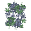
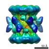
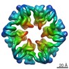
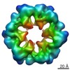
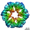
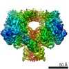
| Title | Architecture and nucleotide-driven conformational states of the Rvb1/Rvb2 dodecamer | |||||||||
 Map data Map data | ATP-state Rvb1/2 dodecamer | |||||||||
 Sample Sample |
| |||||||||
 Keywords Keywords | Rvb1 / Rvb2 | |||||||||
| Function / homology |  Function and homology information Function and homology informationTTT Hsp90 cochaperone complex / R2TP complex / Swr1 complex / Ino80 complex / box C/D snoRNP assembly / NuA4 histone acetyltransferase complex / 3'-5' DNA helicase activity / DNA helicase activity / rRNA processing / 5'-3' DNA helicase activity ...TTT Hsp90 cochaperone complex / R2TP complex / Swr1 complex / Ino80 complex / box C/D snoRNP assembly / NuA4 histone acetyltransferase complex / 3'-5' DNA helicase activity / DNA helicase activity / rRNA processing / 5'-3' DNA helicase activity / DNA helicase / protein stabilization / chromatin remodeling / DNA repair / regulation of transcription by RNA polymerase II / regulation of DNA-templated transcription / chromatin / ATP hydrolysis activity / ATP binding / nucleus Similarity search - Function | |||||||||
| Biological species |  | |||||||||
| Method | single particle reconstruction / cryo EM / Resolution: 9.5 Å | |||||||||
 Authors Authors | Ewens CA / Su M / Zhao L / Houry WA / Southworth DR | |||||||||
 Citation Citation | Journal: Structure / Year: 2016 Title: Architecture and Nucleotide-Dependent Conformational Changes of the Rvb1-Rvb2 AAA+ Complex Revealed by Cryoelectron Microscopy. Authors: Caroline A Ewens / Min Su / Liang Zhao / Nardin Nano / Walid A Houry / Daniel R Southworth /   Abstract: Rvb1 and Rvb2 are essential AAA+ proteins that interact together during the assembly and activity of diverse macromolecules including chromatin remodelers INO80 and SWR-C, and ribonucleoprotein ...Rvb1 and Rvb2 are essential AAA+ proteins that interact together during the assembly and activity of diverse macromolecules including chromatin remodelers INO80 and SWR-C, and ribonucleoprotein complexes including telomerase and snoRNPs. ATP hydrolysis by Rvb1/2 is required for function; however, the mechanism that drives substrate remodeling is unknown. Here we determined the architecture of the yeast Rvb1/2 dodecamer using cryoelectron microscopy and identify that the substrate-binding insertion domain undergoes conformational changes in response to nucleotide state. 2D and 3D classification defines the dodecamer flexibility, revealing distinct arrangements and the hexamer-hexamer interaction interface. Reconstructions of the apo, ATP, and ADP states identify that Rvb1/2 undergoes substantial conformational changes that include a twist in the insertion-domain position and a corresponding rotation of the AAA+ ring. These results reveal how the ATP hydrolysis cycle of the AAA+ domains directs insertion-domain movements that could provide mechanical force during remodeling or helicase activities. | |||||||||
| History |
|
- Structure visualization
| Movie |
Movie viewer |
|---|---|
| Structure viewer | EM map: SurfViewMolmilJmol/JSmol |
| Supplemental images |

- Downloads & links
-EMDB archive
| Map data | emd_3081.map.gz | 5.3 MB | EMDB map data format | |
|---|---|---|---|---|
| Header (meta data) | emd-3081-v30.xmlemd-3081.xml | 10.6 KB 10.6 KB | Display Display | EMDB header |
| Images | 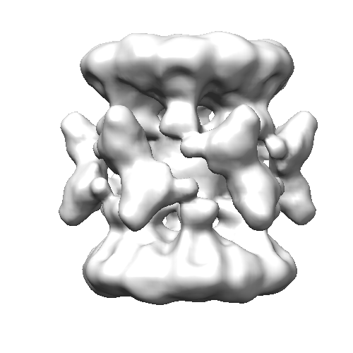 emd_3081.png emd_3081.png | 72.5 KB | ||
| Archive directory |  http://ftp.pdbj.org/pub/emdb/structures/EMD-3081ftp://ftp.pdbj.org/pub/emdb/structures/EMD-3081 http://ftp.pdbj.org/pub/emdb/structures/EMD-3081ftp://ftp.pdbj.org/pub/emdb/structures/EMD-3081 | HTTPS FTP |
-Related structure data


-Links
| EMDB pages | EMDB (EBI/PDBe) / EMDataResource |
|---|
-Map
| File | Download / File: emd_3081.map.gz / Format: CCP4 / Size: 7.8 MB / Type: IMAGE STORED AS FLOATING POINT NUMBER (4 BYTES) | ||||||||||||||||||||||||||||||||||||||||||||||||||||||||||||
|---|---|---|---|---|---|---|---|---|---|---|---|---|---|---|---|---|---|---|---|---|---|---|---|---|---|---|---|---|---|---|---|---|---|---|---|---|---|---|---|---|---|---|---|---|---|---|---|---|---|---|---|---|---|---|---|---|---|---|---|---|---|
| Annotation | ATP-state Rvb1/2 dodecamer | ||||||||||||||||||||||||||||||||||||||||||||||||||||||||||||
| Projections & slices | Image control
Images are generated by Spider. | ||||||||||||||||||||||||||||||||||||||||||||||||||||||||||||
| Voxel size | X=Y=Z: 2 Å | ||||||||||||||||||||||||||||||||||||||||||||||||||||||||||||
| Density |
| ||||||||||||||||||||||||||||||||||||||||||||||||||||||||||||
| Symmetry | Space group: 1 | ||||||||||||||||||||||||||||||||||||||||||||||||||||||||||||
| Details | EMDB XML:
CCP4 map header:
| ||||||||||||||||||||||||||||||||||||||||||||||||||||||||||||
 Z (Sec.)
 Y (Row.)
 X (Col.)


-Supplemental data
- Sample components
-Entire : ATP-state Rvb1/2 dodecamer
| Entire | Name: ATP-state Rvb1/2 dodecamer |
|---|---|
| Components |
|
-Supramolecule #1000: ATP-state Rvb1/2 dodecamer
| Supramolecule | Name: ATP-state Rvb1/2 dodecamer / type: sample / ID: 1000 / Oligomeric state: heterododecamer / Number unique components: 2 |
|---|---|
| Molecular weight | Experimental: 600 KDa / Theoretical: 600 KDa Method: size exclusion chromatography with multiangle light scattering |
-Macromolecule #1: Rvb1
| Macromolecule | Name: Rvb1 / type: protein_or_peptide / ID: 1 / Name.synonym: Pontin / Number of copies: 6 / Oligomeric state: Hexamer / Recombinant expression: Yes |
|---|---|
| Source (natural) | Organism: |
| Molecular weight | Experimental: 50 KDa / Theoretical: 50 KDa |
| Recombinant expression | Organism:  |
| Sequence | UniProtKB: RuvB-like protein 1 |
-Macromolecule #2: Rvb2
| Macromolecule | Name: Rvb2 / type: protein_or_peptide / ID: 2 / Name.synonym: Reptin / Number of copies: 6 / Oligomeric state: Hexamer / Recombinant expression: Yes |
|---|---|
| Source (natural) | Organism: |
| Molecular weight | Experimental: 50 KDa / Theoretical: 50 KDa |
| Recombinant expression | Organism: |
| Sequence | UniProtKB: RuvB-like protein 2 |
-Experimental details
-Structure determination
| Method | cryo EM |
|---|---|
 Processing Processing | single particle reconstruction |
| Aggregation state | particle |
-Sample preparation
| Concentration | 0.3 mg/mL |
|---|---|
| Buffer | pH: 7.4 Details: 25mM HEPES, 150mM KCl, 6mM betaME, 5mM MgCl2, 200microM AMP-PNP |
| Vitrification | Cryogen name: ETHANE / Chamber humidity: 90 % / Instrument: FEI VITROBOT MARK IV / Method: 1s blot |
- Electron microscopy
| Microscope | FEI TITAN KRIOS |
|---|---|
| Date | Apr 23, 2015 |
| Image recording | Category: CCD / Film or detector model: GATAN K2 (4k x 4k) / Number real images: 1600 / Average electron dose: 5 e/Å2 Details: Every image is the average of 30 frames recorded by the direct electron detector |
| Electron beam | Acceleration voltage: 300 kV / Electron source:  FIELD EMISSION GUN FIELD EMISSION GUN |
| Electron optics | Illumination mode: FLOOD BEAM / Imaging mode: DARK FIELD / Cs: 2 mm / Nominal defocus max: 3.0 µm / Nominal defocus min: 1.8 µm / Nominal magnification: 29000 |
| Sample stage | Specimen holder model: FEI TITAN KRIOS AUTOGRID HOLDER |
| Experimental equipment |  Model: Titan Krios / Image courtesy: FEI Company |
-Image processing
| CTF correction | Details: per micrograph |
|---|---|
| Final reconstruction | Applied symmetry - Point group: D6 (2x6 fold dihedral) / Algorithm: OTHER / Resolution.type: BY AUTHOR / Resolution: 9.5 Å / Resolution method: OTHER / Software - Name: relion, boxer, eman2, cftfind, spider / Number images used: 13630 |
-Atomic model buiding 1
| Initial model | PDB ID: Chain - #0 - Chain ID: A / Chain - #1 - Chain ID: B |
|---|---|
| Software | Name: chimera, situs |
| Details | The domains were separately fitted by manual docking |
| Refinement | Space: REAL / Protocol: RIGID BODY FIT / Target criteria: cross correlation |