 Movie
Movie Controller


+ Open data

- Basic information
| Entry | Database: EMDB / ID: EMD-30495 | |||||||||
|---|---|---|---|---|---|---|---|---|---|---|
| Title | Cryo-EM structure of human TMEM120A/TACAN | |||||||||
 Map data Map data | ||||||||||
 Sample Sample |
| |||||||||
 Keywords Keywords | MEMBRANE PROTEIN | |||||||||
| Function / homology |  Function and homology information Function and homology informationcoenzyme A binding / protein heterooligomerization / nuclear inner membrane / monoatomic ion channel activity / detection of mechanical stimulus involved in sensory perception of pain / fat cell differentiation / antiviral innate immune response / bioluminescence / generation of precursor metabolites and energy / protein homooligomerization ...coenzyme A binding / protein heterooligomerization / nuclear inner membrane / monoatomic ion channel activity / detection of mechanical stimulus involved in sensory perception of pain / fat cell differentiation / antiviral innate immune response / bioluminescence / generation of precursor metabolites and energy / protein homooligomerization / monoatomic ion transmembrane transport / endoplasmic reticulum / membrane / plasma membrane Similarity search - Function | |||||||||
| Biological species |  Anaplasma marginale (bacteria) / Anaplasma marginale (bacteria) /  Homo sapiens (human) Homo sapiens (human) | |||||||||
| Method | single particle reconstruction / cryo EM / Resolution: 3.4 Å | |||||||||
 Authors Authors | Yan Z / Wu J | |||||||||
 Citation Citation | Journal: Cell Discov / Year: 2021 Title: Cryo-EM structures of human TMEM120A and TMEM120B. Authors: Meng Ke / Yue Yu / Changjian Zhao / Shirong Lai / Qiang Su / Weidan Yuan / Lina Yang / Dong Deng / Kun Wu / Weizheng Zeng / Jia Geng / Jianping Wu / Zhen Yan /  | |||||||||
| History |
|
- Structure visualization
| Movie |
Movie viewer |
|---|---|
| Structure viewer | EM map: SurfViewMolmilJmol/JSmol |
| Supplemental images |

- Downloads & links
-EMDB archive
| Map data | emd_30495.map.gz | 482.5 KB | EMDB map data format | |
|---|---|---|---|---|
| Header (meta data) | emd-30495-v30.xmlemd-30495.xml | 18.8 KB 18.8 KB | Display Display | EMDB header |
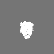
| Images | 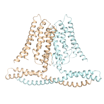 emd_30495.png emd_30495.png | 97.2 KB | ||
| Masks | emd_30495_msk_1.map | 40.6 MB | Mask map | |
| Filedesc metadata | emd-30495.cif.gz | 6.1 KB | ||
| Others | emd_30495_additional_1.map.gzemd_30495_half_map_1.map.gzemd_30495_half_map_2.map.gz | 3.5 MB 37.7 MB 37.7 MB | ||
| Archive directory |  http://ftp.pdbj.org/pub/emdb/structures/EMD-30495ftp://ftp.pdbj.org/pub/emdb/structures/EMD-30495 http://ftp.pdbj.org/pub/emdb/structures/EMD-30495ftp://ftp.pdbj.org/pub/emdb/structures/EMD-30495 | HTTPS FTP |
-Related structure data
| Related structure data |  7cxrMC  7f73C M: atomic model generated by this map C: citing same article ( |
|---|---|
| Similar structure data |


-Links
| EMDB pages | EMDB (EBI/PDBe) / EMDataResource |
|---|---|
| Related items in Molecule of the Month |


-Map
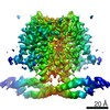
| File | Download / File: emd_30495.map.gz / Format: CCP4 / Size: 40.6 MB / Type: IMAGE STORED AS FLOATING POINT NUMBER (4 BYTES) | ||||||||||||||||||||||||||||||||||||||||||||||||||||||||||||||||||||
|---|---|---|---|---|---|---|---|---|---|---|---|---|---|---|---|---|---|---|---|---|---|---|---|---|---|---|---|---|---|---|---|---|---|---|---|---|---|---|---|---|---|---|---|---|---|---|---|---|---|---|---|---|---|---|---|---|---|---|---|---|---|---|---|---|---|---|---|---|---|
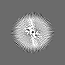
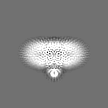
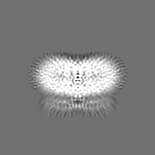
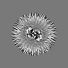
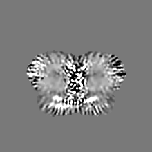
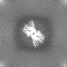
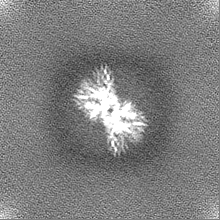
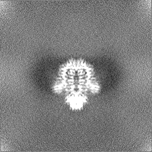
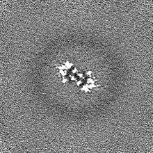
| Projections & slices | Image control
Images are generated by Spider. | ||||||||||||||||||||||||||||||||||||||||||||||||||||||||||||||||||||
| Voxel size | X=Y=Z: 1.087 Å | ||||||||||||||||||||||||||||||||||||||||||||||||||||||||||||||||||||
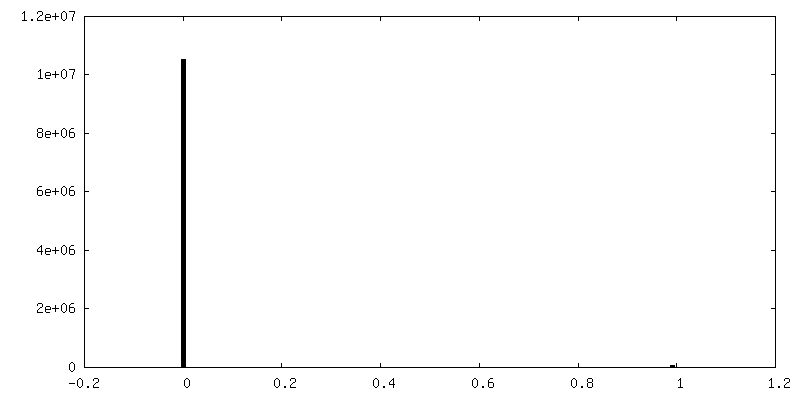
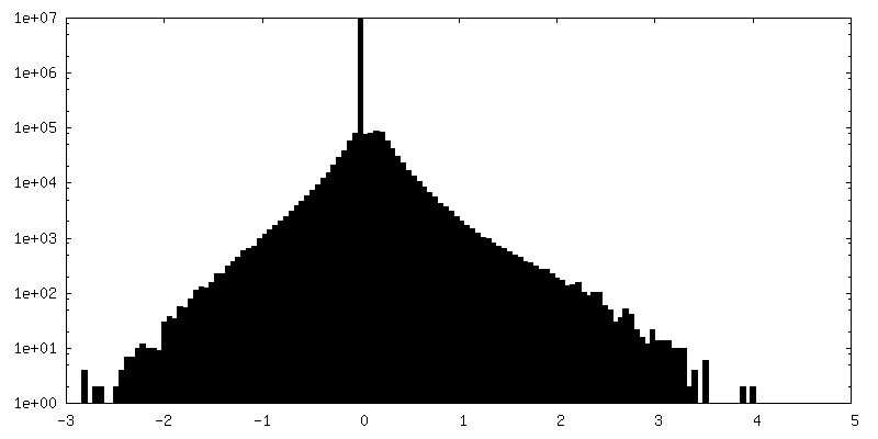
| Density |
| ||||||||||||||||||||||||||||||||||||||||||||||||||||||||||||||||||||
| Symmetry | Space group: 1 | ||||||||||||||||||||||||||||||||||||||||||||||||||||||||||||||||||||
| Details | EMDB XML:
CCP4 map header:
| ||||||||||||||||||||||||||||||||||||||||||||||||||||||||||||||||||||
 Z (Sec.)
 Y (Row.)
 X (Col.)


-Supplemental data
-Mask #1
| File | emd_30495_msk_1.map | ||||||||||||
|---|---|---|---|---|---|---|---|---|---|---|---|---|---|
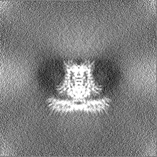
| Projections & Slices |
| ||||||||||||
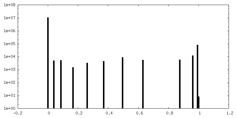
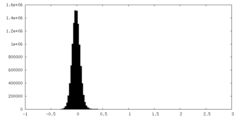
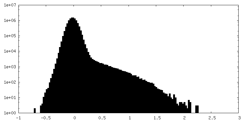
| Density Histograms |

-Additional map: #1
| File | emd_30495_additional_1.map | ||||||||||||
|---|---|---|---|---|---|---|---|---|---|---|---|---|---|
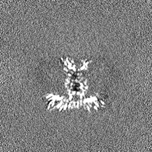
| Projections & Slices |
| ||||||||||||
| Density Histograms |


-Half map: #1
| File | emd_30495_half_map_1.map | ||||||||||||
|---|---|---|---|---|---|---|---|---|---|---|---|---|---|
| Projections & Slices |
| ||||||||||||
| Density Histograms |


-Half map: #2
| File | emd_30495_half_map_2.map | ||||||||||||
|---|---|---|---|---|---|---|---|---|---|---|---|---|---|
| Projections & Slices |
| ||||||||||||
| Density Histograms |


- Sample components
-Entire : Homodimer of human TMEM120A
| Entire | Name: Homodimer of human TMEM120A |
|---|---|
| Components |
|
-Supramolecule #1: Homodimer of human TMEM120A
| Supramolecule | Name: Homodimer of human TMEM120A / type: complex / ID: 1 / Parent: 0 / Macromolecule list: all |
|---|---|
| Source (natural) | Organism: Anaplasma marginale (bacteria) |
-Macromolecule #1: MCherry fluorescent protein,Ion channel TACAN
| Macromolecule | Name: MCherry fluorescent protein,Ion channel TACAN / type: protein_or_peptide / ID: 1 / Number of copies: 2 / Enantiomer: LEVO |
|---|---|
| Source (natural) | Organism: Homo sapiens (human) |
| Molecular weight | Theoretical: 72.643555 KDa |
| Recombinant expression | Organism: Homo sapiens (human) |
| Sequence | String: MDYKDDDDKG SDYKDDDDKG SDYKDDDDKG SDEVDAMVSK GEEDNMAIIK EFMRFKVHME GSVNGHEFEI EGEGEGRPYE GTQTAKLKV TKGGPLPFAW DILSPQFMYG SKAYVKHPAD IPDYLKLSFP EGFKWERVMN FEDGGVVTVT QDSSLQDGEF I YKVKLRGT ...String: MDYKDDDDKG SDYKDDDDKG SDYKDDDDKG SDEVDAMVSK GEEDNMAIIK EFMRFKVHME GSVNGHEFEI EGEGEGRPYE GTQTAKLKV TKGGPLPFAW DILSPQFMYG SKAYVKHPAD IPDYLKLSFP EGFKWERVMN FEDGGVVTVT QDSSLQDGEF I YKVKLRGT NFPSDGPVMQ KKTMGWEASS ERMYPEDGAL KGEIKQRLKL KDGGHYDAEV KTTYKAKKPV QLPGAYNVNI KL DITSHNE DYTIVEQYER AEGRHSTGGM DELYKLEVLF QGPEFMQPPP PGPLGDCLRD WEDLQQDFQN IQETHRLYRL KLE ELTKLQ NNCTSSITRQ KKRLQELALA LKKCKPSLPA EAEGAAQELE NQMKERQGLF FDMEAYLPKK NGLYLSLVLG NVNV TLLSK QAKFAYKDEY EKFKLYLTII LILISFTCRF LLNSRVTDAA FNFLLVWYYC TLTIRESILI NNGSRIKGWW VFHHY VSTF LSGVMLTWPD GLMYQKFRNQ FLSFSMYQSF VQFLQYYYQS GCLYRLRALG ERHTMDLTVE GFQSWMWRGL TFLLPF LFF GHFWQLFNAL TLFNLAQDPQ CKEWQVLMCG FPFLLLFLGN FFTTLRVVHH KFHSQRHGSK KD UniProtKB: MCherry fluorescent protein, Transmembrane protein 120A |
-Experimental details
-Structure determination
| Method | cryo EM |
|---|---|
 Processing Processing | single particle reconstruction |
| Aggregation state | particle |
-Sample preparation
| Buffer | pH: 8 |
|---|---|
| Vitrification | Cryogen name: ETHANE / Instrument: FEI VITROBOT MARK IV |
- Electron microscopy
| Microscope | FEI TITAN KRIOS |
|---|---|
| Image recording | Film or detector model: GATAN K3 (6k x 4k) / Average electron dose: 50.0 e/Å2 |
| Electron beam | Acceleration voltage: 300 kV / Electron source:  FIELD EMISSION GUN FIELD EMISSION GUN |
| Electron optics | Illumination mode: FLOOD BEAM / Imaging mode: BRIGHT FIELD |
| Experimental equipment |  Model: Titan Krios / Image courtesy: FEI Company |
