[English] 日本語
 Yorodumi
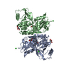
- EMDB-30169: Cryo-EM structure of a human ATP11C-CDC50A flippase in PtdEtn-occ... -
+ Open data
- Basic information
| Entry | Database: EMDB / ID: EMD-30169 | ||||||||||||
|---|---|---|---|---|---|---|---|---|---|---|---|---|---|
| Title | Cryo-EM structure of a human ATP11C-CDC50A flippase in PtdEtn-occluded E2-AlF state | ||||||||||||
 Map data Map data | |||||||||||||
 Sample Sample |
| ||||||||||||
 Keywords Keywords | Flippase / P-type ATPase / P4-ATPase / Phospholipid / MEMBRANE PROTEIN | ||||||||||||
| Function / homology |  Function and homology information Function and homology informationpositive regulation of phospholipid translocation / aminophospholipid flippase activity / aminophospholipid transport / phosphatidylserine flippase activity / protein localization to endosome / ATPase-coupled intramembrane lipid transporter activity / phospholipid-translocating ATPase complex / positive regulation of protein exit from endoplasmic reticulum / phosphatidylserine floppase activity / phosphatidylethanolamine flippase activity ...positive regulation of phospholipid translocation / aminophospholipid flippase activity / aminophospholipid transport / phosphatidylserine flippase activity / protein localization to endosome / ATPase-coupled intramembrane lipid transporter activity / phospholipid-translocating ATPase complex / positive regulation of protein exit from endoplasmic reticulum / phosphatidylserine floppase activity / phosphatidylethanolamine flippase activity / xenobiotic transmembrane transport / P-type phospholipid transporter / phospholipid translocation / azurophil granule membrane / transport vesicle membrane / Ion transport by P-type ATPases / specific granule membrane / recycling endosome / positive regulation of neuron projection development / recycling endosome membrane / late endosome membrane / early endosome membrane / monoatomic ion transmembrane transport / apical plasma membrane / lysosomal membrane / Neutrophil degranulation / endoplasmic reticulum membrane / structural molecule activity / Golgi apparatus / magnesium ion binding / endoplasmic reticulum / ATP hydrolysis activity / ATP binding / membrane / plasma membrane Similarity search - Function | ||||||||||||
| Biological species |  Homo sapiens (human) Homo sapiens (human) | ||||||||||||
| Method | single particle reconstruction / cryo EM / Resolution: 3.9 Å | ||||||||||||
 Authors Authors | Abe K / Nishizawa T | ||||||||||||
| Funding support |  Japan, 3 items Japan, 3 items
| ||||||||||||
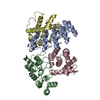
 Citation Citation |  Journal: Cell Rep / Year: 2020 Journal: Cell Rep / Year: 2020Title: Transport Cycle of Plasma Membrane Flippase ATP11C by Cryo-EM. Authors: Hanayo Nakanishi / Tomohiro Nishizawa / Katsumori Segawa / Osamu Nureki / Yoshinori Fujiyoshi / Shigekazu Nagata / Kazuhiro Abe /  Abstract: ATP11C, a plasma membrane phospholipid flippase, maintains the asymmetric distribution of phosphatidylserine accumulated in the inner leaflet. Caspase-dependent inactivation of ATP11C is essential ...ATP11C, a plasma membrane phospholipid flippase, maintains the asymmetric distribution of phosphatidylserine accumulated in the inner leaflet. Caspase-dependent inactivation of ATP11C is essential for an apoptotic "eat me" signal, phosphatidylserine exposure, which prompts phagocytes to engulf cells. We show six cryo-EM structures of ATP11C at 3.0-4.0 Å resolution in five different states of the transport cycle. A structural comparison reveals phosphorylation-driven domain movements coupled with phospholipid binding. Three structures of phospholipid-bound states visualize phospholipid translocation accompanied by the rearrangement of transmembrane helices and an unwound portion at the occlusion site, and thus they detail the basis for head group recognition and the locality of the protein-bound acyl chains in transmembrane grooves. Invariant Lys880 and the surrounding hydrogen-bond network serve as a pivot point for helix bending and precise P domain inclination, which is crucial for dephosphorylation. The structures detail key features of phospholipid translocation by ATP11C, and a common basic mechanism for flippases is emerging. | ||||||||||||
| History |
|
- Structure visualization
| Movie |
 Movie viewer Movie viewer |
|---|---|
| Structure viewer | EM map:  SurfView SurfView Molmil Molmil Jmol/JSmol Jmol/JSmol |
| Supplemental images |
- Downloads & links
-EMDB archive
| Map data |  emd_30169.map.gz emd_30169.map.gz | 2.7 MB |  EMDB map data format EMDB map data format | |
|---|---|---|---|---|
| Header (meta data) |  emd-30169-v30.xml emd-30169-v30.xml emd-30169.xml emd-30169.xml | 17.3 KB 17.3 KB | Display Display |  EMDB header EMDB header |
| Images |  emd_30169.png emd_30169.png | 109.8 KB | ||
| Filedesc metadata |  emd-30169.cif.gz emd-30169.cif.gz | 7.1 KB | ||
| Archive directory |  http://ftp.pdbj.org/pub/emdb/structures/EMD-30169 http://ftp.pdbj.org/pub/emdb/structures/EMD-30169 ftp://ftp.pdbj.org/pub/emdb/structures/EMD-30169 ftp://ftp.pdbj.org/pub/emdb/structures/EMD-30169 | HTTPS FTP |
-Validation report
| Summary document |  emd_30169_validation.pdf.gz emd_30169_validation.pdf.gz | 418.7 KB | Display |  EMDB validaton report EMDB validaton report |
|---|---|---|---|---|
| Full document |  emd_30169_full_validation.pdf.gz emd_30169_full_validation.pdf.gz | 418.3 KB | Display | |
| Data in XML |  emd_30169_validation.xml.gz emd_30169_validation.xml.gz | 5.4 KB | Display | |
| Data in CIF |  emd_30169_validation.cif.gz emd_30169_validation.cif.gz | 6.2 KB | Display | |
| Arichive directory |  https://ftp.pdbj.org/pub/emdb/validation_reports/EMD-30169 https://ftp.pdbj.org/pub/emdb/validation_reports/EMD-30169 ftp://ftp.pdbj.org/pub/emdb/validation_reports/EMD-30169 ftp://ftp.pdbj.org/pub/emdb/validation_reports/EMD-30169 | HTTPS FTP |
-Related structure data
| Related structure data |  7bswMC  7bspC  7bsqC  7bssC  7bsuC  7bsvC M: atomic model generated by this map C: citing same article ( |
|---|---|
| Similar structure data |
- Links
Links
| EMDB pages |  EMDB (EBI/PDBe) / EMDB (EBI/PDBe) /  EMDataResource EMDataResource |
|---|---|
| Related items in Molecule of the Month |
- Map
Map
| File |  Download / File: emd_30169.map.gz / Format: CCP4 / Size: 15.6 MB / Type: IMAGE STORED AS FLOATING POINT NUMBER (4 BYTES) Download / File: emd_30169.map.gz / Format: CCP4 / Size: 15.6 MB / Type: IMAGE STORED AS FLOATING POINT NUMBER (4 BYTES) | ||||||||||||||||||||||||||||||||||||||||||||||||||||||||||||||||||||
|---|---|---|---|---|---|---|---|---|---|---|---|---|---|---|---|---|---|---|---|---|---|---|---|---|---|---|---|---|---|---|---|---|---|---|---|---|---|---|---|---|---|---|---|---|---|---|---|---|---|---|---|---|---|---|---|---|---|---|---|---|---|---|---|---|---|---|---|---|---|
| Projections & slices | Image control
Images are generated by Spider. | ||||||||||||||||||||||||||||||||||||||||||||||||||||||||||||||||||||
| Voxel size | X=Y=Z: 1.35 Å | ||||||||||||||||||||||||||||||||||||||||||||||||||||||||||||||||||||
| Density |
| ||||||||||||||||||||||||||||||||||||||||||||||||||||||||||||||||||||
| Symmetry | Space group: 1 | ||||||||||||||||||||||||||||||||||||||||||||||||||||||||||||||||||||
| Details | EMDB XML:
CCP4 map header:
| ||||||||||||||||||||||||||||||||||||||||||||||||||||||||||||||||||||
-Supplemental data
- Sample components
-Entire : human ATP11C-CDC50A flippase
| Entire | Name: human ATP11C-CDC50A flippase |
|---|---|
| Components |
|
-Supramolecule #1: human ATP11C-CDC50A flippase
| Supramolecule | Name: human ATP11C-CDC50A flippase / type: complex / ID: 1 / Parent: 0 / Macromolecule list: #1-#2 |
|---|---|
| Source (natural) | Organism:  Homo sapiens (human) Homo sapiens (human) |
| Molecular weight | Theoretical: 180 KDa |
-Macromolecule #1: ATP11C
| Macromolecule | Name: ATP11C / type: protein_or_peptide / ID: 1 / Number of copies: 1 / Enantiomer: LEVO |
|---|---|
| Source (natural) | Organism:  Homo sapiens (human) Homo sapiens (human) |
| Molecular weight | Theoretical: 124.403617 KDa |
| Recombinant expression | Organism:  Homo sapiens (human) Homo sapiens (human) |
| Sequence | String: RFCAGEEKRV GTRTVFVGNH PVSETEAYIA QRFCDNRIVS SKYTLWNFLP KNLFEQFRRI ANFYFLIIFL VQVTVDTPTS PVTSGLPLF FVITVTAIKQ GYEDWLRHRA DNEVNKSTVY IIENAKRVRK ESEKIKVGDV VEVQADETFP CDLILLSSCT T DGTCYVTT ...String: RFCAGEEKRV GTRTVFVGNH PVSETEAYIA QRFCDNRIVS SKYTLWNFLP KNLFEQFRRI ANFYFLIIFL VQVTVDTPTS PVTSGLPLF FVITVTAIKQ GYEDWLRHRA DNEVNKSTVY IIENAKRVRK ESEKIKVGDV VEVQADETFP CDLILLSSCT T DGTCYVTT ASLDGESNCK THYAVRDTIA LCTAESIDTL RAAIECEQPQ PDLYKFVGRI NIYSNSLEAV ARSLGPENLL LK GATLKNT EKIYGVAVYT GMETKMALNY QGKSQKRSAV EKSINAFLIV YLFILLTKAA VCTTLKYVWQ STPYNDEPWY NQK TQKERE TLKVLKMFTD FLSFMVLFNF IIPVSMYVTV EMQKFLGSFF ISWDKDFYDE EINEGALVNT SDLNEELGQV DYVF TDKTG TLTENSMEFI ECCIDGHKYK GVTQEVDGLS QTDGTLTYFD KVDKNREELF LRALCLCHTV EIKTNDAVDG ATESA ELTY ISSSPDEIAL VKGAKRYGFT FLGNRNGYMR VENQRKEIEE YELLHTLNFD AVRRRMSVIV KTQEGDILLF CKGADS AVF PRVQNHEIEL TKVHVERNAM DGYRTLCVAF KEIAPDDYER INRQLIEAKM ALQDREEKME KVFDDIETNM NLIGATA VE DKLQDQAAET IEALHAAGLK VWVLTGDKME TAKSTCYACR LFQTNTELLE LTTKTIEESE RKEDRLHELL IEYRKKLL H EFPKSTRSFK KAWTEHQEYG LIIDGSTLSL ILNSSQDSSS NNYKSIFLQI CMKCTAVLCC RMAPLQKAQI VRMVKNLKG SPITLSIGDG ANDVSMILES HVGIGIKGKE GRQAARNSDY SVPKFKHLKK LLLAHGHLYY VRIAHLVQYF FYKNLCFILP QFLYQFFCG FSQQPLYDAA YLTMYNICFT SLPILAYSLL EQHINIDTLT SDPRLYMKIS GNAMLQLGPF LYWTFLAAFE G TVFFFGTY FLFQTASLEE NGKVYGNWTF GTIVFTVLVF TVTLKLALDT RFWTWINHFV IWGSLAFYVF FSFFWGGIIW PF LKQQRMY FVFAQMLSSV STWLAIILLI FISLFPEILL IVLKNVR |
-Macromolecule #2: CDC50A
| Macromolecule | Name: CDC50A / type: protein_or_peptide / ID: 2 / Number of copies: 1 / Enantiomer: LEVO |
|---|---|
| Source (natural) | Organism:  Homo sapiens (human) Homo sapiens (human) |
| Molecular weight | Theoretical: 40.900801 KDa |
| Recombinant expression | Organism:  Homo sapiens (human) Homo sapiens (human) |
| Sequence | String: MAMNYNAKDE VDGGPPCAPG GTAKTRRPDN TAFKQQRLPA WQPILTAGTV LPIFFIIGLI FIPIGIGIFV TSNNIREIEI DYTGTEPSS PCNKCLSPDV TPCFCTINFT LEKSFEGNVF MYYGLSNFYQ NHRRYVKSRD DSQLNGDSSA LLNPSKECEP Y RRNEDKPI ...String: MAMNYNAKDE VDGGPPCAPG GTAKTRRPDN TAFKQQRLPA WQPILTAGTV LPIFFIIGLI FIPIGIGIFV TSNNIREIEI DYTGTEPSS PCNKCLSPDV TPCFCTINFT LEKSFEGNVF MYYGLSNFYQ NHRRYVKSRD DSQLNGDSSA LLNPSKECEP Y RRNEDKPI APCGAIMNSM FNDTLELFLI GQDSYPIPIA LKKKGIAWWT DKNVKFRNPP GGDNLEERFK GTTKPVNWLK PV YMLDSDP DNNGFINEDF IVWMRTAALP TFRKLYRLIE RKSDLHPTLP AGRYWLNVTY NYPVHYFDGR KRMILSTISW MGG KNPFLG IAYIAVGSIS FLLGVVLLVI NHKYRNSSNT ADITI |
-Macromolecule #3: TETRAFLUOROALUMINATE ION
| Macromolecule | Name: TETRAFLUOROALUMINATE ION / type: ligand / ID: 3 / Number of copies: 1 / Formula: ALF |
|---|---|
| Molecular weight | Theoretical: 102.975 Da |
| Chemical component information | 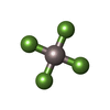 ChemComp-ALF: |
-Macromolecule #4: 1,2-dioleoyl-sn-glycero-3-phosphoethanolamine
| Macromolecule | Name: 1,2-dioleoyl-sn-glycero-3-phosphoethanolamine / type: ligand / ID: 4 / Number of copies: 1 / Formula: PEE |
|---|---|
| Molecular weight | Theoretical: 744.034 Da |
| Chemical component information | 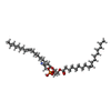 ChemComp-PEE: |
-Macromolecule #5: 2-acetamido-2-deoxy-beta-D-glucopyranose
| Macromolecule | Name: 2-acetamido-2-deoxy-beta-D-glucopyranose / type: ligand / ID: 5 / Number of copies: 4 / Formula: NAG |
|---|---|
| Molecular weight | Theoretical: 221.208 Da |
| Chemical component information |  ChemComp-NAG: |
-Macromolecule #6: alpha-D-mannopyranose
| Macromolecule | Name: alpha-D-mannopyranose / type: ligand / ID: 6 / Number of copies: 1 / Formula: MAN |
|---|---|
| Molecular weight | Theoretical: 180.156 Da |
| Chemical component information | 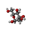 ChemComp-MAN: |
-Experimental details
-Structure determination
| Method | cryo EM |
|---|---|
 Processing Processing | single particle reconstruction |
| Aggregation state | particle |
- Sample preparation
| Concentration | 8 mg/mL |
|---|---|
| Buffer | pH: 6.5 |
| Vitrification | Cryogen name: ETHANE / Chamber humidity: 99 % / Chamber temperature: 298 K / Instrument: FEI VITROBOT MARK IV |
- Electron microscopy
| Microscope | TFS KRIOS |
|---|---|
| Image recording | Film or detector model: GATAN K3 (6k x 4k) / Average electron dose: 64.0 e/Å2 |
| Electron beam | Acceleration voltage: 300 kV / Electron source:  FIELD EMISSION GUN FIELD EMISSION GUN |
| Electron optics | Illumination mode: FLOOD BEAM / Imaging mode: BRIGHT FIELD |
| Experimental equipment |  Model: Titan Krios / Image courtesy: FEI Company |
- Image processing
| Startup model | Type of model: NONE |
|---|---|
| Final reconstruction | Resolution.type: BY AUTHOR / Resolution: 3.9 Å / Resolution method: FSC 0.143 CUT-OFF / Software - Name: RELION (ver. 3) / Number images used: 400000 |
| Initial angle assignment | Type: MAXIMUM LIKELIHOOD |
| Final angle assignment | Type: MAXIMUM LIKELIHOOD |
-Atomic model buiding 1
| Refinement | Protocol: RIGID BODY FIT |
|---|---|
| Output model |  PDB-7bsw: |
 Movie
Movie Controller






 Z (Sec.)
 Y (Row.)
 X (Col.)


