+ データを開く
- 基本情報
基本情報
| 登録情報 | データベース: EMDB / ID: EMD-2455 | |||||||||
|---|---|---|---|---|---|---|---|---|---|---|
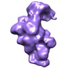
| タイトル | 30S Ribosome Subunit Assembly Intermediates, Intermediate 4c | |||||||||
 マップデータ マップデータ | Assembly Intermediate Group 4c | |||||||||
 試料 試料 |
| |||||||||
 キーワード キーワード | Ribosome Assembly / 30S Ribosome Subunit / 30S Assembly / Time-resolved electron microscopy / Discovery Single-particle Profiling / 30S Ribosome Subunit Assembly Intermediate | |||||||||
| 生物種 |  | |||||||||
| 手法 | 単粒子再構成法 / ネガティブ染色法 / 解像度: 47.07 Å | |||||||||
 データ登録者 データ登録者 | Mulder AM / Yoshioka C / Beck A / Bunner A / Milligan RA / Potter CS / Carragher B / Williamson JR | |||||||||
 引用 引用 |  ジャーナル: Science / 年: 2010 ジャーナル: Science / 年: 2010タイトル: Visualizing ribosome biogenesis: parallel assembly pathways for the 30S subunit. 著者: Anke M Mulder / Craig Yoshioka / Andrea H Beck / Anne E Bunner / Ronald A Milligan / Clinton S Potter / Bridget Carragher / James R Williamson /  要旨: Ribosomes are self-assembling macromolecular machines that translate DNA into proteins, and an understanding of ribosome biogenesis is central to cellular physiology. Previous studies on the ...Ribosomes are self-assembling macromolecular machines that translate DNA into proteins, and an understanding of ribosome biogenesis is central to cellular physiology. Previous studies on the Escherichia coli 30S subunit suggest that ribosome assembly occurs via multiple parallel pathways rather than through a single rate-limiting step, but little mechanistic information is known about this process. Discovery single-particle profiling (DSP), an application of time-resolved electron microscopy, was used to obtain more than 1 million snapshots of assembling 30S subunits, identify and visualize the structures of 14 assembly intermediates, and monitor the population flux of these intermediates over time. DSP results were integrated with mass spectrometry data to construct the first ribosome-assembly mechanism that incorporates binding dependencies, rate constants, and structural characterization of populated intermediates. | |||||||||
| 履歴 |
|
- 構造の表示
構造の表示
| ムービー |
 ムービービューア ムービービューア |
|---|---|
| 構造ビューア | EMマップ:  SurfView SurfView Molmil Molmil Jmol/JSmol Jmol/JSmol |
| 添付画像 |
- ダウンロードとリンク
-EMDBアーカイブ
| マップデータ |  emd_2455.map.gz emd_2455.map.gz | 15 MB |  EMDBマップデータ形式 EMDBマップデータ形式 | |
|---|---|---|---|---|
| ヘッダ (付随情報) |  emd-2455-v30.xml emd-2455-v30.xml emd-2455.xml emd-2455.xml | 7.5 KB 7.5 KB | 表示 表示 |  EMDBヘッダ EMDBヘッダ |
| 画像 |  EMD-2455grp4c.tif EMD-2455grp4c.tif | 155.8 KB | ||
| アーカイブディレクトリ |  http://ftp.pdbj.org/pub/emdb/structures/EMD-2455 http://ftp.pdbj.org/pub/emdb/structures/EMD-2455 ftp://ftp.pdbj.org/pub/emdb/structures/EMD-2455 ftp://ftp.pdbj.org/pub/emdb/structures/EMD-2455 | HTTPS FTP |
-検証レポート
| 文書・要旨 |  emd_2455_validation.pdf.gz emd_2455_validation.pdf.gz | 192.4 KB | 表示 |  EMDB検証レポート EMDB検証レポート |
|---|---|---|---|---|
| 文書・詳細版 |  emd_2455_full_validation.pdf.gz emd_2455_full_validation.pdf.gz | 191.6 KB | 表示 | |
| XML形式データ |  emd_2455_validation.xml.gz emd_2455_validation.xml.gz | 6.6 KB | 表示 | |
| アーカイブディレクトリ |  https://ftp.pdbj.org/pub/emdb/validation_reports/EMD-2455 https://ftp.pdbj.org/pub/emdb/validation_reports/EMD-2455 ftp://ftp.pdbj.org/pub/emdb/validation_reports/EMD-2455 ftp://ftp.pdbj.org/pub/emdb/validation_reports/EMD-2455 | HTTPS FTP |
-関連構造データ
- リンク
リンク
| EMDBのページ |  EMDB (EBI/PDBe) / EMDB (EBI/PDBe) /  EMDataResource EMDataResource |
|---|---|
| 「今月の分子」の関連する項目 |
- マップ
マップ
| ファイル |  ダウンロード / ファイル: emd_2455.map.gz / 形式: CCP4 / 大きさ: 100.6 MB / タイプ: IMAGE STORED AS FLOATING POINT NUMBER (4 BYTES) ダウンロード / ファイル: emd_2455.map.gz / 形式: CCP4 / 大きさ: 100.6 MB / タイプ: IMAGE STORED AS FLOATING POINT NUMBER (4 BYTES) | ||||||||||||||||||||||||||||||||||||||||||||||||||||||||||||||||||||
|---|---|---|---|---|---|---|---|---|---|---|---|---|---|---|---|---|---|---|---|---|---|---|---|---|---|---|---|---|---|---|---|---|---|---|---|---|---|---|---|---|---|---|---|---|---|---|---|---|---|---|---|---|---|---|---|---|---|---|---|---|---|---|---|---|---|---|---|---|---|
| 注釈 | Assembly Intermediate Group 4c | ||||||||||||||||||||||||||||||||||||||||||||||||||||||||||||||||||||
| 投影像・断面図 | 画像のコントロール
画像は Spider により作成 | ||||||||||||||||||||||||||||||||||||||||||||||||||||||||||||||||||||
| ボクセルのサイズ | X=Y=Z: 1.55 Å | ||||||||||||||||||||||||||||||||||||||||||||||||||||||||||||||||||||
| 密度 |
| ||||||||||||||||||||||||||||||||||||||||||||||||||||||||||||||||||||
| 対称性 | 空間群: 1 | ||||||||||||||||||||||||||||||||||||||||||||||||||||||||||||||||||||
| 詳細 | EMDB XML:
CCP4マップ ヘッダ情報:
| ||||||||||||||||||||||||||||||||||||||||||||||||||||||||||||||||||||
-添付データ
- 試料の構成要素
-全体 : 30S Reconstitution Reaction
| 全体 | 名称: 30S Reconstitution Reaction |
|---|---|
| 要素 |
|
-超分子 #1000: 30S Reconstitution Reaction
| 超分子 | 名称: 30S Reconstitution Reaction / タイプ: sample / ID: 1000 / Number unique components: 21 |
|---|---|
| 分子量 | 理論値: 789 KDa |
-超分子 #1: 30S Ribosome Subunit
| 超分子 | 名称: 30S Ribosome Subunit / タイプ: complex / ID: 1 / Name.synonym: 30S Subunit / 組換発現: No / Ribosome-details: ribosome-prokaryote: SSU 30S, PSR16s |
|---|---|
| 由来(天然) | 生物種:  |
| 分子量 | 理論値: 789 KDa |
-実験情報
-構造解析
| 手法 | ネガティブ染色法 |
|---|---|
 解析 解析 | 単粒子再構成法 |
| 試料の集合状態 | particle |
- 試料調製
試料調製
| 緩衝液 | pH: 7.5 / 詳細: 25 mM Tris-HCl, 330 mM KCl, 20 mM MgCl2, 2 mM DTT |
|---|---|
| 染色 | タイプ: NEGATIVE / 詳細: 2% UA |
| 凍結 | 凍結剤: NONE / 装置: OTHER |
- 電子顕微鏡法
| 顕微鏡 | FEI TECNAI F20 |
|---|---|
| 日付 | 2009年1月7日 |
| 撮影 | 実像数: 6635 |
| 電子線 | 加速電圧: 120 kV / 電子線源:  FIELD EMISSION GUN FIELD EMISSION GUN |
| 電子光学系 | 照射モード: SPOT SCAN / 撮影モード: BRIGHT FIELD |
| 試料ステージ | 試料ホルダー: Single tilt / 試料ホルダーモデル: OTHER |
| 実験機器 |  モデル: Tecnai F20 / 画像提供: FEI Company |
- 画像解析
画像解析
| CTF補正 | 詳細: Ace2 |
|---|---|
| 最終 再構成 | 想定した対称性 - 点群: C1 (非対称) / アルゴリズム: OTHER / 解像度のタイプ: BY AUTHOR / 解像度: 47.07 Å / 解像度の算出法: FSC 0.5 CUT-OFF / ソフトウェア - 名称: Appion, Spider, Eman / 使用した粒子像数: 1612 |
 ムービー
ムービー コントローラー


 Z (Sec.)
 Y (Row.)
 X (Col.)


