ムービー
ムービー コントローラー


+ データを開く

- 基本情報
基本情報
| 登録情報 | データベース: EMDB / ID: EMD-23217 | |||||||||
|---|---|---|---|---|---|---|---|---|---|---|
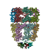
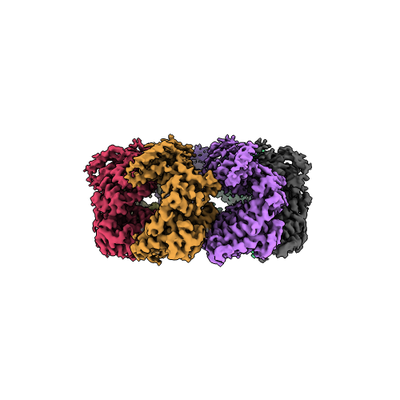
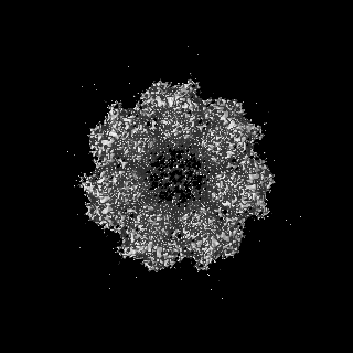
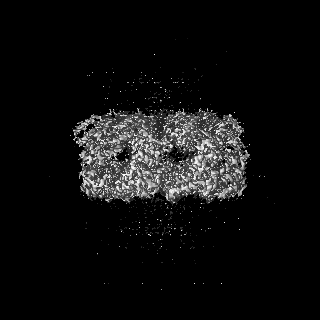
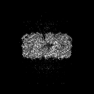
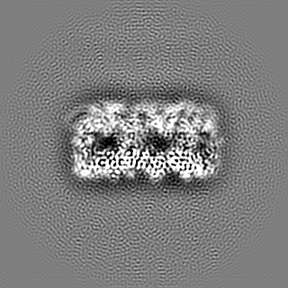
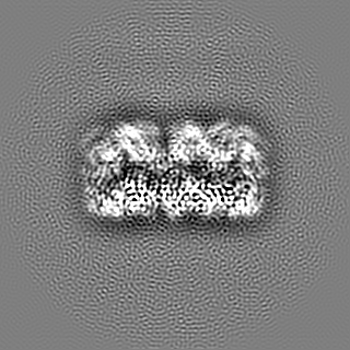
| タイトル | Human mitochondrial chaperonin mHsp60 | |||||||||
 マップデータ マップデータ | Cryo-EM structure of mHsp60 | |||||||||
 試料 試料 |
| |||||||||
 キーワード キーワード | mHsp60 / GroEL / chaperonin / CHAPERONE / ISOMERASE | |||||||||
| 機能・相同性 |  機能・相同性情報 機能・相同性情報coated vesicle / isotype switching to IgG isotypes / mitochondrial unfolded protein response / TFAP2A acts as a transcriptional repressor during retinoic acid induced cell differentiation / apolipoprotein A-I binding / lipopolysaccharide receptor complex / protein import into mitochondrial intermembrane space / high-density lipoprotein particle binding / migrasome / cysteine-type endopeptidase activator activity ...coated vesicle / isotype switching to IgG isotypes / mitochondrial unfolded protein response / TFAP2A acts as a transcriptional repressor during retinoic acid induced cell differentiation / apolipoprotein A-I binding / lipopolysaccharide receptor complex / protein import into mitochondrial intermembrane space / high-density lipoprotein particle binding / migrasome / cysteine-type endopeptidase activator activity / positive regulation of T cell mediated immune response to tumor cell / Mitochondrial protein import / positive regulation of macrophage activation / negative regulation of execution phase of apoptosis / chaperonin ATPase / cellular response to interleukin-7 / MyD88-dependent toll-like receptor signaling pathway / 'de novo' protein folding / biological process involved in interaction with symbiont / sperm plasma membrane / apoptotic mitochondrial changes / B cell proliferation / B cell activation / positive regulation of interferon-alpha production / DNA replication origin binding / positive regulation of interleukin-10 production / apolipoprotein binding / response to unfolded protein / positive regulation of execution phase of apoptosis / Mitochondrial unfolded protein response (UPRmt) / isomerase activity / chaperone-mediated protein complex assembly / positive regulation of interleukin-12 production / clathrin-coated pit / Mitochondrial protein degradation / secretory granule / response to cold / T cell activation / ATP-dependent protein folding chaperone / lipopolysaccharide binding / protein maturation / positive regulation of interleukin-6 production / positive regulation of type II interferon production / positive regulation of T cell activation / p53 binding / : / double-stranded RNA binding / single-stranded DNA binding / protein refolding / protein-folding chaperone binding / protein folding / sperm midpiece / early endosome / mitochondrial inner membrane / protein stabilization / mitochondrial matrix / ubiquitin protein ligase binding / negative regulation of apoptotic process / enzyme binding / cell surface / ATP hydrolysis activity / protein-containing complex / mitochondrion / : / RNA binding / extracellular exosome / ATP binding / membrane / plasma membrane / cytoplasm / cytosol 類似検索 - 分子機能 | |||||||||
| 生物種 |  Homo sapiens (ヒト) Homo sapiens (ヒト) | |||||||||
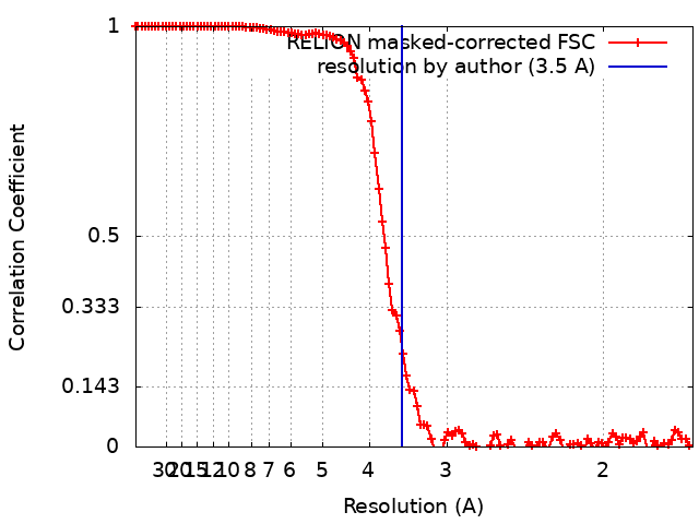
| 手法 | 単粒子再構成法 / クライオ電子顕微鏡法 / 解像度: 3.5 Å | |||||||||
 データ登録者 データ登録者 | Chen L / Wang JCY | |||||||||
| 資金援助 |  米国, 1件 米国, 1件
| |||||||||
 引用 引用 | ジャーナル: Sci Rep / 年: 2021 タイトル: Structural basis for the structural dynamics of human mitochondrial chaperonin mHsp60. 著者: Joseph Che-Yen Wang / Lingling Chen / 要旨: Human mitochondrial chaperonin mHsp60 is essential for mitochondrial function by assisting folding of mitochondrial proteins. Unlike the double-ring bacterial GroEL, mHsp60 exists as a heptameric ...Human mitochondrial chaperonin mHsp60 is essential for mitochondrial function by assisting folding of mitochondrial proteins. Unlike the double-ring bacterial GroEL, mHsp60 exists as a heptameric ring that is unstable and dissociates to subunits. The structural dynamics has been implicated for a unique mechanism of mHsp60. We purified active heptameric mHsp60, and determined a cryo-EM structure of mHsp60 heptamer at 3.4 Å. Of the three domains, the equatorial domains contribute most to the inter-subunit interactions, which include a four-stranded β sheet. Our structural comparison with GroEL shows that mHsp60 contains several unique sequences that directly decrease the sidechain interactions around the β sheet and indirectly shorten β strands by disengaging the backbones of the flanking residues from hydrogen bonding in the β strand conformation. The decreased inter-subunit interactions result in a small inter-subunit interface in mHsp60 compared to GroEL, providing a structural basis for the dynamics of mHsp60 subunit association. Importantly, the unique sequences are conserved among higher eukaryotic mitochondrial chaperonins, suggesting the importance of structural dynamics for eukaryotic chaperonins. Our structural comparison with the single-ring mHsp60-mHsp10 shows that upon mHsp10 binding the shortened inter-subunit β sheet is restored and the overall inter-subunit interface of mHsp60 increases drastically. Our structural basis for the mHsp10 induced stabilization of mHsp60 subunit interaction is consistent with the literature that mHsp10 stabilizes mHsp60 quaternary structure. Together, our studies provide structural bases for structural dynamics of the mHsp60 heptamer and for the stabilizing effect of mHsp10 on mHsp60 subunit association. | |||||||||
| 履歴 |
|
- 構造の表示
構造の表示
| ムービー |
ムービービューア |
|---|---|
| 構造ビューア | EMマップ: SurfViewMolmilJmol/JSmol |
| 添付画像 |
- ダウンロードとリンク
-EMDBアーカイブ
| マップデータ | emd_23217.map.gz | 117 MB | EMDBマップデータ形式 | |
|---|---|---|---|---|
| ヘッダ (付随情報) | emd-23217-v30.xmlemd-23217.xml | 18 KB 18 KB | 表示 表示 | EMDBヘッダ |
| FSC (解像度算出) | emd_23217_fsc.xml | 11.4 KB | 表示 | FSCデータファイル |
| 画像 |  emd_23217.png emd_23217.png | 66.9 KB | ||
| Filedesc metadata | emd-23217.cif.gz | 5.9 KB | ||
| アーカイブディレクトリ |  http://ftp.pdbj.org/pub/emdb/structures/EMD-23217ftp://ftp.pdbj.org/pub/emdb/structures/EMD-23217 http://ftp.pdbj.org/pub/emdb/structures/EMD-23217ftp://ftp.pdbj.org/pub/emdb/structures/EMD-23217 | HTTPS FTP |
-関連構造データ


-リンク
| EMDBのページ | EMDB (EBI/PDBe) / EMDataResource |
|---|---|
| 「今月の分子」の関連する項目 |

-マップ
| ファイル | ダウンロード / ファイル: emd_23217.map.gz / 形式: CCP4 / 大きさ: 125 MB / タイプ: IMAGE STORED AS FLOATING POINT NUMBER (4 BYTES) | ||||||||||||||||||||||||||||||||||||||||||||||||||||||||||||
|---|---|---|---|---|---|---|---|---|---|---|---|---|---|---|---|---|---|---|---|---|---|---|---|---|---|---|---|---|---|---|---|---|---|---|---|---|---|---|---|---|---|---|---|---|---|---|---|---|---|---|---|---|---|---|---|---|---|---|---|---|---|
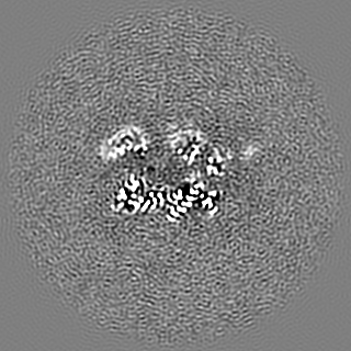
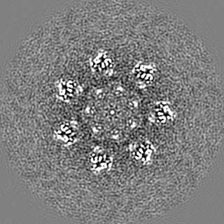
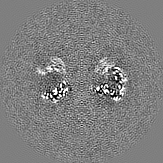
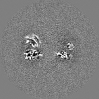
| 注釈 | Cryo-EM structure of mHsp60 | ||||||||||||||||||||||||||||||||||||||||||||||||||||||||||||
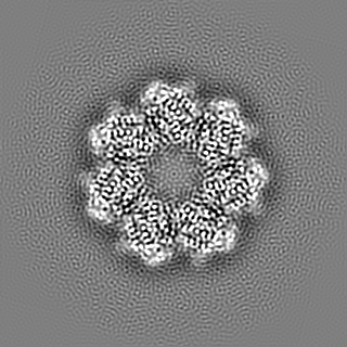
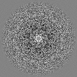
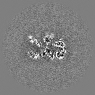
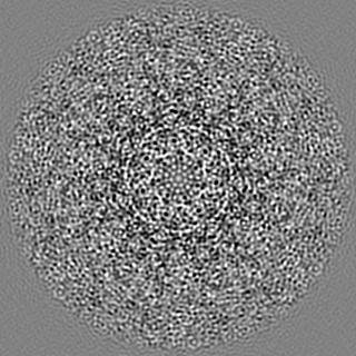
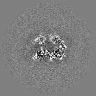
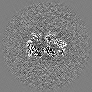
| 投影像・断面図 | 画像のコントロール
画像は Spider により作成 | ||||||||||||||||||||||||||||||||||||||||||||||||||||||||||||
| ボクセルのサイズ | X=Y=Z: 0.84 Å | ||||||||||||||||||||||||||||||||||||||||||||||||||||||||||||
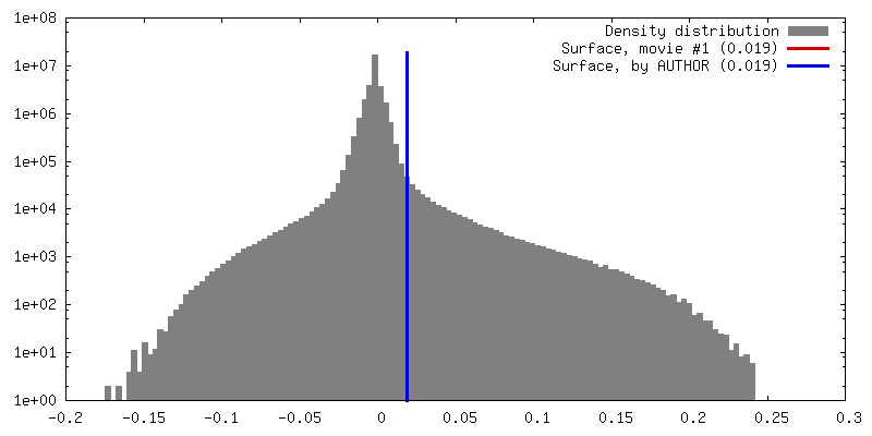
| 密度 |
| ||||||||||||||||||||||||||||||||||||||||||||||||||||||||||||
| 対称性 | 空間群: 1 | ||||||||||||||||||||||||||||||||||||||||||||||||||||||||||||
| 詳細 | EMDB XML:
CCP4マップ ヘッダ情報:
| ||||||||||||||||||||||||||||||||||||||||||||||||||||||||||||
 Z (Sec.)
 Y (Row.)
 X (Col.)


-添付データ
- 試料の構成要素
-全体 : Human mitochondrial chaperonin mHsp60 in a heptameric ring confor...
| 全体 | 名称: Human mitochondrial chaperonin mHsp60 in a heptameric ring conformation |
|---|---|
| 要素 |
|
-超分子 #1: Human mitochondrial chaperonin mHsp60 in a heptameric ring confor...
| 超分子 | 名称: Human mitochondrial chaperonin mHsp60 in a heptameric ring conformation タイプ: complex / ID: 1 / 親要素: 0 / 含まれる分子: all 詳細: Human mitochondrial chaperonin mHsp60 in a heptameric ring conformation |
|---|---|
| 由来(天然) | 生物種: Homo sapiens (ヒト) |
| 分子量 | 理論値: 60 KDa |
-分子 #1: 60 kDa heat shock protein, mitochondrial
| 分子 | 名称: 60 kDa heat shock protein, mitochondrial / タイプ: protein_or_peptide / ID: 1 / コピー数: 7 / 光学異性体: LEVO / EC番号: chaperonin ATPase |
|---|---|
| 由来(天然) | 生物種: Homo sapiens (ヒト) |
| 分子量 | 理論値: 55.861137 KDa |
| 組換発現 | 生物種:  |
| 配列 | 文字列: AKDVKFGADA RALMLQGVDL LADAVAVTMG PKGRTVIIEQ SWGSPKVTKD GVTVAKSIDL KDKYKNIGAK LVQDVANNTN EEAGDGTTT ATVLARSIAK EGFEKISKGA NPVEIRRGVM LAVDAVIAEL KKQSKPVTTP EEIAQVATIS ANGDKEIGNI I SDAMKKVG ...文字列: AKDVKFGADA RALMLQGVDL LADAVAVTMG PKGRTVIIEQ SWGSPKVTKD GVTVAKSIDL KDKYKNIGAK LVQDVANNTN EEAGDGTTT ATVLARSIAK EGFEKISKGA NPVEIRRGVM LAVDAVIAEL KKQSKPVTTP EEIAQVATIS ANGDKEIGNI I SDAMKKVG RKGVITVKDG KTLNDELEII EGMKFDRGYI SPYFINTSKG QKCEFQDAYV LLSEKKISSI QSIVPALEIA NA HRKPLVI IAEDVDGEAL STLVLNRLKV GLQVVAVKAP GFGDNRKNQL KDMAIATGGA VFGEEGLTLN LEDVQPHDLG KVG EVIVTK DDAMLLKGKG DKAQIEKRIQ EIIEQLDVTT SEYEKEKLNE RLAKLSDGVA VLKVGGTSDV EVNEKKDRVT DALN ATRAA VEEGIVLGGG CALLRCIPAL DSLTPANEDQ KIGIEIIKRT LKIPAMTIAK NAGVEGSLIV EKIMQSSSEV GYDAM AGDF VNMVEKGIID PTKVVRTALL DAAGVASLLT TAEVVVTEIP UniProtKB: 60 kDa heat shock protein, mitochondrial |
-実験情報
-構造解析
| 手法 | クライオ電子顕微鏡法 |
|---|---|
 解析 解析 | 単粒子再構成法 |
| 試料の集合状態 | particle |
-試料調製
| 濃度 | 9 mg/mL | ||||||||||||||||||
|---|---|---|---|---|---|---|---|---|---|---|---|---|---|---|---|---|---|---|---|
| 緩衝液 | pH: 7.4 構成要素:
| ||||||||||||||||||
| グリッド | モデル: Quantifoil / 支持フィルム - #0 - Film type ID: 1 / 支持フィルム - #0 - 材質: GOLD / 支持フィルム - #0 - トポロジー: HOLEY / 支持フィルム - #1 - Film type ID: 2 / 支持フィルム - #1 - 材質: CARBON / 支持フィルム - #1 - トポロジー: HOLEY | ||||||||||||||||||
| 凍結 | 凍結剤: ETHANE / チャンバー内湿度: 100 % / 装置: FEI VITROBOT MARK IV | ||||||||||||||||||
| 詳細 | The sample is monodisperse |
- 電子顕微鏡法
| 顕微鏡 | FEI TITAN KRIOS |
|---|---|
| 特殊光学系 | エネルギーフィルター - 名称: GIF Bioquantum / エネルギーフィルター - スリット幅: 30 eV |
| 撮影 | #0 - Image recording ID: 1 #0 - フィルム・検出器のモデル: GATAN K3 BIOQUANTUM (6k x 4k) #0 - 撮影したグリッド数: 1 / #0 - 平均電子線量: 44.0 e/Å2 / #1 - Image recording ID: 2 #1 - フィルム・検出器のモデル: GATAN K3 (6k x 4k) #1 - 撮影したグリッド数: 3 / #1 - 平均電子線量: 53.0 e/Å2 |
| 電子線 | 加速電圧: 300 kV / 電子線源:  FIELD EMISSION GUN FIELD EMISSION GUN |
| 電子光学系 | C2レンズ絞り径: 50.0 µm / 照射モード: FLOOD BEAM / 撮影モード: BRIGHT FIELD / Cs: 2.7 mm |
| 試料ステージ | 試料ホルダーモデル: FEI TITAN KRIOS AUTOGRID HOLDER ホルダー冷却材: NITROGEN |
| 実験機器 |  モデル: Titan Krios / 画像提供: FEI Company |
+画像解析 #1
+画像解析 #2
-原子モデル構築 1
| 初期モデル |
| ||||||||||||||||
|---|---|---|---|---|---|---|---|---|---|---|---|---|---|---|---|---|---|
| 精密化 | 空間: REAL / プロトコル: RIGID BODY FIT | ||||||||||||||||
| 得られたモデル |  PDB-7l7s: |