+ Open data
- Basic information
| Entry | Database: EMDB / ID: EMD-22912 | |||||||||
|---|---|---|---|---|---|---|---|---|---|---|
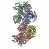
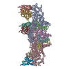
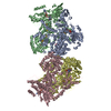
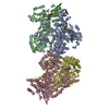
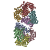
| Title | CRISPR-Cas Type I-D complex with a dsDNA target | |||||||||
 Map data Map data | Unedited final map | |||||||||
 Sample Sample |
| |||||||||
| Biological species |  | |||||||||
| Method | single particle reconstruction / cryo EM / Resolution: 7.2 Å | |||||||||
 Authors Authors | Schwartz EA / Taylor DW | |||||||||
| Funding support |  United States, 1 items United States, 1 items
| |||||||||
 Citation Citation |  Journal: Mol Cell / Year: 2020 Journal: Mol Cell / Year: 2020Title: Diverse CRISPR-Cas Complexes Require Independent Translation of Small and Large Subunits from a Single Gene. Authors: Tess M McBride / Evan A Schwartz / Abhishek Kumar / David W Taylor / Peter C Fineran / Robert D Fagerlund /   Abstract: CRISPR-Cas adaptive immune systems provide prokaryotes with defense against viruses by degradation of specific invading nucleic acids. Despite advances in the biotechnological exploitation of select ...CRISPR-Cas adaptive immune systems provide prokaryotes with defense against viruses by degradation of specific invading nucleic acids. Despite advances in the biotechnological exploitation of select systems, multiple CRISPR-Cas types remain uncharacterized. Here, we investigated the previously uncharacterized type I-D interference complex and revealed that it is a genetic and structural hybrid with similarity to both type I and type III systems. Surprisingly, formation of the functional complex required internal in-frame translation of small subunits from within the large subunit gene. We further show that internal translation to generate small subunits is widespread across diverse type I-D, I-B, and I-C systems, which account for roughly one quarter of CRISPR-Cas systems. Our work reveals the unexpected expansion of protein coding potential from within single cas genes, which has important implications for understanding CRISPR-Cas function and evolution. | |||||||||
| History |
|
- Structure visualization
| Movie |
 Movie viewer Movie viewer |
|---|---|
| Structure viewer | EM map:  SurfView SurfView Molmil Molmil Jmol/JSmol Jmol/JSmol |
| Supplemental images |
- Downloads & links
-EMDB archive
| Map data |  emd_22912.map.gz emd_22912.map.gz | 289.3 MB |  EMDB map data format EMDB map data format | |
|---|---|---|---|---|
| Header (meta data) |  emd-22912-v30.xml emd-22912-v30.xml emd-22912.xml emd-22912.xml | 11 KB 11 KB | Display Display |  EMDB header EMDB header |
| Images |  emd_22912.png emd_22912.png | 105.1 KB | ||
| Archive directory |  http://ftp.pdbj.org/pub/emdb/structures/EMD-22912 http://ftp.pdbj.org/pub/emdb/structures/EMD-22912 ftp://ftp.pdbj.org/pub/emdb/structures/EMD-22912 ftp://ftp.pdbj.org/pub/emdb/structures/EMD-22912 | HTTPS FTP |
-Validation report
| Summary document |  emd_22912_validation.pdf.gz emd_22912_validation.pdf.gz | 312.1 KB | Display |  EMDB validaton report EMDB validaton report |
|---|---|---|---|---|
| Full document |  emd_22912_full_validation.pdf.gz emd_22912_full_validation.pdf.gz | 311.6 KB | Display | |
| Data in XML |  emd_22912_validation.xml.gz emd_22912_validation.xml.gz | 7.4 KB | Display | |
| Arichive directory |  https://ftp.pdbj.org/pub/emdb/validation_reports/EMD-22912 https://ftp.pdbj.org/pub/emdb/validation_reports/EMD-22912 ftp://ftp.pdbj.org/pub/emdb/validation_reports/EMD-22912 ftp://ftp.pdbj.org/pub/emdb/validation_reports/EMD-22912 | HTTPS FTP |
-Related structure data
| Related structure data | C: citing same article ( |
|---|---|
| Similar structure data |
- Links
Links
| EMDB pages |  EMDB (EBI/PDBe) / EMDB (EBI/PDBe) /  EMDataResource EMDataResource |
|---|
- Map
Map
| File |  Download / File: emd_22912.map.gz / Format: CCP4 / Size: 307.5 MB / Type: IMAGE STORED AS FLOATING POINT NUMBER (4 BYTES) Download / File: emd_22912.map.gz / Format: CCP4 / Size: 307.5 MB / Type: IMAGE STORED AS FLOATING POINT NUMBER (4 BYTES) | ||||||||||||||||||||||||||||||||||||||||||||||||||||||||||||
|---|---|---|---|---|---|---|---|---|---|---|---|---|---|---|---|---|---|---|---|---|---|---|---|---|---|---|---|---|---|---|---|---|---|---|---|---|---|---|---|---|---|---|---|---|---|---|---|---|---|---|---|---|---|---|---|---|---|---|---|---|---|
| Annotation | Unedited final map | ||||||||||||||||||||||||||||||||||||||||||||||||||||||||||||
| Projections & slices | Image control
Images are generated by Spider. | ||||||||||||||||||||||||||||||||||||||||||||||||||||||||||||
| Voxel size | X=Y=Z: 1.045 Å | ||||||||||||||||||||||||||||||||||||||||||||||||||||||||||||
| Density |
| ||||||||||||||||||||||||||||||||||||||||||||||||||||||||||||
| Symmetry | Space group: 1 | ||||||||||||||||||||||||||||||||||||||||||||||||||||||||||||
| Details | EMDB XML:
CCP4 map header:
| ||||||||||||||||||||||||||||||||||||||||||||||||||||||||||||
-Supplemental data
- Sample components
-Entire : CRISPR-Cas type I-D complex
| Entire | Name: CRISPR-Cas type I-D complex |
|---|---|
| Components |
|
-Supramolecule #1: CRISPR-Cas type I-D complex
| Supramolecule | Name: CRISPR-Cas type I-D complex / type: complex / ID: 1 / Parent: 0 Details: full complex from Synechocystis expressed and assembled within E. coli. |
|---|---|
| Source (natural) | Organism:  |
| Recombinant expression | Organism:  |
| Molecular weight | Theoretical: 4 kDa/nm |
-Experimental details
-Structure determination
| Method | cryo EM |
|---|---|
 Processing Processing | single particle reconstruction |
| Aggregation state | particle |
- Sample preparation
| Concentration | 0.4 mg/mL | |||||||||||||||
|---|---|---|---|---|---|---|---|---|---|---|---|---|---|---|---|---|
| Buffer | pH: 7.5 Component:
| |||||||||||||||
| Grid | Model: C-flat / Material: COPPER / Mesh: 400 / Pretreatment - Type: PLASMA CLEANING | |||||||||||||||
| Vitrification | Cryogen name: ETHANE / Chamber humidity: 100 % / Chamber temperature: 277 K / Instrument: FEI VITROBOT MARK IV | |||||||||||||||
| Details | Monodisperse sample |
- Electron microscopy
| Microscope | FEI TITAN KRIOS |
|---|---|
| Image recording | Film or detector model: GATAN K3 (6k x 4k) / Number grids imaged: 1 / Average exposure time: 3.0 sec. / Average electron dose: 41.2 e/Å2 |
| Electron beam | Acceleration voltage: 300 kV / Electron source:  FIELD EMISSION GUN FIELD EMISSION GUN |
| Electron optics | C2 aperture diameter: 100.0 µm / Illumination mode: FLOOD BEAM / Imaging mode: BRIGHT FIELD / Cs: 2.7 mm / Nominal magnification: 22500 |
| Sample stage | Specimen holder model: FEI TITAN KRIOS AUTOGRID HOLDER / Cooling holder cryogen: NITROGEN |
| Experimental equipment |  Model: Titan Krios / Image courtesy: FEI Company |
 Movie
Movie Controller




 Z (Sec.)
 Y (Row.)
 X (Col.)


