Movie
Movie Controller

[English] 日本語
 Yorodumi
- EMDB-22596: Focused cryo-EM map of rabbit RyR1 central and transmembrane doma... -
+ Open data

- Basic information
| Entry | Database: EMDB / ID: EMD-22596 | |||||||||||||||||||||
|---|---|---|---|---|---|---|---|---|---|---|---|---|---|---|---|---|---|---|---|---|---|---|
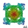
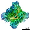
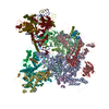
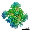
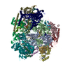
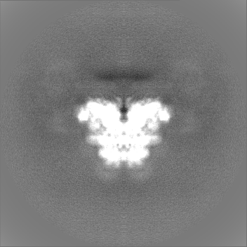
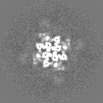
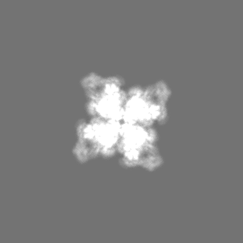
| Title | Focused cryo-EM map of rabbit RyR1 central and transmembrane domains in the presence of Mg2+ and AMP-PCP in nanodisc | |||||||||||||||||||||
 Map data Map data | Masked full map | |||||||||||||||||||||
 Sample Sample |
| |||||||||||||||||||||
| Biological species |  | |||||||||||||||||||||
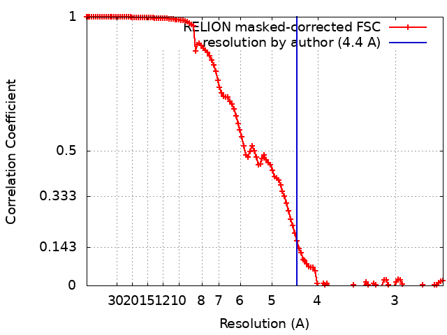
| Method | single particle reconstruction / cryo EM / Resolution: 4.4 Å | |||||||||||||||||||||
 Authors Authors | Nayak AR / Samso M | |||||||||||||||||||||
| Funding support |  United States, 6 items United States, 6 items
| |||||||||||||||||||||
 Citation Citation | Journal: To Be Published Title: Molecular Mechanism of Inhibition of RyR1 by Magnesium Authors: Nayak AR / Will AH / Hartmann PC / Lobo JJ / Rangubpit W / Sompornpisut P / Samso M | |||||||||||||||||||||
| History |
|
- Structure visualization
| Movie |
 Movie viewer Movie viewer |
|---|---|
| Structure viewer | EM map: SurfViewMolmilJmol/JSmol |
| Supplemental images |

- Downloads & links
-EMDB archive
| Map data | emd_22596.map.gz | 9.3 MB | EMDB map data format | |
|---|---|---|---|---|
| Header (meta data) | emd-22596-v30.xmlemd-22596.xml | 25.1 KB 25.1 KB | Display Display | EMDB header |
| FSC (resolution estimation) | emd_22596_fsc.xml | 12.5 KB | Display | FSC data file |
| Images | 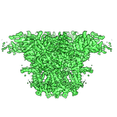 emd_22596.png emd_22596.png | 166.8 KB | ||
| Masks | emd_22596_msk_1.map | 166.4 MB | Mask map | |
| Others | emd_22596_additional_1.map.gzemd_22596_additional_2.map.gzemd_22596_half_map_1.map.gzemd_22596_half_map_2.map.gz | 127.3 MB 127.2 MB 8.3 MB 8.3 MB | ||
| Archive directory |  http://ftp.pdbj.org/pub/emdb/structures/EMD-22596ftp://ftp.pdbj.org/pub/emdb/structures/EMD-22596 http://ftp.pdbj.org/pub/emdb/structures/EMD-22596ftp://ftp.pdbj.org/pub/emdb/structures/EMD-22596 | HTTPS FTP |
-Related structure data
| Related structure data | |
|---|---|
| Similar structure data |


-Links
| EMDB pages | EMDB (EBI/PDBe) / EMDataResource |
|---|
-Map
| File | Download / File: emd_22596.map.gz / Format: CCP4 / Size: 166.4 MB / Type: IMAGE STORED AS FLOATING POINT NUMBER (4 BYTES) | ||||||||||||||||||||||||||||||||||||||||||||||||||||||||||||
|---|---|---|---|---|---|---|---|---|---|---|---|---|---|---|---|---|---|---|---|---|---|---|---|---|---|---|---|---|---|---|---|---|---|---|---|---|---|---|---|---|---|---|---|---|---|---|---|---|---|---|---|---|---|---|---|---|---|---|---|---|---|
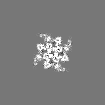
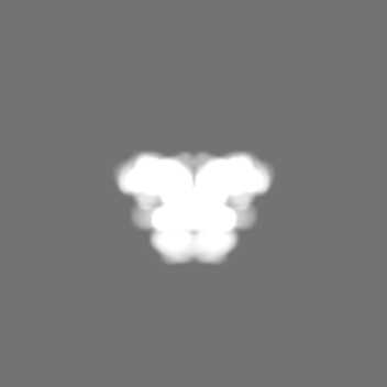
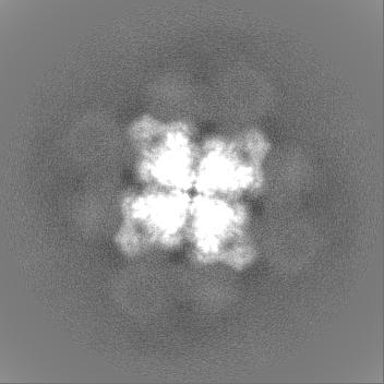
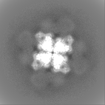
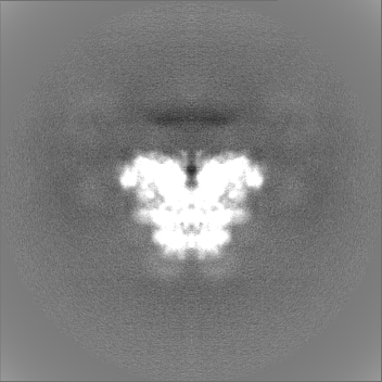
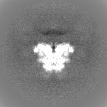
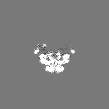
| Annotation | Masked full map | ||||||||||||||||||||||||||||||||||||||||||||||||||||||||||||
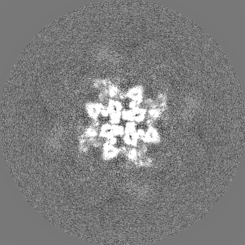
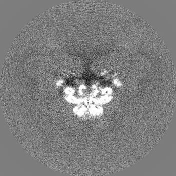
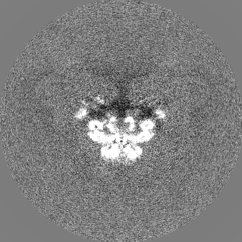
| Projections & slices | Image control
Images are generated by Spider. | ||||||||||||||||||||||||||||||||||||||||||||||||||||||||||||
| Voxel size | X=Y=Z: 1.36 Å | ||||||||||||||||||||||||||||||||||||||||||||||||||||||||||||
| Density |
| ||||||||||||||||||||||||||||||||||||||||||||||||||||||||||||
| Symmetry | Space group: 1 | ||||||||||||||||||||||||||||||||||||||||||||||||||||||||||||
| Details | EMDB XML:
CCP4 map header:
| ||||||||||||||||||||||||||||||||||||||||||||||||||||||||||||
 Z (Sec.)
 Y (Row.)
 X (Col.)


-Supplemental data
-Mask #1
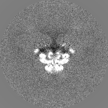
| File | emd_22596_msk_1.map | ||||||||||||
|---|---|---|---|---|---|---|---|---|---|---|---|---|---|
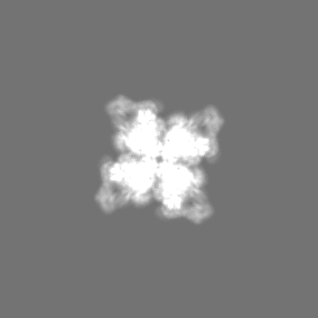
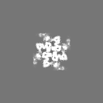
| Projections & Slices |
| ||||||||||||
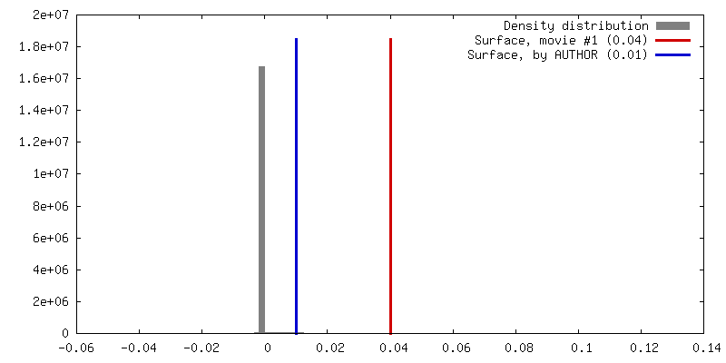
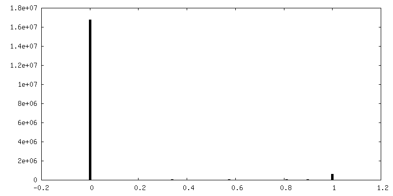
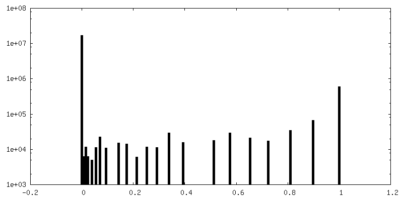
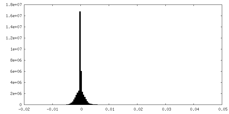
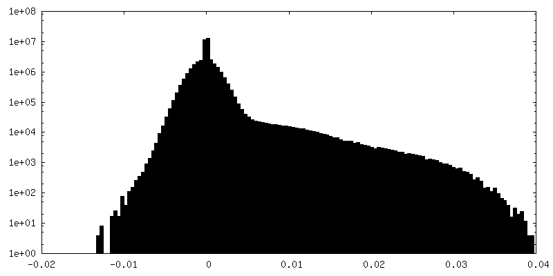
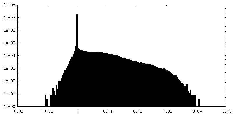
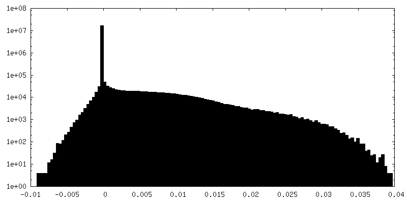
| Density Histograms |


-Additional map: Unfiltered and unmasked half map1 up to Nyquist frequency
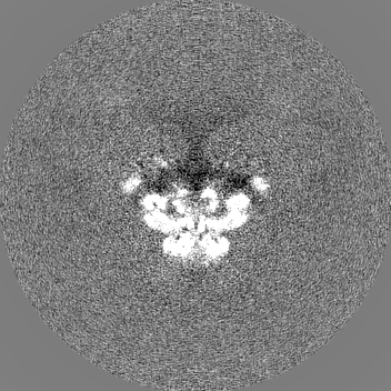
| File | emd_22596_additional_1.map | ||||||||||||
|---|---|---|---|---|---|---|---|---|---|---|---|---|---|
| Annotation | Unfiltered and unmasked half map1 up to Nyquist frequency | ||||||||||||
| Projections & Slices |
| ||||||||||||
| Density Histograms |


-Additional map: Unfiltered and unmasked half map2 up to Nyquist frequency
| File | emd_22596_additional_2.map | ||||||||||||
|---|---|---|---|---|---|---|---|---|---|---|---|---|---|
| Annotation | Unfiltered and unmasked half map2 up to Nyquist frequency | ||||||||||||
| Projections & Slices |
| ||||||||||||
| Density Histograms |

-Half map: Masked half map1
| File | emd_22596_half_map_1.map | ||||||||||||
|---|---|---|---|---|---|---|---|---|---|---|---|---|---|
| Annotation | Masked half map1 | ||||||||||||
| Projections & Slices |
| ||||||||||||
| Density Histograms |


-Half map: Masked half map2
| File | emd_22596_half_map_2.map | ||||||||||||
|---|---|---|---|---|---|---|---|---|---|---|---|---|---|
| Annotation | Masked half map2 | ||||||||||||
| Projections & Slices |
| ||||||||||||
| Density Histograms |


- Sample components
-Entire : Rabbit RyR1 with Mg2+ and AMP-PCP
| Entire | Name: Rabbit RyR1 with Mg2+ and AMP-PCP |
|---|---|
| Components |
|
-Supramolecule #1: Rabbit RyR1 with Mg2+ and AMP-PCP
| Supramolecule | Name: Rabbit RyR1 with Mg2+ and AMP-PCP / type: complex / ID: 1 / Parent: 0 / Macromolecule list: all Details: Purified RyR1 was reconstituted with membrane scaffold protein, MSP1E3D1, and POPC. |
|---|---|
| Source (natural) | Organism: |
| Molecular weight | Experimental: 2.26 MDa |
-Macromolecule #1: RyR1
| Macromolecule | Name: RyR1 / type: protein_or_peptide / ID: 1 Details: Rabbit RyR1 corresponding to amino acid sequence 3668-5037 Enantiomer: LEVO |
|---|---|
| Source (natural) | Organism: |
| Sequence | String: SFEDRMIDDL SKAGEQEEEE EEVEEKKPDP LHQLVLHFSR TALTEKSKLD EDYLYMAYAD IMAKSCHLEE GGENEEVEV SFEEKEMEKQ RLLYQQSRLH TRGAAEMVLQ MISACKGETG AMVSSTLKLG ISILNGGNAE V QQKMLDYL KDKKEVGFFQ SIQALMQTCS ...String: SFEDRMIDDL SKAGEQEEEE EEVEEKKPDP LHQLVLHFSR TALTEKSKLD EDYLYMAYAD IMAKSCHLEE GGENEEVEV SFEEKEMEKQ RLLYQQSRLH TRGAAEMVLQ MISACKGETG AMVSSTLKLG ISILNGGNAE V QQKMLDYL KDKKEVGFFQ SIQALMQTCS VLDLNAFERQ NKAEGLGMVN EDGTVINRQN GEKVMADDEF TQ DLFRFLQ LLCEGHNNDF QNYLRTQTGN TTTINIIICT VDYLLRLQES ISDFYWYYSG KDVIEEQGKR NFS KAMSVA KQVFNSLTEY IQGPCTGNQQ SLAHSRLWDA VVGFLHVFAH MMMKLAQDSS QIELLKELLD LQKD MVVML LSLLEGNVVN GMIARQMVDM LVESSSNVEM ILKFFDMFLK LKDIVGSEAF QDYVTDPRGL ISKKD FQKA MDSQKQFTGP EIQFLLSCSE ADENEMINFE EFANRFQEPA RDIGFNVAVL LTNLSEHV P HDPRLRNFLE LAESILEYFR PYLGRAEIMG ASRRIERIYF EISETNRAQW EMPQVKESKR QFIFDVVNE GGEAEKMELF VSFCEDTIFE MQIAAQISEF WGELEVQRVK FLNYLSRNFY TLRFLALFLA FAINFILLF YKVSDSPPAN MVYYFLEEST GYMEPALWCL SLLHTLVAFL CIIGYNCLKV PLVIFKREKE LARKLEFDGL YITEQPGDD DVKGQWDRLV LNTPSFPSNY WDKFVKRKVL DKHGDIFGRE RIAELLGMDL ASLEITAHNE R KPDPPPGL LTWLMSIDVK YQIWKFGVIF TDNSFLYLGW YMVMSLLGHY NNFFFAAHLL DIAMGVKTLR TI LSSVTHN GKQLVMTVGL LAVVVYLYTV VAFNFFRKFY NKSEDEDEPD MKCDDMMTCY LFHMYVGVRA GGG IGDEIE DPAGDEYELY RVVFDITFFF FVIVILLAII QGLIIDAFGE LRDQQEQVKE DMETKCFICG IGSD YFDTT PHGFETHTLE EHNLANYMFF LMYLINKDET EHTGQESYVW KMYQERCWDF FPAGDCFRKQ YEDQL S |
-Experimental details
-Structure determination
| Method | cryo EM |
|---|---|
 Processing Processing | single particle reconstruction |
| Aggregation state | particle |
-Sample preparation
| Concentration | 4.2 mg/mL | |||||||||||||||||||||
|---|---|---|---|---|---|---|---|---|---|---|---|---|---|---|---|---|---|---|---|---|---|---|
| Buffer | pH: 7.4 Component:
| |||||||||||||||||||||
| Grid | Model: C-flat-2/1 / Material: GOLD / Mesh: 300 / Support film - Material: CARBON / Support film - topology: HOLEY / Pretreatment - Type: GLOW DISCHARGE / Pretreatment - Atmosphere: AIR | |||||||||||||||||||||
| Vitrification | Cryogen name: ETHANE / Chamber humidity: 95 % / Chamber temperature: 277 K / Instrument: FEI VITROBOT MARK IV Details: Sample was blotted for 1 second on both sides with Whatman hardened ashless filter paper with blot force 2.. | |||||||||||||||||||||
| Details | Purified RyR1 was reconstituted with membrane scaffold protein, MSP1E3D1, and POPC in 1:2:50 molar ratio. |
- Electron microscopy
| Microscope | FEI TITAN KRIOS |
|---|---|
| Specialist optics | Energy filter - Slit width: 20 eV |
| Image recording | Film or detector model: GATAN K2 SUMMIT (4k x 4k) / Detector mode: SUPER-RESOLUTION / Number grids imaged: 2 / Number real images: 3371 / Average electron dose: 60.0 e/Å2 |
| Electron beam | Acceleration voltage: 300 kV / Electron source:  FIELD EMISSION GUN FIELD EMISSION GUN |
| Electron optics | C2 aperture diameter: 100.0 µm / Illumination mode: FLOOD BEAM / Imaging mode: BRIGHT FIELD / Cs: 2.7 mm / Nominal defocus max: 2.5 µm / Nominal defocus min: 1.0 µm / Nominal magnification: 105000 |
| Sample stage | Cooling holder cryogen: NITROGEN |
| Experimental equipment |  Model: Titan Krios / Image courtesy: FEI Company |