 Movie
Movie Controller

[English] 日本語
 Yorodumi
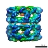
- PDB-7liv: Structure of human transfer RNA visualized in the cytomegalovirus... -
+ Open data

- Basic information
| Entry | Database: PDB / ID: 7liv | |||||||||||||||||||||||||||
|---|---|---|---|---|---|---|---|---|---|---|---|---|---|---|---|---|---|---|---|---|---|---|---|---|---|---|---|---|
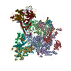
| Title | Structure of human transfer RNA visualized in the cytomegalovirus, a DNA virus | |||||||||||||||||||||||||||
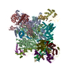
 Components Components |
| |||||||||||||||||||||||||||
 Keywords Keywords | VIRUS / HCMV / tRNA | |||||||||||||||||||||||||||
| Function / homology |  Function and homology information Function and homology informationhost cell viral assembly compartment / T=16 icosahedral viral capsid / viral tegument / viral capsid assembly / host cell cytoplasmic vesicle / viral process / viral capsid / host cell perinuclear region of cytoplasm / host cell nucleus / structural molecule activity / DNA binding Similarity search - Function | |||||||||||||||||||||||||||
| Biological species |   Human cytomegalovirus Human cytomegalovirus | |||||||||||||||||||||||||||
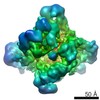
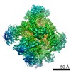
| Method | ELECTRON MICROSCOPY / single particle reconstruction / cryo EM / Resolution: 3.6 Å | |||||||||||||||||||||||||||
 Authors Authors | Liu, Y.T. / Strugatsky, D. / Liu, W. / Zhou, Z.H. | |||||||||||||||||||||||||||
| Funding support |  United States, 5items United States, 5items
| |||||||||||||||||||||||||||
 Citation Citation | Journal: Nat Commun / Year: 2021 Title: Structure of human cytomegalovirus virion reveals host tRNA binding to capsid-associated tegument protein pp150. Authors: Yun-Tao Liu / David Strugatsky / Wei Liu / Z Hong Zhou /  Abstract: Under the Baltimore nucleic acid-based virus classification scheme, the herpesvirus human cytomegalovirus (HCMV) is a Class I virus, meaning that it contains a double-stranded DNA genome-and no RNA. ...Under the Baltimore nucleic acid-based virus classification scheme, the herpesvirus human cytomegalovirus (HCMV) is a Class I virus, meaning that it contains a double-stranded DNA genome-and no RNA. Here, we report sub-particle cryoEM reconstructions of HCMV virions at 2.9 Å resolution revealing structures resembling non-coding transfer RNAs (tRNAs) associated with the virion's capsid-bound tegument protein, pp150. Through deep sequencing, we show that these RNA sequences match human tRNAs, and we built atomic models using the most abundant tRNA species. Based on our models, tRNA recruitment is mediated by the electrostatic interactions between tRNA phosphate groups and the helix-loop-helix motif of HCMV pp150. The specificity of these interactions may explain the absence of such tRNA densities in murine cytomegalovirus and other human herpesviruses. | |||||||||||||||||||||||||||
| History |
|
- Structure visualization
| Movie |
Movie viewer |
|---|---|
| Structure viewer | Molecule: MolmilJmol/JSmol |
- Downloads & links
-Download
| PDBx/mmCIF format | 7liv.cif.gz | 1.1 MB | Display | PDBx/mmCIF format |
|---|---|---|---|---|
| PDB format | pdb7liv.ent.gz | 948.4 KB | Display | PDB format |
| PDBx/mmJSON format | 7liv.json.gz | Tree view | PDBx/mmJSON format | |
| Others |  Other downloads Other downloads |
-Validation report
| Arichive directory | https://data.pdbj.org/pub/pdb/validation_reports/li/7livftp://data.pdbj.org/pub/pdb/validation_reports/li/7liv | HTTPS FTP |
|---|
-Related structure data
| Related structure data |  23386MC 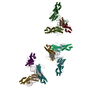 7lj3C M: map data used to model this data C: citing same article ( |
|---|---|
| Similar structure data |


-Links
 PDBj
PDBj
- Assembly
| Deposited unit | 
|
|---|---|
| 1 |
|
-Components
| #1: Protein | Mass: 154048.906 Da / Num. of mol.: 3 / Source method: isolated from a natural source / Source: (natural)  Human cytomegalovirus (strain AD169) / Strain: AD169 / References: UniProt: P16729 Human cytomegalovirus (strain AD169) / Strain: AD169 / References: UniProt: P16729#2: Protein | Mass: 33232.418 Da / Num. of mol.: 3 / Source method: isolated from a natural source / Source: (natural) Human cytomegalovirus (strain AD169) / Strain: AD169 / References: UniProt: P08318#3: Protein | Mass: 8495.924 Da / Num. of mol.: 3 / Source method: isolated from a natural source / Source: (natural) Human cytomegalovirus (strain AD169) / Strain: AD169 / References: UniProt: Q7M6N6#4: Protein | | Mass: 33071.270 Da / Num. of mol.: 1 / Source method: isolated from a natural source / Source: (natural) Human cytomegalovirus (strain AD169) / Strain: AD169 / References: UniProt: P16783#5: Protein | Mass: 34635.750 Da / Num. of mol.: 2 / Source method: isolated from a natural source / Source: (natural) Human cytomegalovirus (strain AD169) / Strain: AD169 / References: UniProt: P16728Has protein modification | Y | |
|---|
-Experimental details
-Experiment
| Experiment | Method: ELECTRON MICROSCOPY |
|---|---|
| EM experiment | Aggregation state: PARTICLE / 3D reconstruction method: single particle reconstruction |
- Sample preparation
| Component | Name: Human cytomegalovirus (strain AD169) / Type: VIRUS / Entity ID: all / Source: NATURAL |
|---|---|
| Source (natural) | Organism: Human cytomegalovirus (strain AD169) / Strain: AD169 |
| Details of virus | Empty: NO / Enveloped: YES / Isolate: STRAIN / Type: VIRION |
| Buffer solution | pH: 7.4 |
| Specimen | Embedding applied: NO / Shadowing applied: NO / Staining applied: NO / Vitrification applied: YES |
| Vitrification | Cryogen name: ETHANE |
- Electron microscopy imaging
| Experimental equipment |  Model: Titan Krios / Image courtesy: FEI Company |
|---|---|
| Microscopy | Model: FEI TITAN KRIOS |
| Electron gun | Electron source:  FIELD EMISSION GUN / Accelerating voltage: 300 kV / Illumination mode: FLOOD BEAM FIELD EMISSION GUN / Accelerating voltage: 300 kV / Illumination mode: FLOOD BEAM |
| Electron lens | Mode: BRIGHT FIELD |
| Image recording | Electron dose: 18.9 e/Å2 / Film or detector model: GATAN K2 SUMMIT (4k x 4k) |
- Processing
| Software | Name: PHENIX / Version: 1.16_3549: / Classification: refinement | ||||||||||||||||||||||||
|---|---|---|---|---|---|---|---|---|---|---|---|---|---|---|---|---|---|---|---|---|---|---|---|---|---|
| EM software | Name: PHENIX / Category: model refinement | ||||||||||||||||||||||||
| CTF correction | Type: NONE | ||||||||||||||||||||||||
| 3D reconstruction | Resolution: 3.6 Å / Resolution method: FSC 0.143 CUT-OFF / Num. of particles: 97166 / Symmetry type: POINT | ||||||||||||||||||||||||
| Refine LS restraints |
|
