- EMDB-23376: Structure of human transfer RNA visualized in the cytomegalovirus... -
+
Open data
ID or keywords:
Loading...
-
Basic information
Entry
Database: EMDB / ID: EMD-23376
Title
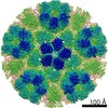
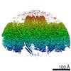
Structure of human transfer RNA visualized in the cytomegalovirus, a DNA virus
Map data
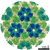
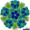
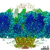
5-fold sub-particle reconstruction of HCMV virion particles
Sample
transfer RNA visualized in the cytomegalovirus != Human herpesvirus 5 strain AD169
transfer RNA visualized in the cytomegalovirus
Virus: Human herpesvirus 5 strain AD169
Function / homology
Function and homology information
host cell viral assembly compartment / T=16 icosahedral viral capsid / viral tegument / viral capsid assembly / host cell cytoplasmic vesicle / viral process / viral capsid / host cell perinuclear region of cytoplasm / host cell nucleus / structural molecule activity / DNA binding Similarity search - Function
Herpesvirus UL11/UL32 / Herpesvirus UL32 N-terminal domain / Small capsid protein, Herpesviridae / Small capsid protein of Herpesviridae / Herpesvirus capsid shell protein 1 / Herpesvirus capsid shell protein VP19C / Herpesvirus capsid protein 2 / Herpesvirus VP23 like capsid protein / Herpesvirus major capsid protein / Herpesvirus major capsid protein, upper domain superfamily / Herpes virus major capsid protein Similarity search - Domain/homology
Large structural phosphoprotein / Triplex capsid protein 2 / Major capsid protein / Triplex capsid protein 1 / Small capsomere-interacting protein Similarity search - Component
Biological species
Human herpesvirus 5 strain AD169
Method
single particle reconstruction / cryo EM / Resolution: 3.2 Å
National Institutes of Health/National Institute of Dental and Craniofacial Research (NIH/NIDCR)
DE028583/DE025567
United States
National Institutes of Health/National Institute Of Allergy and Infectious Diseases (NIH/NIAID)
AI094386
United States
National Institutes of Health/National Institute of General Medical Sciences (NIH/NIGMS)
1U24GM116792
United States
National Institutes of Health/National Institute of General Medical Sciences (NIH/NIGMS)
1S10OD018111
United States
National Science Foundation (NSF, United States)
DBI-1338135, DMR-1548924
United States
Citation
Journal: Nat Commun / Year: 2021 Title: Structure of human cytomegalovirus virion reveals host tRNA binding to capsid-associated tegument protein pp150. Authors: Yun-Tao Liu / David Strugatsky / Wei Liu / Z Hong Zhou / Abstract: Under the Baltimore nucleic acid-based virus classification scheme, the herpesvirus human cytomegalovirus (HCMV) is a Class I virus, meaning that it contains a double-stranded DNA genome-and no RNA. ...Under the Baltimore nucleic acid-based virus classification scheme, the herpesvirus human cytomegalovirus (HCMV) is a Class I virus, meaning that it contains a double-stranded DNA genome-and no RNA. Here, we report sub-particle cryoEM reconstructions of HCMV virions at 2.9 Å resolution revealing structures resembling non-coding transfer RNAs (tRNAs) associated with the virion's capsid-bound tegument protein, pp150. Through deep sequencing, we show that these RNA sequences match human tRNAs, and we built atomic models using the most abundant tRNA species. Based on our models, tRNA recruitment is mediated by the electrostatic interactions between tRNA phosphate groups and the helix-loop-helix motif of HCMV pp150. The specificity of these interactions may explain the absence of such tRNA densities in murine cytomegalovirus and other human herpesviruses.
History
Deposition
Jan 27, 2021
-
Header (metadata) release
Sep 15, 2021
-
Map release
Sep 15, 2021
-
Update
Sep 15, 2021
-
Current status
Sep 15, 2021
Processing site: RCSB / Status: Released
-
Structure visualization
Movie
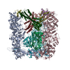
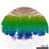
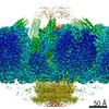
Surface view with section colored by density value
In the structure databanks used in Yorodumi, some data are registered as the other names, "COVID-19 virus" and "2019-nCoV". Here are the details of the virus and the list of structure data.
Jan 31, 2019. EMDB accession codes are about to change! (news from PDBe EMDB page)
EMDB accession codes are about to change! (news from PDBe EMDB page)
The allocation of 4 digits for EMDB accession codes will soon come to an end. Whilst these codes will remain in use, new EMDB accession codes will include an additional digit and will expand incrementally as the available range of codes is exhausted. The current 4-digit format prefixed with “EMD-” (i.e. EMD-XXXX) will advance to a 5-digit format (i.e. EMD-XXXXX), and so on. It is currently estimated that the 4-digit codes will be depleted around Spring 2019, at which point the 5-digit format will come into force.
The EM Navigator/Yorodumi systems omit the EMD- prefix.
Related info.:Q: What is EMD? / ID/Accession-code notation in Yorodumi/EM Navigator
Yorodumi is a browser for structure data from EMDB, PDB, SASBDB, etc.
This page is also the successor to EM Navigator detail page, and also detail information page/front-end page for Omokage search.
The word "yorodu" (or yorozu) is an old Japanese word meaning "ten thousand". "mi" (miru) is to see.
Related info.:EMDB / PDB / SASBDB / Comparison of 3 databanks / Yorodumi Search / Aug 31, 2016. New EM Navigator & Yorodumi / Yorodumi Papers / Jmol/JSmol / Function and homology information / Changes in new EM Navigator and Yorodumi
 Movie
Movie Controller

 Yorodumi
 Open data
 Basic information
 Map data
 Sample
 Function and homology information
 Human herpesvirus 5 strain AD169
 Authors
 United States, 5 items
  Citation
 Structure visualization
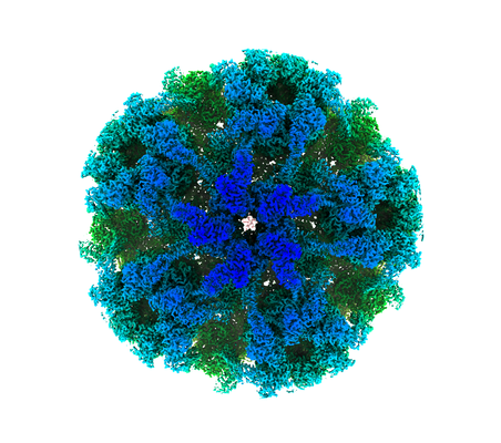
 Downloads & links
 emd_23376.png
 http://ftp.pdbj.org/pub/emdb/structures/EMD-23376


 Z (Sec.)
 Y (Row.)
 X (Col.)


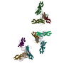
 Sample components
 Processing
 Electron microscopy
 FIELD EMISSION GUN
