 Movie
Movie Controller

[English] 日本語
 Yorodumi
- EMDB-20044: High-resolution filamentous structures of in vitro polymerized Pr... -
+ Open data

- Basic information
| Entry | Database: EMDB / ID: EMD-20044 | |||||||||
|---|---|---|---|---|---|---|---|---|---|---|
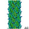
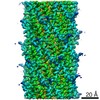
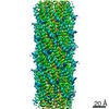
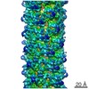
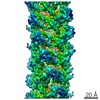
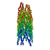
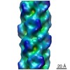
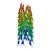
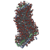
| Title | High-resolution filamentous structures of in vitro polymerized PrgI needle | |||||||||
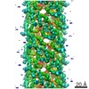
 Map data Map data | EM map of in vitro polymerized PrgI filaments | |||||||||
 Sample Sample |
| |||||||||
 Keywords Keywords | helical reconstruction / Salmonella / Type III secretion system / PROTEIN TRANSPORT | |||||||||
| Function / homology |  Function and homology information Function and homology informationtype III protein secretion system complex / protein secretion by the type III secretion system / cell surface / extracellular region / identical protein binding Similarity search - Function | |||||||||
| Biological species |  Salmonella enterica subsp. enterica serovar Typhimurium str. LT2 (bacteria) / Salmonella typhimurium (strain SL1344) (bacteria) Salmonella enterica subsp. enterica serovar Typhimurium str. LT2 (bacteria) / Salmonella typhimurium (strain SL1344) (bacteria) | |||||||||
| Method | helical reconstruction / cryo EM / Resolution: 3.2 Å | |||||||||
 Authors Authors | Guo EZ / Galan JE | |||||||||
| Funding support |  United States, 1 items United States, 1 items
| |||||||||
 Citation Citation | Journal: PLoS Biol / Year: 2019 Title: A polymorphic helix of a Salmonella needle protein relays signals defining distinct steps in type III secretion. Authors: Emily Z Guo / Daniel C Desrosiers / Jan Zalesak / James Tolchard / Mélanie Berbon / Birgit Habenstein / Thomas Marlovits / Antoine Loquet / Jorge E Galán /    Abstract: Type III protein-secretion machines are essential for the interactions of many pathogenic or symbiotic bacterial species with their respective eukaryotic hosts. The core component of these machines ...Type III protein-secretion machines are essential for the interactions of many pathogenic or symbiotic bacterial species with their respective eukaryotic hosts. The core component of these machines is the injectisome, a multiprotein complex that mediates the selection of substrates, their passage through the bacterial envelope, and ultimately their delivery into eukaryotic target cells. The injectisome is composed of a large cytoplasmic complex or sorting platform, a multiring base embedded in the bacterial envelope, and a needle-like filament that protrudes several nanometers from the bacterial surface and is capped at its distal end by the tip complex. A characteristic feature of these machines is that their activity is stimulated by contact with target host cells. The sensing of target cells, thought to be mediated by the distal tip of the needle filament, generates an activating signal that must be transduced to the secretion machine by the needle filament. Here, through a multidisciplinary approach, including solid-state NMR (SSNMR) and cryo electron microscopy (cryo-EM) analyses, we have identified critical residues of the needle filament protein of a Salmonella Typhimurium type III secretion system that are involved in the regulation of the activity of the secretion machine. We found that mutations in the needle filament protein result in various specific phenotypes associated with different steps in the type III secretion process. More specifically, these studies reveal an important role for a polymorphic helix of the needle filament protein and the residues that line the lumen of its central channel in the control of type III secretion. | |||||||||
| History |
|
- Structure visualization
| Movie |
Movie viewer |
|---|---|
| Structure viewer | EM map: SurfViewMolmilJmol/JSmol |
| Supplemental images |

- Downloads & links
-EMDB archive
| Map data | emd_20044.map.gz | 5.4 MB | EMDB map data format | |
|---|---|---|---|---|
| Header (meta data) | emd-20044-v30.xmlemd-20044.xml | 13.1 KB 13.1 KB | Display Display | EMDB header |
| Images | 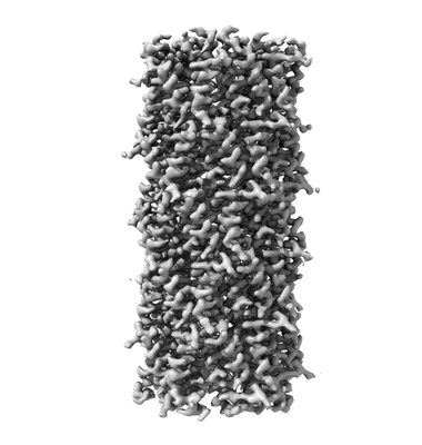 emd_20044.png emd_20044.png | 51.9 KB | ||
| Filedesc metadata | emd-20044.cif.gz | 5.9 KB | ||
| Archive directory |  http://ftp.pdbj.org/pub/emdb/structures/EMD-20044ftp://ftp.pdbj.org/pub/emdb/structures/EMD-20044 http://ftp.pdbj.org/pub/emdb/structures/EMD-20044ftp://ftp.pdbj.org/pub/emdb/structures/EMD-20044 | HTTPS FTP |
-Related structure data
| Related structure data | 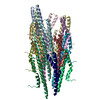 6offMC 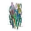 6ofeC 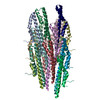 6ofgC  6ofhC C: citing same article ( M: atomic model generated by this map |
|---|---|
| Similar structure data |
-Links
| EMDB pages | EMDB (EBI/PDBe) / EMDataResource |
|---|
-Map
| File | Download / File: emd_20044.map.gz / Format: CCP4 / Size: 64 MB / Type: IMAGE STORED AS FLOATING POINT NUMBER (4 BYTES) | ||||||||||||||||||||||||||||||||||||||||||||||||||||||||||||
|---|---|---|---|---|---|---|---|---|---|---|---|---|---|---|---|---|---|---|---|---|---|---|---|---|---|---|---|---|---|---|---|---|---|---|---|---|---|---|---|---|---|---|---|---|---|---|---|---|---|---|---|---|---|---|---|---|---|---|---|---|---|
| Annotation | EM map of in vitro polymerized PrgI filaments | ||||||||||||||||||||||||||||||||||||||||||||||||||||||||||||
| Projections & slices | Image control
Images are generated by Spider. | ||||||||||||||||||||||||||||||||||||||||||||||||||||||||||||
| Voxel size | X=Y=Z: 1.045 Å | ||||||||||||||||||||||||||||||||||||||||||||||||||||||||||||
| Density |
| ||||||||||||||||||||||||||||||||||||||||||||||||||||||||||||
| Symmetry | Space group: 1 | ||||||||||||||||||||||||||||||||||||||||||||||||||||||||||||
| Details | EMDB XML:
CCP4 map header:
| ||||||||||||||||||||||||||||||||||||||||||||||||||||||||||||
 Z (Sec.)
 Y (Row.)
 X (Col.)


-Supplemental data
- Sample components
-Entire : PrgI
| Entire | Name: PrgI |
|---|---|
| Components |
|
-Supramolecule #1: PrgI
| Supramolecule | Name: PrgI / type: complex / ID: 1 / Parent: 0 / Macromolecule list: all |
|---|---|
| Source (natural) | Organism: Salmonella enterica subsp. enterica serovar Typhimurium str. LT2 (bacteria) |
-Macromolecule #1: Protein PrgI
| Macromolecule | Name: Protein PrgI / type: protein_or_peptide / ID: 1 / Number of copies: 18 / Enantiomer: LEVO |
|---|---|
| Source (natural) | Organism: Salmonella typhimurium (strain SL1344) (bacteria) / Strain: SL1344 |
| Molecular weight | Theoretical: 9.147145 KDa |
| Recombinant expression | Organism: |
| Sequence | String: GSHMATPWSG YLDDVSAKFD TGVDNLQTQV TEALDKLAAK PSDPALLAAY QSKLSEYNLY RNAQSNTVKV FKDIDAAIIQ NFR UniProtKB: Type III secretion system apparatus |
-Experimental details
-Structure determination
| Method | cryo EM |
|---|---|
 Processing Processing | helical reconstruction |
| Aggregation state | filament |
-Sample preparation
| Concentration | 1.5 mg/mL |
|---|---|
| Buffer | pH: 7.5 |
| Grid | Details: unspecified |
| Vitrification | Cryogen name: ETHANE |
- Electron microscopy
| Microscope | FEI TITAN KRIOS |
|---|---|
| Image recording | Film or detector model: GATAN K2 SUMMIT (4k x 4k) / Average electron dose: 47.2 e/Å2 |
| Electron beam | Acceleration voltage: 300 kV / Electron source:  FIELD EMISSION GUN FIELD EMISSION GUN |
| Electron optics | Illumination mode: FLOOD BEAM / Imaging mode: BRIGHT FIELD |
| Experimental equipment |  Model: Titan Krios / Image courtesy: FEI Company |
-Image processing
| Final reconstruction | Applied symmetry - Helical parameters - Δz: 4.25 Å Applied symmetry - Helical parameters - Δ&Phi: 63.34 ° Applied symmetry - Helical parameters - Axial symmetry: C1 (asymmetric) Resolution.type: BY AUTHOR / Resolution: 3.2 Å / Resolution method: FSC 0.143 CUT-OFF / Number images used: 10201 |
|---|---|
| CTF correction | Type: PHASE FLIPPING AND AMPLITUDE CORRECTION |
| Startup model | Type of model: OTHER |
| Final angle assignment | Type: NOT APPLICABLE |
