 Movie
Movie Controller


+ Open data

- Basic information
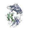
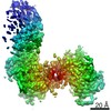
| Entry | Database: PDB / ID: 1de4 | ||||||
|---|---|---|---|---|---|---|---|
| Title | HEMOCHROMATOSIS PROTEIN HFE COMPLEXED WITH TRANSFERRIN RECEPTOR | ||||||
 Components Components |
| ||||||
 Keywords Keywords | METAL TRANSPORT INHIBITOR/RECEPTOR / HFE /  HEREDITARY HEMOCHROMATOSIS / MHC CLASS I / TRANSFERRIN RECEPTOR / METAL TRANSPORT INHIBITOR-RECEPTOR COMPLEX HEREDITARY HEMOCHROMATOSIS / MHC CLASS I / TRANSFERRIN RECEPTOR / METAL TRANSPORT INHIBITOR-RECEPTOR COMPLEX | ||||||
| Function / homology |  Function and homology information Function and homology informationnegative regulation of antigen processing and presentation of endogenous peptide antigen via MHC class I / response to iron ion starvation / transferrin receptor activity / negative regulation of T cell cytokine production / hormone biosynthetic process / negative regulation of mitochondrial fusion / negative regulation of CD8-positive, alpha-beta T cell activation / transferrin transport / co-receptor binding / Transferrin endocytosis and recycling ...negative regulation of antigen processing and presentation of endogenous peptide antigen via MHC class I / response to iron ion starvation / transferrin receptor activity / negative regulation of T cell cytokine production / hormone biosynthetic process / negative regulation of mitochondrial fusion / negative regulation of CD8-positive, alpha-beta T cell activation / transferrin transport / co-receptor binding / Transferrin endocytosis and recycling / transferrin receptor binding / positive regulation of isotype switching / regulation of protein localization to cell surface / basal part of cell / positive regulation of signaling receptor activity / response to iron ion / response to copper ion / response to manganese ion / RND1 GTPase cycle / RND2 GTPase cycle / RHOB GTPase cycle / Golgi Associated Vesicle Biogenesis / RHOJ GTPase cycle / RHOC GTPase cycle / RHOQ GTPase cycle / positive regulation of peptide hormone secretion / positive regulation of SMAD protein signal transduction / RHOH GTPase cycle / CDC42 GTPase cycle / transport across blood-brain barrier / RHOG GTPase cycle / negative regulation of proteasomal ubiquitin-dependent protein catabolic process / RHOA GTPase cycle / RAC2 GTPase cycle / RAC3 GTPase cycle / positive regulation of bone resorption / BMP signaling pathway / beta-2-microglobulin binding / response to retinoic acid / positive regulation of T cell proliferation / clathrin-coated pit / negative regulation of signaling receptor activity / positive regulation of B cell proliferation / Hsp70 protein binding / RAC1 GTPase cycle / negative regulation of ubiquitin-dependent protein catabolic process / response to nutrient / osteoclast differentiation / antigen processing and presentation of endogenous peptide antigen via MHC class I via ER pathway, TAP-independent / antigen processing and presentation of endogenous peptide antigen via MHC class Ib / positive regulation of ferrous iron binding / positive regulation of transferrin receptor binding / cellular response to leukemia inhibitory factor / positive regulation of receptor binding / early endosome lumen / Nef mediated downregulation of MHC class I complex cell surface expression / DAP12 interactions / negative regulation of receptor binding / acute-phase response / Endosomal/Vacuolar pathway / Antigen Presentation: Folding, assembly and peptide loading of class I MHC / clathrin-coated endocytic vesicle membrane / cellular response to iron(III) ion / antigen processing and presentation of exogenous protein antigen via MHC class Ib, TAP-dependent / positive regulation of protein-containing complex assembly / negative regulation of forebrain neuron differentiation / ER to Golgi transport vesicle membrane / regulation of erythrocyte differentiation / peptide antigen assembly with MHC class I protein complex / response to molecule of bacterial origin / regulation of iron ion transport / MHC class I peptide loading complex / HFE-transferrin receptor complex / receptor internalization / T cell mediated cytotoxicity / recycling endosome / cellular response to iron ion / antigen processing and presentation of endogenous peptide antigen via MHC class I / positive regulation of T cell cytokine production / MHC class I protein complex / multicellular organismal-level iron ion homeostasis / negative regulation of neurogenesis / peptide antigen assembly with MHC class II protein complex / positive regulation of receptor-mediated endocytosis / positive regulation of T cell mediated cytotoxicity / MHC class II protein complex / cellular response to nicotine / positive regulation of protein localization to nucleus / recycling endosome membrane / specific granule lumen / phagocytic vesicle membrane / peptide antigen binding / positive regulation of cellular senescence / antigen processing and presentation of exogenous peptide antigen via MHC class II / negative regulation of epithelial cell proliferation / Immunoregulatory interactions between a Lymphoid and a non-Lymphoid cell / Interferon gamma signaling / positive regulation of immune response / Modulation by Mtb of host immune system / double-stranded RNA bindingSimilarity search - Function | ||||||
| Biological species |  Homo sapiens (human) Homo sapiens (human) | ||||||
| Method | X-RAY DIFFRACTION / SYNCHROTRON / MIRAS / Resolution: 2.8 Å | ||||||
 Authors Authors | Bennett, M.J. / Lebron, J.A. / Bjorkman, P.J. | ||||||
 Citation Citation | Journal: Nature / Year: 2000 Title: Crystal structure of the hereditary haemochromatosis protein HFE complexed with transferrin receptor. Authors: Bennett, M.J. / Lebron, J.A. / Bjorkman, P.J. | ||||||
| History |
|
- Structure visualization
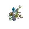
| Structure viewer | Molecule: MolmilJmol/JSmol |
|---|
- Downloads & links
-Download
| PDBx/mmCIF format | 1de4.cif.gz | 599.1 KB | Display | PDBx/mmCIF format |
|---|---|---|---|---|
| PDB format | pdb1de4.ent.gz | 493.6 KB | Display | PDB format |
| PDBx/mmJSON format | 1de4.json.gz | Tree view | PDBx/mmJSON format | |
| Others |  Other downloads Other downloads |
-Validation report
| Arichive directory | https://data.pdbj.org/pub/pdb/validation_reports/de/1de4ftp://data.pdbj.org/pub/pdb/validation_reports/de/1de4 | HTTPS FTP |
|---|
-Related structure data
| Related structure data | |
|---|---|
| Similar structure data |


-Links
 PDBj
PDBj


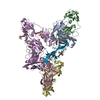
- Assembly
| Deposited unit | 
| ||||||||
|---|---|---|---|---|---|---|---|---|---|
| 1 | 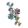
| ||||||||
| 2 | 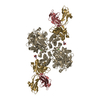
| ||||||||
| Unit cell |
|
-Components
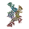
-Protein , 3 types, 9 molecules ADGBEHCFI
| #1: Protein | Mass: 32339.434 Da / Num. of mol.: 3 / Fragment: ECTODOMAIN Source method: isolated from a genetically manipulated source Source: (gene. exp.) Homo sapiens (human) / Plasmid: PBJ5-GS / Cell (production host): OVARY CELLS / Production host:   Cricetulus griseus (Chinese hamster) / References: UniProt: Q30201 Cricetulus griseus (Chinese hamster) / References: UniProt: Q30201#2: Protein | Beta-2 microglobulinMass: 11748.160 Da / Num. of mol.: 3 Source method: isolated from a genetically manipulated source Source: (gene. exp.) Homo sapiens (human) / Cell (production host): OVARY CELLS / Production host: Cricetulus griseus (Chinese hamster) / References: UniProt: P61769#3: Protein | Mass: 71807.258 Da / Num. of mol.: 3 / Fragment: ECTODOMAIN Source method: isolated from a genetically manipulated source Source: (gene. exp.) Homo sapiens (human) / Plasmid: PACGP67A / Cell (production host): HIGH 5 INSECT CELLS / Production host:  Trichoplusia ni (cabbage looper) / References: UniProt: P02786 Trichoplusia ni (cabbage looper) / References: UniProt: P02786 |
|---|
-Sugars , 1 types, 3 molecules 
| #4: Sugar | N-Acetylglucosamine Type: D-saccharide, beta linking / Mass: 221.208 Da / Num. of mol.: 3 Type: D-saccharide, beta linking / Mass: 221.208 Da / Num. of mol.: 3Source method: isolated from a genetically manipulated source Formula: C8H15NO6 |
|---|
-Non-polymers , 3 types, 13 molecules 


| #5: Chemical |  Mass: 40.078 Da / Num. of mol.: 3 / Source method: obtained synthetically / Formula: Ca Mass: 40.078 Da / Num. of mol.: 3 / Source method: obtained synthetically / Formula: Ca#6: Chemical | ChemComp-GOL / | Glycerol Mass: 92.094 Da / Num. of mol.: 1 / Source method: obtained synthetically / Formula: C3H8O3 Mass: 92.094 Da / Num. of mol.: 1 / Source method: obtained synthetically / Formula: C3H8O3#7: Water | ChemComp-HOH / | WaterMass: 18.015 Da / Num. of mol.: 9 / Source method: isolated from a natural source / Formula: H2O |
|---|
-Experimental details
-Experiment
| Experiment | Method: X-RAY DIFFRACTION / Number of used crystals: 1 |
|---|
- Sample preparation
| Crystal | Density Matthews: 3.74 Å3/Da / Density % sol: 61 % | ||||||||||||||||||||
|---|---|---|---|---|---|---|---|---|---|---|---|---|---|---|---|---|---|---|---|---|---|
| Crystal grow | Temperature: 298 K / Method: vapor diffusion, hanging drop / pH: 8 Details: PEG 8000, TRIS, TRIMETHYLAMINE HCL, pH 8.0, VAPOR DIFFUSION, HANGING DROP, temperature 298K | ||||||||||||||||||||
| Crystal grow | *PLUS | ||||||||||||||||||||
| Components of the solutions | *PLUS
|
-Data collection
| Diffraction | Mean temperature: 100 K |
|---|---|
| Diffraction source | Source: SYNCHROTRON / Site: SSRL  / Beamline: BL9-1 / Wavelength: 0.98 / Beamline: BL9-1 / Wavelength: 0.98 |
| Detector | Type: MARRESEARCH / Detector: IMAGE PLATE / Date: Jan 7, 1999 |
| Radiation | Protocol: SINGLE WAVELENGTH / Monochromatic (M) / Laue (L): M / Scattering type: x-ray |
| Radiation wavelength | Wavelength: 0.98 Å / Relative weight: 1 |
| Reflection | Resolution: 2.8→30 Å / Num. all: 122846 / Num. obs: 122846 / % possible obs: 98.1 % / Observed criterion σ(I): -3 / Redundancy: 2.8 % / Rmerge(I) obs: 0.07 / Net I/σ(I): 18 |
| Reflection shell | Resolution: 2.8→2.85 Å / Redundancy: 2.9 % / Rmerge(I) obs: 0.341 / Mean I/σ(I) obs: 3.3 / % possible all: 99.8 |
| Reflection | *PLUS |
| Reflection shell | *PLUS % possible obs: 99.8 % |
- Processing
| Software |
| ||||||||||||||||||||||||||||||||||||||||||||||||||||||||||||||||||||||||||||||||
|---|---|---|---|---|---|---|---|---|---|---|---|---|---|---|---|---|---|---|---|---|---|---|---|---|---|---|---|---|---|---|---|---|---|---|---|---|---|---|---|---|---|---|---|---|---|---|---|---|---|---|---|---|---|---|---|---|---|---|---|---|---|---|---|---|---|---|---|---|---|---|---|---|---|---|---|---|---|---|---|---|---|
| Refinement | Method to determine structure: MIRAS / Resolution: 2.8→30 Å / Rfactor Rfree error: 0.002 / Data cutoff high absF: 685830.33 / Data cutoff low absF: 0 / Isotropic thermal model: RESTRAINED / Cross valid method: THROUGHOUT / σ(F): 0 / Stereochemistry target values: ENGH AND HUBER Details: SEVERAL SIDECHAINS ARE MODELED AS ALA AS IN UNCOMPLEXED HFE. A POSITIVE DIFFERENCE PEAK NEAR TFR CHAINS C,F,I WHICH LACKED PROTEIN LIGANDS WAS MODELED AS A WATER MOLECULE (WATERS 4,6,8). A ...Details: SEVERAL SIDECHAINS ARE MODELED AS ALA AS IN UNCOMPLEXED HFE. A POSITIVE DIFFERENCE PEAK NEAR TFR CHAINS C,F,I WHICH LACKED PROTEIN LIGANDS WAS MODELED AS A WATER MOLECULE (WATERS 4,6,8). A POSITIVE DIFFERENCE PEAK NEAR THE HFE CHAIN G PLATFORM WAS MODELED AS A GLYCEROL. SEVERAL LOOPS HAVE RESIDUES WITH LOW CORRELATIONS AGAINST THE FINAL MAP: HFE PLATFORM LOOPS FROM STRAND 1 (S1) TO STRAND 2 (S2), S4-ALPHA1 HELIX; HFE ALPHA3 DOMAIN LOOPS S1-S2, S3-S4,S6-S7; BETA-2-MICROGLOBULIN LOOPS S1-S2, S3-S4, S5-S6, C-TERMINUS.
| ||||||||||||||||||||||||||||||||||||||||||||||||||||||||||||||||||||||||||||||||
| Solvent computation | Solvent model: FLAT MODEL / Bsol: 30.72 Å2 / ksol: 0.303 e/Å3 | ||||||||||||||||||||||||||||||||||||||||||||||||||||||||||||||||||||||||||||||||
| Displacement parameters | Biso mean: 66.3 Å2
| ||||||||||||||||||||||||||||||||||||||||||||||||||||||||||||||||||||||||||||||||
| Refinement step | Cycle: LAST / Resolution: 2.8→30 Å
| ||||||||||||||||||||||||||||||||||||||||||||||||||||||||||||||||||||||||||||||||
| Refine LS restraints |
| ||||||||||||||||||||||||||||||||||||||||||||||||||||||||||||||||||||||||||||||||
| Refine LS restraints NCS | NCS model details: RESTRAINED | ||||||||||||||||||||||||||||||||||||||||||||||||||||||||||||||||||||||||||||||||
| LS refinement shell | Resolution: 2.8→2.98 Å / Rfactor Rfree error: 0.009 / Total num. of bins used: 6
| ||||||||||||||||||||||||||||||||||||||||||||||||||||||||||||||||||||||||||||||||
| Xplor file |
| ||||||||||||||||||||||||||||||||||||||||||||||||||||||||||||||||||||||||||||||||
| Software | *PLUS Name: CNS / Version: 0.5 / Classification: refinement | ||||||||||||||||||||||||||||||||||||||||||||||||||||||||||||||||||||||||||||||||
| Refine LS restraints | *PLUS
|
