[English] 日本語
 Yorodumi
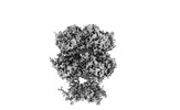
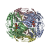
- EMDB-19550: cryoEM structure of purified Acs1 filament from meiotic yeast cells -
+ Open data
- Basic information
| Entry |  | |||||||||
|---|---|---|---|---|---|---|---|---|---|---|
| Title | cryoEM structure of purified Acs1 filament from meiotic yeast cells | |||||||||
 Map data Map data | ||||||||||
 Sample Sample |
| |||||||||
 Keywords Keywords | metabolic enzyme / filament / cryoEM / CYTOSOLIC PROTEIN | |||||||||
| Biological species |   | |||||||||
| Method | single particle reconstruction / cryo EM / Resolution: 3.6 Å | |||||||||
 Authors Authors | Hugener J / Xu J / Wettstein R / Ioannidi L / Velikov D / Wollweber F / Henggeler A / Matos J / Pilhofer M | |||||||||
| Funding support |  Switzerland, European Union, 2 items Switzerland, European Union, 2 items
| |||||||||
 Citation Citation |  Journal: Cell / Year: 2024 Journal: Cell / Year: 2024Title: FilamentID reveals the composition and function of metabolic enzyme polymers during gametogenesis. Authors: Jannik Hugener / Jingwei Xu / Rahel Wettstein / Lydia Ioannidi / Daniel Velikov / Florian Wollweber / Adrian Henggeler / Joao Matos / Martin Pilhofer /   Abstract: Gamete formation and subsequent offspring development often involve extended phases of suspended cellular development or even dormancy. How cells adapt to recover and resume growth remains poorly ...Gamete formation and subsequent offspring development often involve extended phases of suspended cellular development or even dormancy. How cells adapt to recover and resume growth remains poorly understood. Here, we visualized budding yeast cells undergoing meiosis by cryo-electron tomography (cryoET) and discovered elaborate filamentous assemblies decorating the nucleus, cytoplasm, and mitochondria. To determine filament composition, we developed a "filament identification" (FilamentID) workflow that combines multiscale cryoET/cryo-electron microscopy (cryoEM) analyses of partially lysed cells or organelles. FilamentID identified the mitochondrial filaments as being composed of the conserved aldehyde dehydrogenase Ald4 and the nucleoplasmic/cytoplasmic filaments as consisting of acetyl-coenzyme A (CoA) synthetase Acs1. Structural characterization further revealed the mechanism underlying polymerization and enabled us to genetically perturb filament formation. Acs1 polymerization facilitates the recovery of chronologically aged spores and, more generally, the cell cycle re-entry of starved cells. FilamentID is broadly applicable to characterize filaments of unknown identity in diverse cellular contexts. | |||||||||
| History |
|
- Structure visualization
| Supplemental images |
|---|
- Downloads & links
-EMDB archive
| Map data |  emd_19550.map.gz emd_19550.map.gz | 49.9 MB |  EMDB map data format EMDB map data format | |
|---|---|---|---|---|
| Header (meta data) |  emd-19550-v30.xml emd-19550-v30.xml emd-19550.xml emd-19550.xml | 14 KB 14 KB | Display Display |  EMDB header EMDB header |
| Images |  emd_19550.png emd_19550.png | 42.4 KB | ||
| Masks |  emd_19550_msk_1.map emd_19550_msk_1.map | 52.7 MB |  Mask map Mask map | |
| Filedesc metadata |  emd-19550.cif.gz emd-19550.cif.gz | 4.9 KB | ||
| Others |  emd_19550_half_map_1.map.gz emd_19550_half_map_1.map.gz emd_19550_half_map_2.map.gz emd_19550_half_map_2.map.gz | 49 MB 49 MB | ||
| Archive directory |  http://ftp.pdbj.org/pub/emdb/structures/EMD-19550 http://ftp.pdbj.org/pub/emdb/structures/EMD-19550 ftp://ftp.pdbj.org/pub/emdb/structures/EMD-19550 ftp://ftp.pdbj.org/pub/emdb/structures/EMD-19550 | HTTPS FTP |
-Validation report
| Summary document |  emd_19550_validation.pdf.gz emd_19550_validation.pdf.gz | 1 MB | Display |  EMDB validaton report EMDB validaton report |
|---|---|---|---|---|
| Full document |  emd_19550_full_validation.pdf.gz emd_19550_full_validation.pdf.gz | 1 MB | Display | |
| Data in XML |  emd_19550_validation.xml.gz emd_19550_validation.xml.gz | 11.8 KB | Display | |
| Data in CIF |  emd_19550_validation.cif.gz emd_19550_validation.cif.gz | 13.9 KB | Display | |
| Arichive directory |  https://ftp.pdbj.org/pub/emdb/validation_reports/EMD-19550 https://ftp.pdbj.org/pub/emdb/validation_reports/EMD-19550 ftp://ftp.pdbj.org/pub/emdb/validation_reports/EMD-19550 ftp://ftp.pdbj.org/pub/emdb/validation_reports/EMD-19550 | HTTPS FTP |
-Related structure data
- Links
Links
| EMDB pages |  EMDB (EBI/PDBe) / EMDB (EBI/PDBe) /  EMDataResource EMDataResource |
|---|
- Map
Map
| File |  Download / File: emd_19550.map.gz / Format: CCP4 / Size: 52.7 MB / Type: IMAGE STORED AS FLOATING POINT NUMBER (4 BYTES) Download / File: emd_19550.map.gz / Format: CCP4 / Size: 52.7 MB / Type: IMAGE STORED AS FLOATING POINT NUMBER (4 BYTES) | ||||||||||||||||||||||||||||||||||||
|---|---|---|---|---|---|---|---|---|---|---|---|---|---|---|---|---|---|---|---|---|---|---|---|---|---|---|---|---|---|---|---|---|---|---|---|---|---|
| Projections & slices | Image control
Images are generated by Spider. | ||||||||||||||||||||||||||||||||||||
| Voxel size | X=Y=Z: 1.3 Å | ||||||||||||||||||||||||||||||||||||
| Density |
| ||||||||||||||||||||||||||||||||||||
| Symmetry | Space group: 1 | ||||||||||||||||||||||||||||||||||||
| Details | EMDB XML:
|
-Supplemental data
-Mask #1
| File |  emd_19550_msk_1.map emd_19550_msk_1.map | ||||||||||||
|---|---|---|---|---|---|---|---|---|---|---|---|---|---|
| Projections & Slices |
| ||||||||||||
| Density Histograms |
-Half map: #1
| File | emd_19550_half_map_1.map | ||||||||||||
|---|---|---|---|---|---|---|---|---|---|---|---|---|---|
| Projections & Slices |
| ||||||||||||
| Density Histograms |
-Half map: #2
| File | emd_19550_half_map_2.map | ||||||||||||
|---|---|---|---|---|---|---|---|---|---|---|---|---|---|
| Projections & Slices |
| ||||||||||||
| Density Histograms |
- Sample components
-Entire : Purified Acetyl-CoA synthetase 1 filament from meiotic yeast cells
| Entire | Name: Purified Acetyl-CoA synthetase 1 filament from meiotic yeast cells |
|---|---|
| Components |
|
-Supramolecule #1: Purified Acetyl-CoA synthetase 1 filament from meiotic yeast cells
| Supramolecule | Name: Purified Acetyl-CoA synthetase 1 filament from meiotic yeast cells type: complex / ID: 1 / Parent: 0 / Macromolecule list: all |
|---|---|
| Source (natural) | Organism:  |
-Macromolecule #1: Acetyl-CoA synthetase 1 from yeast SK1 strain
| Macromolecule | Name: Acetyl-CoA synthetase 1 from yeast SK1 strain / type: protein_or_peptide / ID: 1 / Enantiomer: LEVO |
|---|---|
| Source (natural) | Organism:  |
| Sequence | String: MSPSAVQSSK LEEQSSEID K LKAKMSQS AS TAQQKKE HEY EHLTSV KIVP QRPIS DRLQP AIAT HYSPHL DGL QDYQRLH KE SIEDPAKF F GSKATQFLN WSKPFDKVFI PDSKTGRPS F QNNAWFLN GQ LNACYNC VDR HALKTP NKKA IIFEG ...String: MSPSAVQSSK LEEQSSEID K LKAKMSQS AS TAQQKKE HEY EHLTSV KIVP QRPIS DRLQP AIAT HYSPHL DGL QDYQRLH KE SIEDPAKF F GSKATQFLN WSKPFDKVFI PDSKTGRPS F QNNAWFLN GQ LNACYNC VDR HALKTP NKKA IIFEG DEPGQ GYSI TYKELL EEV CQVAQVL TY SMGVRKGD T VAVYMPMVP EAIITLLAIS RIGAIHSVV F AGFSSNSL RD RINDGDS KVV ITTDES NRGG KVIET KRIVD DALR ETPGVR HVL VYRKTNN PS VAFHAPRD L DWATEKKKY KTYYPCTPVD SEDPLFLLY T SGSTGAPK GV QHSTAGY LLG ALLTMR YTFD THQED VFFTA GDIG WITGHT YVV YGPLLYG CA TLVFEGTP A YPNYSRYWD IIDEHKVTQF YVAPTALRL L KRAGDSYI EN HSLKSLR CLG SVGEPI AAEV WEWYS EKIGK NEIP IVDTYW QTE SGSHLVT PL AGGVTPMK P GSASFPFFG IDAVVLDPNT GEELNTSHA E GVLAVKAA WP SFARTIW KNH DRYLDT YLNP YPGYY FTGDG AAKD KDGYIW ILG RVDDVVN VS GHRLSTAE I EAAIIEDPI VAECAVVGFN DDLTGQAVA A FVVLKNKS NW STATDDE LQD IKKHLV FTVR KDIGP FAAPK LIIL VDDLPK TRS GKIMRRI LR KILAGESD Q LGDVSTLSN PGIVRHLIDS VKL |
-Experimental details
-Structure determination
| Method | cryo EM |
|---|---|
 Processing Processing | single particle reconstruction |
| Aggregation state | filament |
- Sample preparation
| Buffer | pH: 6.4 |
|---|---|
| Vitrification | Cryogen name: ETHANE-PROPANE |
- Electron microscopy
| Microscope | FEI TITAN KRIOS |
|---|---|
| Image recording | Film or detector model: GATAN K3 (6k x 4k) / Average electron dose: 60.0 e/Å2 |
| Electron beam | Acceleration voltage: 300 kV / Electron source:  FIELD EMISSION GUN FIELD EMISSION GUN |
| Electron optics | Illumination mode: FLOOD BEAM / Imaging mode: BRIGHT FIELD / Nominal defocus max: 2.0 µm / Nominal defocus min: 1.0 µm |
| Experimental equipment |  Model: Titan Krios / Image courtesy: FEI Company |
- Image processing
| Startup model | Type of model: OTHER Details: the initial model is determined from cryoEM structure of Acs1 determined by filamentID (D_1292136168) |
|---|---|
| Final reconstruction | Applied symmetry - Point group: C3 (3 fold cyclic) / Resolution.type: BY AUTHOR / Resolution: 3.6 Å / Resolution method: FSC 0.143 CUT-OFF / Software - Name: cryoSPARC / Number images used: 7310 |
| Initial angle assignment | Type: NOT APPLICABLE |
| Final angle assignment | Type: NOT APPLICABLE |
 Movie
Movie Controller


 Z (Sec.)
 Y (Row.)
 X (Col.)


