[English] 日本語
 Yorodumi
- EMDB-1953: Structure of the CS1 pilus of enterotoxigenic Escherichia coli re... -
+ Open data
- Basic information
| Entry | Database: EMDB / ID: EMD-1953 | |||||||||
|---|---|---|---|---|---|---|---|---|---|---|
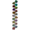
| Title | Structure of the CS1 pilus of enterotoxigenic Escherichia coli reveals structural polymorphism | |||||||||
 Map data Map data | CS1-Group 3 | |||||||||
 Sample Sample |
| |||||||||
 Keywords Keywords | pili / bacterial pathogenesis / colonization / virulence factor / donor strand complementarity / image reconstruction / helical symmetry / adhesion | |||||||||
| Biological species |  | |||||||||
| Method | helical reconstruction / negative staining / Resolution: 20.0 Å | |||||||||
 Authors Authors | Galkin VE / Kolappan S / Ng D / Zong Z / Li J / Yu X / Egelman EH / Craig L | |||||||||
 Citation Citation |  Journal: J Bacteriol / Year: 2013 Journal: J Bacteriol / Year: 2013Title: The structure of the CS1 pilus of enterotoxigenic Escherichia coli reveals structural polymorphism. Authors: Vitold E Galkin / Subramaniapillai Kolappan / Dixon Ng / ZuSheng Zong / Juliana Li / Xiong Yu / Edward H Egelman / Lisa Craig /  Abstract: Enterotoxigenic Escherichia coli (ETEC) is a bacterial pathogen that causes diarrhea in children and travelers in developing countries. ETEC adheres to host epithelial cells in the small intestine ...Enterotoxigenic Escherichia coli (ETEC) is a bacterial pathogen that causes diarrhea in children and travelers in developing countries. ETEC adheres to host epithelial cells in the small intestine via a variety of different pili. The CS1 pilus is a prototype for a family of related pili, including the CFA/I pili, present on ETEC and other Gram-negative bacterial pathogens. These pili are assembled by an outer membrane usher protein that catalyzes subunit polymerization via donor strand complementation, in which the N terminus of each incoming pilin subunit fits into a hydrophobic groove in the terminal subunit, completing a β-sheet in the Ig fold. Here we determined a crystal structure of the CS1 major pilin subunit, CooA, to a 1.6-Å resolution. CooA is a globular protein with an Ig fold and is similar in structure to the CFA/I major pilin CfaB. We determined three distinct negative-stain electron microscopic reconstructions of the CS1 pilus and generated pseudoatomic-resolution pilus structures using the CooA crystal structure. CS1 pili adopt multiple structural states with differences in subunit orientations and packing. We propose that the structural perturbations are accommodated by flexibility in the N-terminal donor strand of CooA and by plasticity in interactions between exposed flexible loops on adjacent subunits. Our results suggest that CS1 and other pili of this class are extensible filaments that can be stretched in response to mechanical stress encountered during colonization. | |||||||||
| History |
|
- Structure visualization
| Movie |
 Movie viewer Movie viewer |
|---|---|
| Structure viewer | EM map:  SurfView SurfView Molmil Molmil Jmol/JSmol Jmol/JSmol |
| Supplemental images |
- Downloads & links
-EMDB archive
| Map data |  emd_1953.map.gz emd_1953.map.gz | 112.9 KB |  EMDB map data format EMDB map data format | |
|---|---|---|---|---|
| Header (meta data) |  emd-1953-v30.xml emd-1953-v30.xml emd-1953.xml emd-1953.xml | 10.6 KB 10.6 KB | Display Display |  EMDB header EMDB header |
| Images |  EMD-1953.png EMD-1953.png | 50.3 KB | ||
| Archive directory |  http://ftp.pdbj.org/pub/emdb/structures/EMD-1953 http://ftp.pdbj.org/pub/emdb/structures/EMD-1953 ftp://ftp.pdbj.org/pub/emdb/structures/EMD-1953 ftp://ftp.pdbj.org/pub/emdb/structures/EMD-1953 | HTTPS FTP |
-Validation report
| Summary document |  emd_1953_validation.pdf.gz emd_1953_validation.pdf.gz | 199 KB | Display |  EMDB validaton report EMDB validaton report |
|---|---|---|---|---|
| Full document |  emd_1953_full_validation.pdf.gz emd_1953_full_validation.pdf.gz | 198.1 KB | Display | |
| Data in XML |  emd_1953_validation.xml.gz emd_1953_validation.xml.gz | 5.3 KB | Display | |
| Arichive directory |  https://ftp.pdbj.org/pub/emdb/validation_reports/EMD-1953 https://ftp.pdbj.org/pub/emdb/validation_reports/EMD-1953 ftp://ftp.pdbj.org/pub/emdb/validation_reports/EMD-1953 ftp://ftp.pdbj.org/pub/emdb/validation_reports/EMD-1953 | HTTPS FTP |
-Related structure data
| Related structure data |  3s0v  1951C  1952C  4hjiC M: atomic model generated by this map C: citing same article ( |
|---|---|
| Similar structure data |
- Links
Links
| EMDB pages |  EMDB (EBI/PDBe) / EMDB (EBI/PDBe) /  EMDataResource EMDataResource |
|---|
- Map
Map
| File |  Download / File: emd_1953.map.gz / Format: CCP4 / Size: 3.7 MB / Type: IMAGE STORED AS FLOATING POINT NUMBER (4 BYTES) Download / File: emd_1953.map.gz / Format: CCP4 / Size: 3.7 MB / Type: IMAGE STORED AS FLOATING POINT NUMBER (4 BYTES) | ||||||||||||||||||||||||||||||||||||||||||||||||||||||||||||
|---|---|---|---|---|---|---|---|---|---|---|---|---|---|---|---|---|---|---|---|---|---|---|---|---|---|---|---|---|---|---|---|---|---|---|---|---|---|---|---|---|---|---|---|---|---|---|---|---|---|---|---|---|---|---|---|---|---|---|---|---|---|
| Annotation | CS1-Group 3 | ||||||||||||||||||||||||||||||||||||||||||||||||||||||||||||
| Projections & slices | Image control
Images are generated by Spider. | ||||||||||||||||||||||||||||||||||||||||||||||||||||||||||||
| Voxel size | X=Y=Z: 4.16 Å | ||||||||||||||||||||||||||||||||||||||||||||||||||||||||||||
| Density |
| ||||||||||||||||||||||||||||||||||||||||||||||||||||||||||||
| Symmetry | Space group: 1 | ||||||||||||||||||||||||||||||||||||||||||||||||||||||||||||
| Details | EMDB XML:
CCP4 map header:
| ||||||||||||||||||||||||||||||||||||||||||||||||||||||||||||
-Supplemental data
- Sample components
-Entire : CS1 pilus
| Entire | Name: CS1 pilus |
|---|---|
| Components |
|
-Supramolecule #1000: CS1 pilus
| Supramolecule | Name: CS1 pilus / type: sample / ID: 1000 / Oligomeric state: Oligomer / Number unique components: 1 |
|---|
-Supramolecule #1: CooA
| Supramolecule | Name: CooA / type: organelle_or_cellular_component / ID: 1 / Name.synonym: CS1 pilin / Oligomeric state: oligomer / Recombinant expression: No / Database: NCBI |
|---|---|
| Source (natural) | Organism:  |
| Molecular weight | Experimental: 15 KDa |
-Experimental details
-Structure determination
| Method | negative staining |
|---|---|
 Processing Processing | helical reconstruction |
| Aggregation state | filament |
- Sample preparation
| Concentration | 0.2 mg/mL |
|---|---|
| Buffer | pH: 7.4 Details: 137 mM NaCl, 2.7 mM KCl, 10 mM Na2HPO4, 2 mM KH2PO4, 10 mM EDTA |
| Staining | Type: NEGATIVE / Details: 2% uranyl acetate |
| Grid | Details: glow discharged continuous carbon coated grids |
| Vitrification | Cryogen name: NONE / Instrument: OTHER |
- Electron microscopy
| Microscope | FEI TECNAI 12 |
|---|---|
| Alignment procedure | Legacy - Astigmatism: objective lens astigmatism was corrected at 100,000 times magnification |
| Details | Low dose mode |
| Image recording | Category: FILM / Film or detector model: KODAK SO-163 FILM / Digitization - Scanner: NIKON COOLSCAN / Digitization - Sampling interval: 4.16 µm / Number real images: 10 / Average electron dose: 10 e/Å2 |
| Electron beam | Acceleration voltage: 80 kV / Electron source: LAB6 |
| Electron optics | Calibrated magnification: 30000 / Illumination mode: OTHER / Imaging mode: BRIGHT FIELD / Cs: 2 mm / Nominal defocus max: 2.0 µm / Nominal defocus min: 1.0 µm / Nominal magnification: 30000 |
| Sample stage | Specimen holder: Eucentric / Specimen holder model: SIDE ENTRY, EUCENTRIC |
- Image processing
| Details | Pilus segments were selected using the helixboxer program in SPIDER. |
|---|---|
| Final reconstruction | Applied symmetry - Helical parameters - Δz: 10.0 Å Applied symmetry - Helical parameters - Δ&Phi: 111.9 ° Algorithm: OTHER / Resolution.type: BY AUTHOR / Resolution: 20.0 Å / Resolution method: FSC 0.5 CUT-OFF / Software - Name: SPIDER, IHRSR |
-Atomic model buiding 1
| Initial model | PDB ID:  3s0v Chain - Chain ID: A |
|---|---|
| Details | PDBEntryID_givenInChain. Protocol: Rigid body. A single subunit was manually docked using CHIMERA and the filament was built by applying the symmetry parameters for the reconstruction. |
| Refinement | Space: REAL / Protocol: RIGID BODY FIT |
| Output model | 
PDB-3s0v: |
 Movie
Movie Controller


 UCSF Chimera


 Z (Sec.)
 Y (Row.)
 X (Col.)


