[English] 日本語
 Yorodumi
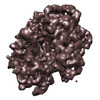
- EMDB-1726: Ribosome dynamics and tRNA movement as visualized by time-resolve... -
+ Open data
- Basic information
| Entry | Database: EMDB / ID: EMD-1726 | |||||||||
|---|---|---|---|---|---|---|---|---|---|---|
| Title | Ribosome dynamics and tRNA movement as visualized by time-resolved electron cryomicroscopy | |||||||||
 Map data Map data | Average map of E. coli 70S-fMetVal-tRNAVal post-translocation complex prepared at 4 degrees C | |||||||||
 Sample Sample |
| |||||||||
 Keywords Keywords | Ribosome / translation / translocation / tRNA | |||||||||
| Biological species |  | |||||||||
| Method | single particle reconstruction / cryo EM / Resolution: 11.0 Å | |||||||||
 Authors Authors | Fischer N / Konevega AL / Wintermeyer W / Rodnina MV / Stark H | |||||||||
 Citation Citation |  Journal: Nature / Year: 2010 Journal: Nature / Year: 2010Title: Ribosome dynamics and tRNA movement by time-resolved electron cryomicroscopy. Authors: Niels Fischer / Andrey L Konevega / Wolfgang Wintermeyer / Marina V Rodnina / Holger Stark /  Abstract: The translocation step of protein synthesis entails large-scale rearrangements of the ribosome-transfer RNA (tRNA) complex. Here we have followed tRNA movement through the ribosome during ...The translocation step of protein synthesis entails large-scale rearrangements of the ribosome-transfer RNA (tRNA) complex. Here we have followed tRNA movement through the ribosome during translocation by time-resolved single-particle electron cryomicroscopy (cryo-EM). Unbiased computational sorting of cryo-EM images yielded 50 distinct three-dimensional reconstructions, showing the tRNAs in classical, hybrid and various novel intermediate states that provide trajectories and kinetic information about tRNA movement through the ribosome. The structures indicate how tRNA movement is coupled with global and local conformational changes of the ribosome, in particular of the head and body of the small ribosomal subunit, and show that dynamic interactions between tRNAs and ribosomal residues confine the path of the tRNAs through the ribosome. The temperature dependence of ribosome dynamics reveals a surprisingly flat energy landscape of conformational variations at physiological temperature. The ribosome functions as a Brownian machine that couples spontaneous conformational changes driven by thermal energy to directed movement. | |||||||||
| History |
|
- Structure visualization
| Movie |
 Movie viewer Movie viewer |
|---|---|
| Structure viewer | EM map:  SurfView SurfView Molmil Molmil Jmol/JSmol Jmol/JSmol |
| Supplemental images |
- Downloads & links
-EMDB archive
| Map data |  emd_1726.map.gz emd_1726.map.gz | 25 MB |  EMDB map data format EMDB map data format | |
|---|---|---|---|---|
| Header (meta data) |  emd-1726-v30.xml emd-1726-v30.xml emd-1726.xml emd-1726.xml | 9.5 KB 9.5 KB | Display Display |  EMDB header EMDB header |
| Images |  emd_1726.png emd_1726.png | 183.5 KB | ||
| Archive directory |  http://ftp.pdbj.org/pub/emdb/structures/EMD-1726 http://ftp.pdbj.org/pub/emdb/structures/EMD-1726 ftp://ftp.pdbj.org/pub/emdb/structures/EMD-1726 ftp://ftp.pdbj.org/pub/emdb/structures/EMD-1726 | HTTPS FTP |
-Validation report
| Summary document |  emd_1726_validation.pdf.gz emd_1726_validation.pdf.gz | 257.2 KB | Display |  EMDB validaton report EMDB validaton report |
|---|---|---|---|---|
| Full document |  emd_1726_full_validation.pdf.gz emd_1726_full_validation.pdf.gz | 256.3 KB | Display | |
| Data in XML |  emd_1726_validation.xml.gz emd_1726_validation.xml.gz | 6.5 KB | Display | |
| Arichive directory |  https://ftp.pdbj.org/pub/emdb/validation_reports/EMD-1726 https://ftp.pdbj.org/pub/emdb/validation_reports/EMD-1726 ftp://ftp.pdbj.org/pub/emdb/validation_reports/EMD-1726 ftp://ftp.pdbj.org/pub/emdb/validation_reports/EMD-1726 | HTTPS FTP |
-Related structure data
| Related structure data |  1716C  1717C  1718C  1719C  1720C  1721C  1722C  1723C  1724C  1725C  1727C C: citing same article ( |
|---|---|
| Similar structure data |
- Links
Links
| EMDB pages |  EMDB (EBI/PDBe) / EMDB (EBI/PDBe) /  EMDataResource EMDataResource |
|---|---|
| Related items in Molecule of the Month |
- Map
Map
| File |  Download / File: emd_1726.map.gz / Format: CCP4 / Size: 26.4 MB / Type: IMAGE STORED AS FLOATING POINT NUMBER (4 BYTES) Download / File: emd_1726.map.gz / Format: CCP4 / Size: 26.4 MB / Type: IMAGE STORED AS FLOATING POINT NUMBER (4 BYTES) | ||||||||||||||||||||||||||||||||||||||||||||||||||||||||||||||||||||
|---|---|---|---|---|---|---|---|---|---|---|---|---|---|---|---|---|---|---|---|---|---|---|---|---|---|---|---|---|---|---|---|---|---|---|---|---|---|---|---|---|---|---|---|---|---|---|---|---|---|---|---|---|---|---|---|---|---|---|---|---|---|---|---|---|---|---|---|---|---|
| Annotation | Average map of E. coli 70S-fMetVal-tRNAVal post-translocation complex prepared at 4 degrees C | ||||||||||||||||||||||||||||||||||||||||||||||||||||||||||||||||||||
| Projections & slices | Image control
Images are generated by Spider. | ||||||||||||||||||||||||||||||||||||||||||||||||||||||||||||||||||||
| Voxel size | X=Y=Z: 1.87 Å | ||||||||||||||||||||||||||||||||||||||||||||||||||||||||||||||||||||
| Density |
| ||||||||||||||||||||||||||||||||||||||||||||||||||||||||||||||||||||
| Symmetry | Space group: 1 | ||||||||||||||||||||||||||||||||||||||||||||||||||||||||||||||||||||
| Details | EMDB XML:
CCP4 map header:
| ||||||||||||||||||||||||||||||||||||||||||||||||||||||||||||||||||||
-Supplemental data
- Sample components
-Entire : E. coli 70S-fMetVal-tRNAVal post-translocation complex at 4 degrees C
| Entire | Name: E. coli 70S-fMetVal-tRNAVal post-translocation complex at 4 degrees C |
|---|---|
| Components |
|
-Supramolecule #1000: E. coli 70S-fMetVal-tRNAVal post-translocation complex at 4 degrees C
| Supramolecule | Name: E. coli 70S-fMetVal-tRNAVal post-translocation complex at 4 degrees C type: sample / ID: 1000 / Number unique components: 3 |
|---|---|
| Molecular weight | Theoretical: 2.5 MDa |
-Supramolecule #1: Ribosome
| Supramolecule | Name: Ribosome / type: complex / ID: 1 / Name.synonym: E. coli 70S / Recombinant expression: No / Ribosome-details: ribosome-prokaryote: ALL |
|---|---|
| Source (natural) | Organism:  |
| Molecular weight | Theoretical: 2.5 MDa |
-Macromolecule #1: fMetVal-tRNAVal
| Macromolecule | Name: fMetVal-tRNAVal / type: rna / ID: 1 / Name.synonym: peptidyl tRNA / Classification: TRANSFER / Structure: DOUBLE HELIX / Synthetic?: No |
|---|---|
| Source (natural) | Organism:  |
| Molecular weight | Theoretical: 25 KDa |
-Macromolecule #2: m022 mRNA
| Macromolecule | Name: m022 mRNA / type: rna / ID: 2 / Name.synonym: mRNA / Details: coding sequence AUGGUU / Classification: OTHER / Structure: SINGLE STRANDED / Synthetic?: Yes |
|---|---|
| Source (natural) | Organism: synthetic construct (others) |
-Experimental details
-Structure determination
| Method | cryo EM |
|---|---|
 Processing Processing | single particle reconstruction |
| Aggregation state | particle |
- Sample preparation
| Buffer | pH: 7.5 Details: 50 mM Tris-HCl, 70 mM NH4Cl, 30 mM KCl, 7 mM MgCl2, 0.6 mM spermine, 0.4 mM spermidine |
|---|---|
| Vitrification | Cryogen name: ETHANE / Chamber humidity: 40 % / Chamber temperature: 77 K / Instrument: HOMEMADE PLUNGER Details: Vitrification instrument: Custom-built CEVS. Dew-point temperature (temperature on the grid) adjusted to 4 degrees C Method: Manual blotting for about 2 seconds |
- Electron microscopy
| Microscope | FEI/PHILIPS CM200FEG |
|---|---|
| Temperature | Average: 77 K |
| Alignment procedure | Legacy - Astigmatism: Objective lens astigmatism was corrected at 200,000 times magnification |
| Image recording | Category: CCD / Film or detector model: GENERIC TVIPS (4k x 4k) / Average electron dose: 20 e/Å2 |
| Electron beam | Acceleration voltage: 160 kV / Electron source:  FIELD EMISSION GUN FIELD EMISSION GUN |
| Electron optics | Calibrated magnification: 162740 / Illumination mode: SPOT SCAN / Imaging mode: BRIGHT FIELD / Cs: 2.0 mm / Nominal defocus max: 2.0 µm / Nominal defocus min: 0.5 µm / Nominal magnification: 161000 |
| Sample stage | Specimen holder: Eucentric / Specimen holder model: GATAN LIQUID NITROGEN |
- Image processing
| CTF correction | Details: Local |
|---|---|
| Final reconstruction | Applied symmetry - Point group: C1 (asymmetric) / Algorithm: OTHER / Resolution.type: BY AUTHOR / Resolution: 11.0 Å / Resolution method: FSC 0.5 CUT-OFF / Software - Name: IMAGIC, custom, Spider / Number images used: 16893 |
 Movie
Movie Controller


 Z (Sec.)
 Y (Row.)
 X (Col.)


