 Movie
Movie Controller

[English] 日本語
 Yorodumi
- EMDB-15952: Map from local refinement with a mask around T3 SAM lyase from th... -
+ Open data

- Basic information
| Entry |  | |||||||||
|---|---|---|---|---|---|---|---|---|---|---|
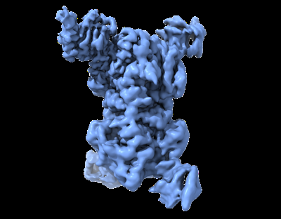
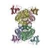
| Title | Map from local refinement with a mask around T3 SAM lyase from the complex of T3 SAM lyase with MetK. | |||||||||
 Map data Map data | ||||||||||
 Sample Sample |
| |||||||||
 Keywords Keywords | SAM lyase / complex / LYASE | |||||||||
| Function / homology |  Function and homology information Function and homology informationmethionine adenosyltransferase / methionine adenosyltransferase activity / S-adenosylmethionine biosynthetic process / L-methionine cycle / potassium ion binding / one-carbon metabolic process / magnesium ion binding / ATP binding / identical protein binding / cytosol Similarity search - Function | |||||||||
| Biological species |  Enterobacteria phage T3 (virus) / Enterobacteria phage T3 (virus) /  | |||||||||
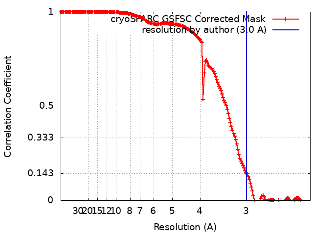
| Method | single particle reconstruction / cryo EM / Resolution: 3.0 Å | |||||||||
 Authors Authors | Triguis S / Selmer M | |||||||||
| Funding support |  Sweden, 2 items Sweden, 2 items
| |||||||||
 Citation Citation | Journal: Cell Rep / Year: 2023 Title: Phage T3 overcomes the BREX defense through SAM cleavage and inhibition of SAM synthesis by SAM lyase. Authors: Aleksandr Andriianov / Silvia Trigüis / Alena Drobiazko / Nicolas Sierro / Nikolai V Ivanov / Maria Selmer / Konstantin Severinov / Artem Isaev /    Abstract: Bacteriophage T3 encodes a SAMase that, through cleavage of S-adenosyl methionine (SAM), circumvents the SAM-dependent type I restriction-modification (R-M) defense. We show that SAMase also allows ...Bacteriophage T3 encodes a SAMase that, through cleavage of S-adenosyl methionine (SAM), circumvents the SAM-dependent type I restriction-modification (R-M) defense. We show that SAMase also allows T3 to evade the BREX defense. Although SAM depletion weakly affects BREX methylation, it completely inhibits the defensive function of BREX, suggesting that SAM could be a co-factor for BREX-mediated exclusion of phage DNA, similar to its anti-defense role in type I R-M. The anti-BREX activity of T3 SAMase is mediated not just by enzymatic degradation of SAM but also by direct inhibition of MetK, the host SAM synthase. We present a 2.8 Å cryoelectron microscopy (cryo-EM) structure of the eight-subunit T3 SAMase-MetK complex. Structure-guided mutagenesis reveals that this interaction stabilizes T3 SAMase in vivo, further stimulating its anti-BREX activity. This work provides insights in the versatility of bacteriophage counterdefense mechanisms and highlights the role of SAM as a co-factor of diverse bacterial immunity systems. | |||||||||
| History |
|
- Structure visualization
| Supplemental images |
|---|
- Downloads & links
-EMDB archive
| Map data | emd_15952.map.gz | 212.1 MB | EMDB map data format | |
|---|---|---|---|---|
| Header (meta data) | emd-15952-v30.xmlemd-15952.xml | 22 KB 22 KB | Display Display | EMDB header |
| FSC (resolution estimation) | emd_15952_fsc.xml | 17.8 KB | Display | FSC data file |
| Images |  emd_15952.png emd_15952.png | 77.9 KB | ||
| Filedesc metadata | emd-15952.cif.gz | 6.2 KB | ||
| Others | emd_15952_half_map_1.map.gzemd_15952_half_map_2.map.gz | 390.9 MB 390.9 MB | ||
| Archive directory |  http://ftp.pdbj.org/pub/emdb/structures/EMD-15952ftp://ftp.pdbj.org/pub/emdb/structures/EMD-15952 http://ftp.pdbj.org/pub/emdb/structures/EMD-15952ftp://ftp.pdbj.org/pub/emdb/structures/EMD-15952 | HTTPS FTP |
-Related structure data

-Links
| EMDB pages | EMDB (EBI/PDBe) / EMDataResource |
|---|
-Map
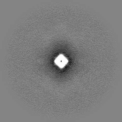
| File | Download / File: emd_15952.map.gz / Format: CCP4 / Size: 421.9 MB / Type: IMAGE STORED AS FLOATING POINT NUMBER (4 BYTES) | ||||||||||||||||||||||||||||||||||||
|---|---|---|---|---|---|---|---|---|---|---|---|---|---|---|---|---|---|---|---|---|---|---|---|---|---|---|---|---|---|---|---|---|---|---|---|---|---|
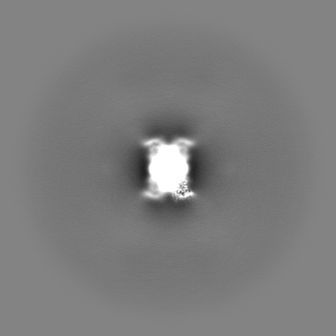
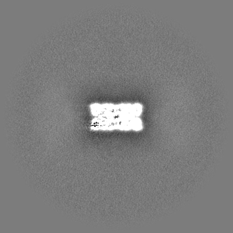
| Projections & slices | Image control
Images are generated by Spider. | ||||||||||||||||||||||||||||||||||||
| Voxel size | X=Y=Z: 1.1125 Å | ||||||||||||||||||||||||||||||||||||
| Density |
| ||||||||||||||||||||||||||||||||||||
| Symmetry | Space group: 1 | ||||||||||||||||||||||||||||||||||||
| Details | EMDB XML:
|
 Z (Sec.)
 Y (Row.)
 X (Col.)


-Supplemental data
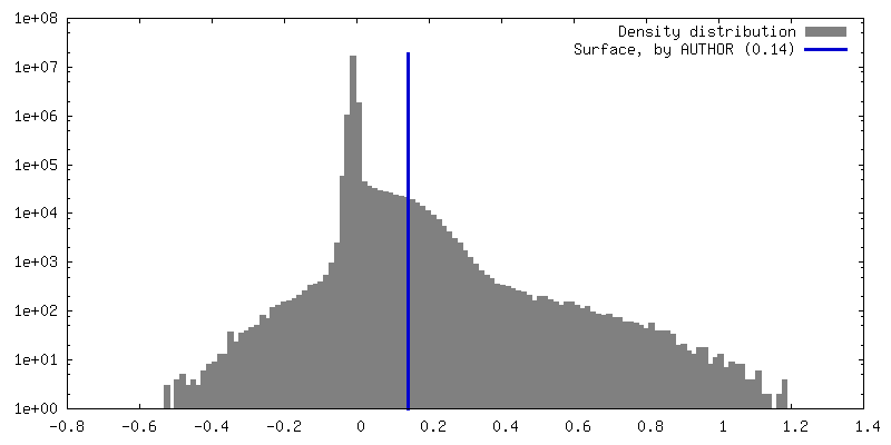
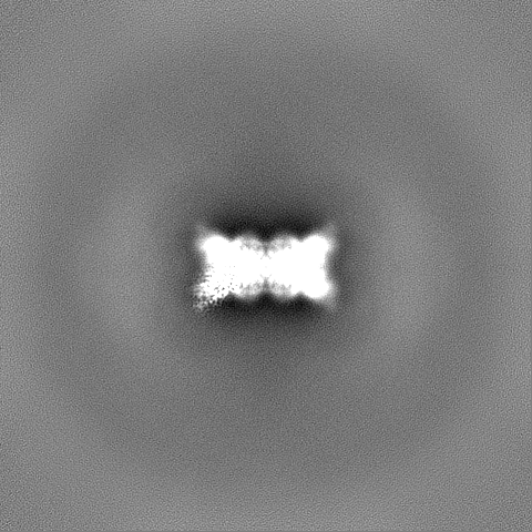
-Half map: #1
| File | emd_15952_half_map_1.map | ||||||||||||
|---|---|---|---|---|---|---|---|---|---|---|---|---|---|
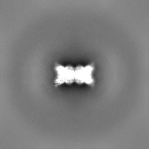
| Projections & Slices |
| ||||||||||||
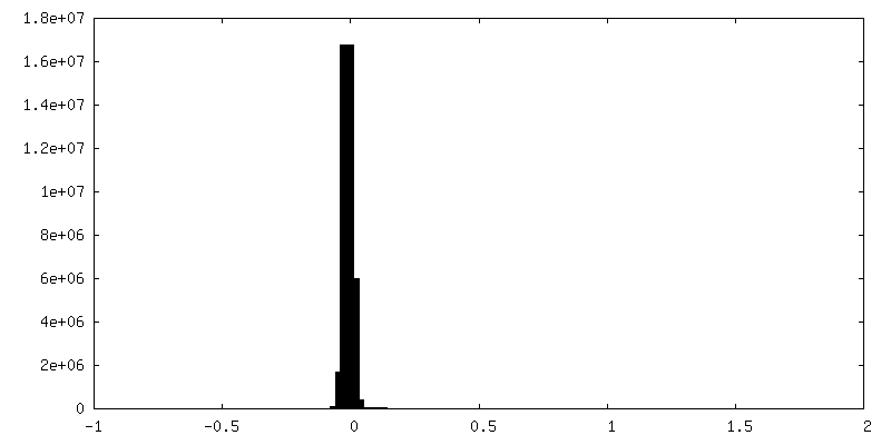
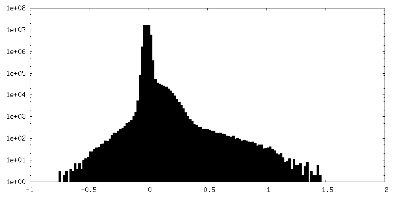
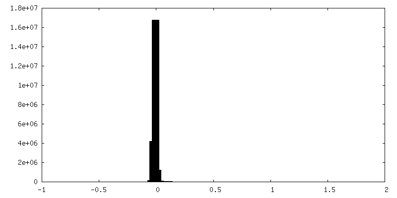
| Density Histograms |
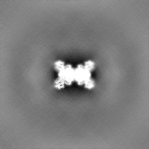
-Half map: #2
| File | emd_15952_half_map_2.map | ||||||||||||
|---|---|---|---|---|---|---|---|---|---|---|---|---|---|
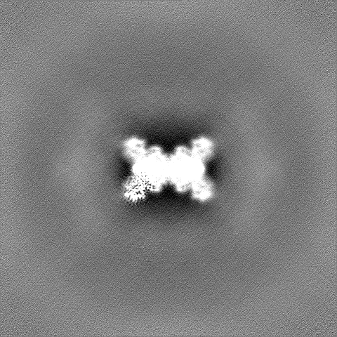
| Projections & Slices |
| ||||||||||||
| Density Histograms |

- Sample components
-Entire : T3 SAM lyase in complex with E. coli S-adenosylmethionine synthase.
| Entire | Name: T3 SAM lyase in complex with E. coli S-adenosylmethionine synthase. |
|---|---|
| Components |
|
-Supramolecule #1: T3 SAM lyase in complex with E. coli S-adenosylmethionine synthase.
| Supramolecule | Name: T3 SAM lyase in complex with E. coli S-adenosylmethionine synthase. type: complex / ID: 1 / Parent: 0 / Macromolecule list: all Details: T3 SAM lyase was recombinantly expressed in E. coli Top10 and co-purified in complex with S-adenosylmethionine synthase from the host. |
|---|---|
| Source (natural) | Organism: Enterobacteria phage T3 (virus) |
| Molecular weight | Theoretical: 239 KDa |
-Supramolecule #2: S-adenosylmethionine synthase in complex with T3 SAM lyase.
| Supramolecule | Name: S-adenosylmethionine synthase in complex with T3 SAM lyase. type: complex / ID: 2 / Parent: 1 / Macromolecule list: all Details: T3 SAM lyase was recombinantly expressed in E. coli Top10 and co-purified in complex with S-adenosylmethionine synthase from the host. |
|---|---|
| Source (natural) | Organism: |
-Supramolecule #3: T3 SAM lyase in complex with S-adenosylmethionine synthase.
| Supramolecule | Name: T3 SAM lyase in complex with S-adenosylmethionine synthase. type: complex / ID: 3 / Parent: 1 / Macromolecule list: all Details: T3 SAM lyase was recombinantly expressed in E. coli Top10 and co-purified in complex with S-adenosylmethionine synthase from the host. |
|---|---|
| Source (natural) | Organism: Enterobacteria phage T3 (virus) |
-Macromolecule #1: S-adenosylmethionine synthase
| Macromolecule | Name: S-adenosylmethionine synthase / type: protein_or_peptide / ID: 1 / Enantiomer: LEVO / EC number: methionine adenosyltransferase |
|---|---|
| Source (natural) | Organism: |
| Sequence | String: MAKHLFTSES VSEGHPDKIA DQISDAVLDA ILEQDPKARV ACETYVKTGM VLVGGEITTS AWVDIEEITR NTVREIGYVH SDMGFDANS CAVLSAIGKQ SPDINQGVDR ADPLEQGAGD QGLMFGYATN ETDVLMPAPI TYAHRLVQRQ AEVRKNGTLP W LRPDAKSQ ...String: MAKHLFTSES VSEGHPDKIA DQISDAVLDA ILEQDPKARV ACETYVKTGM VLVGGEITTS AWVDIEEITR NTVREIGYVH SDMGFDANS CAVLSAIGKQ SPDINQGVDR ADPLEQGAGD QGLMFGYATN ETDVLMPAPI TYAHRLVQRQ AEVRKNGTLP W LRPDAKSQ VTFQYDDGKI VGIDAVVLST QHSEEIDQKS LQEAVMEEII KPILPAEWLT SATKFFINPT GRFVIGGPMG DC GLTGRKI IVDTYGGMAR HGGGAFSGKD PSKVDRSAAY AARYVAKNIV AAGLADRCEI QVSYAIGVAE PTSIMVETFG TEK VPSEQL TLLVREFFDL RPYGLIQMLD LLHPIYKETA AYGHFGREHF PWEKTDKAQL LRDAAGLK UniProtKB: S-adenosylmethionine synthase |
-Macromolecule #2: T3 SAM lyase
| Macromolecule | Name: T3 SAM lyase / type: protein_or_peptide / ID: 2 / Enantiomer: LEVO / EC number: ec: 3.3.1.2 |
|---|---|
| Source (natural) | Organism: Enterobacteria phage T3 (virus) |
| Recombinant expression | Organism: |
| Sequence | String: MIFTKEPANV FYVLVSAFRS NLCDEVNMSR HRHMVSTLRA APGLYGSVES TDLTGCYREA ISSAPTEEKT VRVRCKDKAQ ALNVARLAC NEWEQDCVLV YKSQTHTAGL VYAKGIDGYK AERLPGSFQE VPKGAPLQGC FTIDEFGRRW QVQHHHHHH GENBANK: GENBANK: NP_523296.1 |
-Experimental details
-Structure determination
| Method | cryo EM |
|---|---|
 Processing Processing | single particle reconstruction |
| Aggregation state | particle |
-Sample preparation
| Concentration | 0.125 mg/mL | ||||||||||||
|---|---|---|---|---|---|---|---|---|---|---|---|---|---|
| Buffer | pH: 8 Component:
| ||||||||||||
| Grid | Model: C-flat-1.2/1.3 / Material: COPPER / Mesh: 300 / Support film - Material: CARBON / Support film - topology: HOLEY / Pretreatment - Type: GLOW DISCHARGE / Pretreatment - Time: 60 sec. / Pretreatment - Atmosphere: AIR / Pretreatment - Pressure: 0.039 kPa | ||||||||||||
| Vitrification | Cryogen name: ETHANE / Chamber humidity: 100 % / Chamber temperature: 277 K / Instrument: FEI VITROBOT MARK IV / Details: Blotting for 3 seconds before plunging.. |
- Electron microscopy
| Microscope | TFS KRIOS |
|---|---|
| Specialist optics | Energy filter - Name: GIF Bioquantum / Energy filter - Slit width: 20 eV |
| Image recording | Film or detector model: GATAN K3 BIOQUANTUM (6k x 4k) / Digitization - Dimensions - Width: 5760 pixel / Digitization - Dimensions - Height: 4092 pixel / Number grids imaged: 1 / Number real images: 5447 / Average exposure time: 3.0 sec. / Average electron dose: 47.75 e/Å2 |
| Electron beam | Acceleration voltage: 300 kV / Electron source:  FIELD EMISSION GUN FIELD EMISSION GUN |
| Electron optics | C2 aperture diameter: 50.0 µm / Calibrated defocus max: 2.1 µm / Calibrated defocus min: 0.35000000000000003 µm / Illumination mode: FLOOD BEAM / Imaging mode: BRIGHT FIELD / Cs: 2.7 mm / Nominal defocus max: 2.6 µm / Nominal defocus min: 0.6 µm / Nominal magnification: 105000 |
| Sample stage | Specimen holder model: FEI TITAN KRIOS AUTOGRID HOLDER / Cooling holder cryogen: NITROGEN |
| Experimental equipment |  Model: Titan Krios / Image courtesy: FEI Company |
+Image processing
-Atomic model buiding 1
| Initial model | PDB ID: Chain - Source name: PDB / Chain - Initial model type: experimental model |
|---|---|
| Refinement | Space: REAL / Protocol: OTHER |

