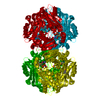
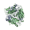
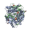
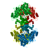
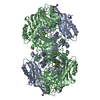
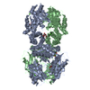
HETEROGEN S-adenosylmethionine synthetase catalyzes the reaction of: AMPPNP + Met -----> SAM + PPNP ...HETEROGEN S-adenosylmethionine synthetase catalyzes the reaction of: AMPPNP + Met -----> SAM + PPNP In the crystal structure, the authors found that two subunits (A, B) contain the substrates (AMPPNP and Met) and the other two subunits (C, D) have the products (SAM and PPNP) in their active sites. In the PDB file, the authors used ANP as AMPPNP, SAM as S-adenosylmethionine, and PPK as PPNP.
The enzyme is a homo-tetramer, and the asymmetric unit contains a tetramer.
-
Components
-
Protein , 1 types, 4 molecules ABCD
#1: Protein
S-adenosylmethioninesynthetase / Methionine adenosyltransferase / AdoMet synthetase / MAT
Mass: 41867.309 Da / Num. of mol.: 4 Source method: isolated from a genetically manipulated source Source: (gene. exp.) Escherichia coli (E. coli) Gene: METK OR METX OR B2942 OR C3528 OR Z4287 OR ECS3818 OR SF2933 Production host: Escherichia coli (E. coli) / References: UniProt: P0A817, methionine adenosyltransferase
In the structure databanks used in Yorodumi, some data are registered as the other names, "COVID-19 virus" and "2019-nCoV". Here are the details of the virus and the list of structure data.
Jan 31, 2019. EMDB accession codes are about to change! (news from PDBe EMDB page)
EMDB accession codes are about to change! (news from PDBe EMDB page)
The allocation of 4 digits for EMDB accession codes will soon come to an end. Whilst these codes will remain in use, new EMDB accession codes will include an additional digit and will expand incrementally as the available range of codes is exhausted. The current 4-digit format prefixed with “EMD-” (i.e. EMD-XXXX) will advance to a 5-digit format (i.e. EMD-XXXXX), and so on. It is currently estimated that the 4-digit codes will be depleted around Spring 2019, at which point the 5-digit format will come into force.
The EM Navigator/Yorodumi systems omit the EMD- prefix.
Related info.:Q: What is EMD? / ID/Accession-code notation in Yorodumi/EM Navigator
Yorodumi is a browser for structure data from EMDB, PDB, SASBDB, etc.
This page is also the successor to EM Navigator detail page, and also detail information page/front-end page for Omokage search.
The word "yorodu" (or yorozu) is an old Japanese word meaning "ten thousand". "mi" (miru) is to see.
Related info.:EMDB / PDB / SASBDB / Comparison of 3 databanks / Yorodumi Search / Aug 31, 2016. New EM Navigator & Yorodumi / Yorodumi Papers / Jmol/JSmol / Function and homology information / Changes in new EM Navigator and Yorodumi
 Movie
Movie Controller


 Open data
 Basic information
 Components
 Keywords
 Function and homology information
 X-RAY DIFFRACTION /
  Authors
 Citation
 Structure visualization
 Downloads & links
 Other downloads
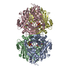


 PDBj
PDBj


 Assembly


 Mass: 39.098 Da / Num. of mol.: 4 / Source method: obtained synthetically / Formula: K
 Mass: 24.305 Da / Num. of mol.: 8 / Source method: obtained synthetically / Formula: Mg
 Mass: 506.196 Da / Num. of mol.: 2 / Source method: obtained synthetically / Formula: C10H17N6O12P3 / Comment: AMP-PNP, energy-carrying molecule analogue*YM
 Type: L-peptide linking / Mass: 149.211 Da / Num. of mol.: 2 / Source method: obtained synthetically / Formula: C5H11NO2S
 Mass: 398.437 Da / Num. of mol.: 2 / Source method: obtained synthetically / Formula: C15H22N6O5S
 Mass: 256.970 Da / Num. of mol.: 2 / Source method: obtained synthetically / Formula: H6NO9P3
 Sample preparation
 Processing