Protein or peptide: Double-strand break repair protein MRE11
Protein or peptide: Nibrin
Ligand: MANGANESE (II) ION
Keywords
DNA repair / complex / HYDROLASE
Function / homology
Function and homology information
chromosomal region / telomeric 3' overhang formation / mitochondrial double-strand break repair via homologous recombination / telomere maintenance via telomere trimming / Mre11 complex / negative regulation of double-strand break repair via nonhomologous end joining / negative regulation of telomere capping / BRCA1-C complex / blastocyst growth / meiotic DNA double-strand break formation ...chromosomal region / telomeric 3' overhang formation / mitochondrial double-strand break repair via homologous recombination / telomere maintenance via telomere trimming / Mre11 complex / negative regulation of double-strand break repair via nonhomologous end joining / negative regulation of telomere capping / BRCA1-C complex / blastocyst growth / meiotic DNA double-strand break formation / Sensing of DNA Double Strand Breaks / regulation of mitotic recombination / protection from non-homologous end joining at telomere / R-loop processing / single-stranded DNA endonuclease activity / phosphorylation-dependent protein binding / homologous chromosome pairing at meiosis / DNA strand resection involved in replication fork processing / nuclear inclusion body / homologous recombination / t-circle formation / nuclease activity / DNA double-strand break processing / 3'-5'-DNA exonuclease activity / double-strand break repair via alternative nonhomologous end joining / mitotic G2/M transition checkpoint / isotype switching / Cytosolic sensors of pathogen-associated DNA / Impaired BRCA2 binding to PALB2 / protein localization to site of double-strand break / HDR through MMEJ (alt-NHEJ) / IRF3-mediated induction of type I IFN / chromatin-protein adaptor activity / mitotic intra-S DNA damage checkpoint signaling / reciprocal meiotic recombination / regulation of DNA-templated DNA replication initiation / neuromuscular process controlling balance / sister chromatid cohesion / HDR through Single Strand Annealing (SSA) / positive regulation of double-strand break repair / Homologous DNA Pairing and Strand Exchange / Defective homologous recombination repair (HRR) due to BRCA1 loss of function / Defective HDR through Homologous Recombination Repair (HRR) due to PALB2 loss of BRCA1 binding function / Defective HDR through Homologous Recombination Repair (HRR) due to PALB2 loss of BRCA2/RAD51/RAD51C binding function / Resolution of D-loop Structures through Synthesis-Dependent Strand Annealing (SDSA) / Resolution of D-loop Structures through Holliday Junction Intermediates / mitotic G2 DNA damage checkpoint signaling / telomere maintenance in response to DNA damage / Impaired BRCA2 binding to RAD51 / positive regulation of telomere maintenance / neuroblast proliferation / Presynaptic phase of homologous DNA pairing and strand exchange / protein K63-linked ubiquitination / telomere maintenance via telomerase / positive regulation of double-strand break repair via homologous recombination / 3'-5' exonuclease activity / intrinsic apoptotic signaling pathway / telomere maintenance / DNA damage checkpoint signaling / replication fork / protein serine/threonine kinase activator activity / meiotic cell cycle / DNA endonuclease activity / DNA damage response, signal transduction by p53 class mediator / Nonhomologous End-Joining (NHEJ) / G2/M DNA damage checkpoint / PML body / DNA Damage/Telomere Stress Induced Senescence / double-strand break repair via homologous recombination / Meiotic recombination / double-strand break repair via nonhomologous end joining / HDR through Homologous Recombination (HRR) / double-strand break repair / manganese ion binding / Recruitment and ATM-mediated phosphorylation of repair and signaling proteins at DNA double strand breaks / site of double-strand break / Processing of DNA double-strand break ends / double-stranded DNA binding / DNA recombination / histone binding / Regulation of TP53 Activity through Phosphorylation / DNA-binding transcription factor binding / damaged DNA binding / Hydrolases; Acting on ester bonds / chromosome, telomeric region / cell population proliferation / regulation of cell cycle / cadherin binding / DNA repair / DNA damage response / negative regulation of apoptotic process / nucleolus / Golgi apparatus / nucleoplasm / identical protein binding / nucleus / cytoplasm / cytosol Similarity search - Function
Nibrin, C-terminal / Nibrin / DNA damage repair protein Nbs1 / DNA damage repair protein Nbs1 / Nibrin, second BRCT domain / Nibrin, second BRCT domain superfamily / Second BRCT domain on Nijmegen syndrome breakage protein / Nibrin-related / DNA double-strand break repair protein Mre11 / Mre11, DNA-binding ...Nibrin, C-terminal / Nibrin / DNA damage repair protein Nbs1 / DNA damage repair protein Nbs1 / Nibrin, second BRCT domain / Nibrin, second BRCT domain superfamily / Second BRCT domain on Nijmegen syndrome breakage protein / Nibrin-related / DNA double-strand break repair protein Mre11 / Mre11, DNA-binding / Mre11, capping domain / Mre11 DNA-binding presumed domain / Mre11 DNA-binding presumed domain / Mre11 nuclease, N-terminal metallophosphatase domain / Forkhead associated domain / Forkhead-associated (FHA) domain profile. / FHA domain / Forkhead-associated (FHA) domain / SMAD/FHA domain superfamily / BRCA1 C Terminus (BRCT) domain / Calcineurin-like phosphoesterase domain, ApaH type / Calcineurin-like phosphoesterase / Metallo-dependent phosphatase-like / BRCT domain / BRCT domain superfamily Similarity search - Domain/homology
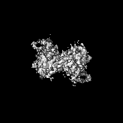
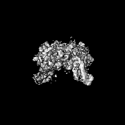
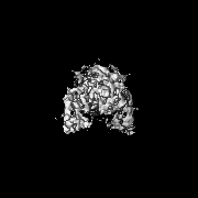
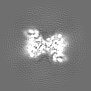
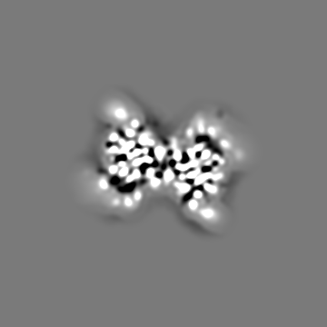
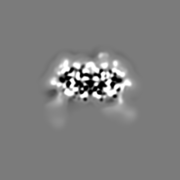
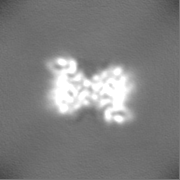
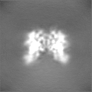
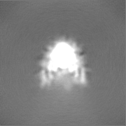
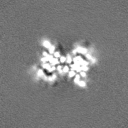
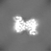
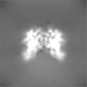
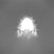
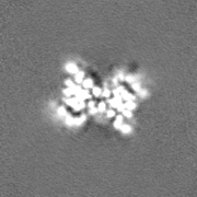
Journal: Mol Cell / Year: 2023 Title: Cryo-EM structure of the Mre11-Rad50-Nbs1 complex reveals the molecular mechanism of scaffolding functions. Authors: Matthias Rotheneder / Kristina Stakyte / Erik van de Logt / Joseph D Bartho / Katja Lammens / Yilan Fan / Aaron Alt / Brigitte Kessler / Christophe Jung / Wynand P Roos / Barbara ...Authors: Matthias Rotheneder / Kristina Stakyte / Erik van de Logt / Joseph D Bartho / Katja Lammens / Yilan Fan / Aaron Alt / Brigitte Kessler / Christophe Jung / Wynand P Roos / Barbara Steigenberger / Karl-Peter Hopfner / Abstract: The DNA double-strand break repair complex Mre11-Rad50-Nbs1 (MRN) detects and nucleolytically processes DNA ends, activates the ATM kinase, and tethers DNA at break sites. How MRN can act both as ...The DNA double-strand break repair complex Mre11-Rad50-Nbs1 (MRN) detects and nucleolytically processes DNA ends, activates the ATM kinase, and tethers DNA at break sites. How MRN can act both as nuclease and scaffold protein is not well understood. The cryo-EM structure of MRN from Chaetomium thermophilum reveals a 2:2:1 complex with a single Nbs1 wrapping around the autoinhibited Mre11 nuclease dimer. MRN has two DNA-binding modes, one ATP-dependent mode for loading onto DNA ends and one ATP-independent mode through Mre11's C terminus, suggesting how it may interact with DSBs and intact DNA. MRNs two 60-nm-long coiled-coil domains form a linear rod structure, the apex of which is assembled by the two joined zinc-hook motifs. Apices from two MRN complexes can further dimerize, forming 120-nm spanning MRN-MRN structures. Our results illustrate the architecture of MRN and suggest how it mechanistically integrates catalytic and tethering functions.
Name: MANGANESE (II) ION / type: ligand / ID: 3 / Number of copies: 4 / Formula: MN
Molecular weight
Theoretical: 54.938 Da
-
Experimental details
-
Structure determination
Method
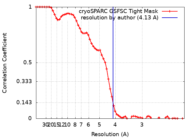
cryo EM
Processing
single particle reconstruction
Aggregation state
particle
-
Sample preparation
Concentration
0.29 mg/mL
Buffer
pH: 7 Component:
Concentration
Formula
Name
20.0 mM
C8H18N2O4S
HEPES
140.0 mM
NaCl
Sodium chloride
5.0 mM
MgCl2
Magnesium chloride
1.0 mM
MnCl2
Manganese chloride
0.02 mM
ZnCl2
Zinc chloride
0.2 mM
C9H15O6P
TCEP
2.0 mM
C10H16N5O12P3S
ATPgS
0.05 percent
C14H28O6
Octyl beta-D-glucopyranoside
Details: 20 mM HEPES (pH 7.0), 140 mM NaCl, 5 mM MgCl2, 1 mM MnCl2, 0.020 mM ZnCl2, 0.2 mM TCEP, 2 mM ATPgS, plus 0.05 percent beta-OG
Grid
Model: Quantifoil R2/1 / Material: COPPER / Mesh: 200 / Support film - Material: CARBON / Support film - topology: HOLEY / Pretreatment - Type: GLOW DISCHARGE / Pretreatment - Time: 7 sec. / Pretreatment - Atmosphere: AIR
Vitrification
Cryogen name: ETHANE / Chamber humidity: 95 % / Chamber temperature: 288 K / Instrument: LEICA EM GP
-
Electron microscopy
Microscope
FEI TITAN KRIOS
Specialist optics
Energy filter - Name: GIF Bioquantum / Energy filter - Slit width: 20 eV
Image recording
Film or detector model: GATAN K2 SUMMIT (4k x 4k) / Detector mode: COUNTING / Number grids imaged: 3 / Number real images: 11325 / Average exposure time: 10.0 sec. / Average electron dose: 43.0 e/Å2
Electron beam
Acceleration voltage: 300 kV / Electron source: FIELD EMISSION GUN
In the structure databanks used in Yorodumi, some data are registered as the other names, "COVID-19 virus" and "2019-nCoV". Here are the details of the virus and the list of structure data.
Jan 31, 2019. EMDB accession codes are about to change! (news from PDBe EMDB page)
EMDB accession codes are about to change! (news from PDBe EMDB page)
The allocation of 4 digits for EMDB accession codes will soon come to an end. Whilst these codes will remain in use, new EMDB accession codes will include an additional digit and will expand incrementally as the available range of codes is exhausted. The current 4-digit format prefixed with “EMD-” (i.e. EMD-XXXX) will advance to a 5-digit format (i.e. EMD-XXXXX), and so on. It is currently estimated that the 4-digit codes will be depleted around Spring 2019, at which point the 5-digit format will come into force.
The EM Navigator/Yorodumi systems omit the EMD- prefix.
Related info.:Q: What is EMD? / ID/Accession-code notation in Yorodumi/EM Navigator
Yorodumi is a browser for structure data from EMDB, PDB, SASBDB, etc.
This page is also the successor to EM Navigator detail page, and also detail information page/front-end page for Omokage search.
The word "yorodu" (or yorozu) is an old Japanese word meaning "ten thousand". "mi" (miru) is to see.
Related info.:EMDB / PDB / SASBDB / Comparison of 3 databanks / Yorodumi Search / Aug 31, 2016. New EM Navigator & Yorodumi / Yorodumi Papers / Jmol/JSmol / Function and homology information / Changes in new EM Navigator and Yorodumi
 Movie
Movie Controller


 Open data
 Basic information
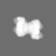
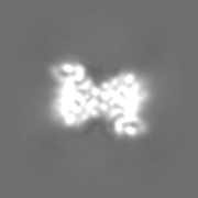
 Map data
 Sample
 Keywords
 Function and homology information
 Homo sapiens (human)
 Authors
 Germany, 4 items
  Citation
 Structure visualization
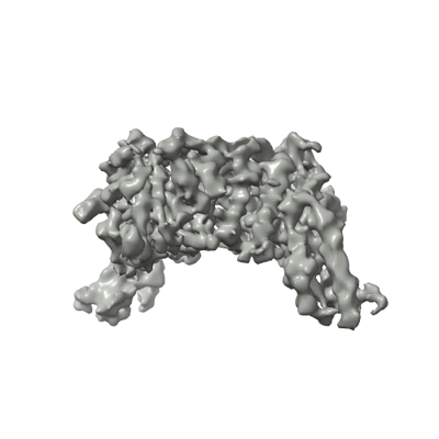
 Downloads & links
 emd_15948.png
 http://ftp.pdbj.org/pub/emdb/structures/EMD-15948


 Z (Sec.)
 Y (Row.)
 X (Col.)


 Sample components
 Processing
 Electron microscopy
 FIELD EMISSION GUN