 Movie
Movie Controller

[English] 日本語
 Yorodumi
- PDB-7zr1: Chaetomium thermophilum Mre11-Rad50-Nbs1 complex bound to ATPyS (... -
+ Open data

- Basic information
| Entry | Database: PDB / ID: 7zr1 | ||||||||||||
|---|---|---|---|---|---|---|---|---|---|---|---|---|---|
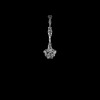
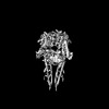
| Title | Chaetomium thermophilum Mre11-Rad50-Nbs1 complex bound to ATPyS (composite structure) | ||||||||||||
 Components Components |
| ||||||||||||
 Keywords Keywords | HYDROLASE / DNA repair / complex / ATPase / coiled-coils | ||||||||||||
| Function / homology |  Function and homology information Function and homology informationmitochondrial double-strand break repair via homologous recombination / Mre11 complex / meiotic DNA double-strand break formation / Hydrolases; Acting on acid anhydrides / single-stranded DNA endonuclease activity / chromosome organization involved in meiotic cell cycle / double-stranded telomeric DNA binding / G-quadruplex DNA binding / single-stranded telomeric DNA binding / 3'-5'-DNA exonuclease activity ...mitochondrial double-strand break repair via homologous recombination / Mre11 complex / meiotic DNA double-strand break formation / Hydrolases; Acting on acid anhydrides / single-stranded DNA endonuclease activity / chromosome organization involved in meiotic cell cycle / double-stranded telomeric DNA binding / G-quadruplex DNA binding / single-stranded telomeric DNA binding / 3'-5'-DNA exonuclease activity / telomere maintenance via recombination / mitotic intra-S DNA damage checkpoint signaling / mitotic G2 DNA damage checkpoint signaling / telomere maintenance via telomerase / guanyl-nucleotide exchange factor activity / telomere maintenance / condensed nuclear chromosome / double-strand break repair via homologous recombination / double-strand break repair via nonhomologous end joining / manganese ion binding / double-strand break repair / chromosome / site of double-strand break / double-stranded DNA binding / Hydrolases; Acting on ester bonds / damaged DNA binding / chromosome, telomeric region / ATP hydrolysis activity / metal ion binding Similarity search - Function | ||||||||||||
| Biological species |  Thermochaetoides thermophila (fungus) Thermochaetoides thermophila (fungus) | ||||||||||||
| Method | ELECTRON MICROSCOPY / single particle reconstruction / cryo EM / Resolution: 4 Å | ||||||||||||
 Authors Authors | Bartho, J.D. / Rotheneder, M. / Stakyte, K. / Lammens, K. / Hopfner, K.P. | ||||||||||||
| Funding support |  Germany, 3items Germany, 3items
| ||||||||||||
 Citation Citation | Journal: Mol Cell / Year: 2023 Title: Cryo-EM structure of the Mre11-Rad50-Nbs1 complex reveals the molecular mechanism of scaffolding functions. Authors: Matthias Rotheneder / Kristina Stakyte / Erik van de Logt / Joseph D Bartho / Katja Lammens / Yilan Fan / Aaron Alt / Brigitte Kessler / Christophe Jung / Wynand P Roos / Barbara ...Authors: Matthias Rotheneder / Kristina Stakyte / Erik van de Logt / Joseph D Bartho / Katja Lammens / Yilan Fan / Aaron Alt / Brigitte Kessler / Christophe Jung / Wynand P Roos / Barbara Steigenberger / Karl-Peter Hopfner / Abstract: The DNA double-strand break repair complex Mre11-Rad50-Nbs1 (MRN) detects and nucleolytically processes DNA ends, activates the ATM kinase, and tethers DNA at break sites. How MRN can act both as ...The DNA double-strand break repair complex Mre11-Rad50-Nbs1 (MRN) detects and nucleolytically processes DNA ends, activates the ATM kinase, and tethers DNA at break sites. How MRN can act both as nuclease and scaffold protein is not well understood. The cryo-EM structure of MRN from Chaetomium thermophilum reveals a 2:2:1 complex with a single Nbs1 wrapping around the autoinhibited Mre11 nuclease dimer. MRN has two DNA-binding modes, one ATP-dependent mode for loading onto DNA ends and one ATP-independent mode through Mre11's C terminus, suggesting how it may interact with DSBs and intact DNA. MRNs two 60-nm-long coiled-coil domains form a linear rod structure, the apex of which is assembled by the two joined zinc-hook motifs. Apices from two MRN complexes can further dimerize, forming 120-nm spanning MRN-MRN structures. Our results illustrate the architecture of MRN and suggest how it mechanistically integrates catalytic and tethering functions. | ||||||||||||
| History |
|
- Structure visualization
| Structure viewer | Molecule: MolmilJmol/JSmol |
|---|
- Downloads & links
-Download
| PDBx/mmCIF format | 7zr1.cif.gz | 613.2 KB | Display | PDBx/mmCIF format |
|---|---|---|---|---|
| PDB format | pdb7zr1.ent.gz | 465.1 KB | Display | PDB format |
| PDBx/mmJSON format | 7zr1.json.gz | Tree view | PDBx/mmJSON format | |
| Others |  Other downloads Other downloads |
-Validation report
| Arichive directory | https://data.pdbj.org/pub/pdb/validation_reports/zr/7zr1ftp://data.pdbj.org/pub/pdb/validation_reports/zr/7zr1 | HTTPS FTP |
|---|
-Related structure data
| Related structure data |  14881MC  7zqyC 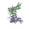 8bahC C: citing same article ( M: map data used to model this data |
|---|---|
| Similar structure data |


-Links
 PDBj
PDBj


- Assembly
| Deposited unit | 
|
|---|---|
| 1 |
|
-Components
-Protein , 3 types, 5 molecules ABCDE
| #1: Protein | Mass: 83036.273 Da / Num. of mol.: 2 Source method: isolated from a genetically manipulated source Source: (gene. exp.) Thermochaetoides thermophila (fungus) / Strain: DSM 1495 / CBS 144.50 / IMI 039719 / Gene: CTHT_0007600 / Production host:  Trichoplusia ni (cabbage looper) / References: UniProt: G0RYR3 Trichoplusia ni (cabbage looper) / References: UniProt: G0RYR3#2: Protein | Mass: 152229.906 Da / Num. of mol.: 2 Source method: isolated from a genetically manipulated source Source: (gene. exp.) Thermochaetoides thermophila (fungus) / Strain: DSM 1495 / CBS 144.50 / IMI 039719 / Gene: CTHT_0073630 / Production host: Trichoplusia ni (cabbage looper) / References: UniProt: G0SHW7#3: Protein | | Mass: 105980.812 Da / Num. of mol.: 1 Source method: isolated from a genetically manipulated source Source: (gene. exp.) Thermochaetoides thermophila (fungus) / Strain: DSM 1495 / CBS 144.50 / IMI 039719 / Gene: CTHT_0047880 / Production host: Trichoplusia ni (cabbage looper) / References: UniProt: G0SAV1 |
|---|
-Non-polymers , 3 types, 8 molecules 


| #4: Chemical | ChemComp-MN /  Mass: 54.938 Da / Num. of mol.: 4 / Source method: obtained synthetically / Formula: Mn / Feature type: SUBJECT OF INVESTIGATION Mass: 54.938 Da / Num. of mol.: 4 / Source method: obtained synthetically / Formula: Mn / Feature type: SUBJECT OF INVESTIGATION#5: Chemical |  Mass: 24.305 Da / Num. of mol.: 2 / Source method: obtained synthetically / Formula: Mg / Feature type: SUBJECT OF INVESTIGATION Mass: 24.305 Da / Num. of mol.: 2 / Source method: obtained synthetically / Formula: Mg / Feature type: SUBJECT OF INVESTIGATION#6: Chemical |  Mass: 523.247 Da / Num. of mol.: 2 / Source method: obtained synthetically / Formula: C10H16N5O12P3S / Feature type: SUBJECT OF INVESTIGATION / Comment: ATP-gamma-S, energy-carrying molecule analogue*YM Mass: 523.247 Da / Num. of mol.: 2 / Source method: obtained synthetically / Formula: C10H16N5O12P3S / Feature type: SUBJECT OF INVESTIGATION / Comment: ATP-gamma-S, energy-carrying molecule analogue*YM |
|---|
-Details
| Has ligand of interest | Y |
|---|
-Experimental details
-Experiment
| Experiment | Method: ELECTRON MICROSCOPY |
|---|---|
| EM experiment | Aggregation state: PARTICLE / 3D reconstruction method: single particle reconstruction |
- Sample preparation
| Component | Name: Chaetomium thermophilum Mre11-Rad50-Nbs1 complex bound to ATPyS Type: COMPLEX / Entity ID: #1-#3 / Source: RECOMBINANT |
|---|---|
| Molecular weight | Experimental value: NO |
| Source (natural) | Organism: Chaetomium thermophilum (fungus) |
| Source (recombinant) | Organism: Trichoplusia ni (cabbage looper) |
| Buffer solution | pH: 7.6 |
| Specimen | Conc.: 0.27 mg/ml / Embedding applied: NO / Shadowing applied: NO / Staining applied: NO / Vitrification applied: YES |
| Specimen support | Grid material: COPPER / Grid mesh size: 200 divisions/in. / Grid type: Quantifoil R2/1 |
| Vitrification | Cryogen name: ETHANE |
- Electron microscopy imaging
| Experimental equipment |  Model: Titan Krios / Image courtesy: FEI Company |
|---|---|
| Microscopy | Model: FEI TITAN KRIOS |
| Electron gun | Electron source:  FIELD EMISSION GUN / Accelerating voltage: 300 kV / Illumination mode: FLOOD BEAM FIELD EMISSION GUN / Accelerating voltage: 300 kV / Illumination mode: FLOOD BEAM |
| Electron lens | Mode: BRIGHT FIELD / Nominal defocus max: 2800 nm / Nominal defocus min: 1000 nm / Cs: 2.7 mm / C2 aperture diameter: 50 µm |
| Specimen holder | Cryogen: NITROGEN / Specimen holder model: FEI TITAN KRIOS AUTOGRID HOLDER |
| Image recording | Electron dose: 43 e/Å2 / Detector mode: COUNTING / Film or detector model: GATAN K2 SUMMIT (4k x 4k) |
| EM imaging optics | Energyfilter name: GIF Bioquantum / Energyfilter slit width: 20 eV |
- Processing
| EM software | Name: EPU / Category: image acquisition |
|---|---|
| CTF correction | Type: PHASE FLIPPING AND AMPLITUDE CORRECTION |
| 3D reconstruction | Resolution: 4 Å / Resolution method: FSC 0.143 CUT-OFF / Num. of particles: 288443 / Symmetry type: POINT |
