[English] 日本語
Yorodumi
- PDB-7zqy: Chaetomium thermophilum Rad50 Zn hook -

+
Open data


ID or keywords:

Loading...

-
Basic information

Entry
Database: PDB / ID: 7zqy
TitleChaetomium thermophilum Rad50 Zn hook
ComponentsDH domain-containing protein
KeywordsHYDROLASE / DNA repair / ATPase / coiled coil / Zn-hook
Function / homology
Function and homology information


Mre11 complex / telomere maintenance / guanyl-nucleotide exchange factor activity / double-strand break repair / ATP hydrolysis activity / metal ion binding
Similarity search - Function
DNA repair protein Rad50, eukaryotes / SbcC/RAD50-like, Walker B motif / Rad50/SbcC-type AAA domain / AAA domain / Dbl homology (DH) domain / Dbl homology (DH) domain profile. / P-loop containing nucleoside triphosphate hydrolase
Similarity search - Domain/homology
DH domain-containing protein
Similarity search - Component
Biological speciesThermochaetoides thermophila (fungus)
MethodX-RAY DIFFRACTION / SYNCHROTRON / MOLECULAR REPLACEMENT / Resolution: 2.51 Å
AuthorsLammens, K. / Rotheneder, M. / Stakyte, K.
Funding support Germany, 1items
OrganizationGrant numberCountry
German Research Foundation (DFG) Germany
Citation
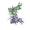
Journal: Mol Cell / Year: 2023
Title: Cryo-EM structure of the Mre11-Rad50-Nbs1 complex reveals the molecular mechanism of scaffolding functions.
Authors: Matthias Rotheneder / Kristina Stakyte / Erik van de Logt / Joseph D Bartho / Katja Lammens / Yilan Fan / Aaron Alt / Brigitte Kessler / Christophe Jung / Wynand P Roos / Barbara ...Authors: Matthias Rotheneder / Kristina Stakyte / Erik van de Logt / Joseph D Bartho / Katja Lammens / Yilan Fan / Aaron Alt / Brigitte Kessler / Christophe Jung / Wynand P Roos / Barbara Steigenberger / Karl-Peter Hopfner /
Abstract: The DNA double-strand break repair complex Mre11-Rad50-Nbs1 (MRN) detects and nucleolytically processes DNA ends, activates the ATM kinase, and tethers DNA at break sites. How MRN can act both as ...The DNA double-strand break repair complex Mre11-Rad50-Nbs1 (MRN) detects and nucleolytically processes DNA ends, activates the ATM kinase, and tethers DNA at break sites. How MRN can act both as nuclease and scaffold protein is not well understood. The cryo-EM structure of MRN from Chaetomium thermophilum reveals a 2:2:1 complex with a single Nbs1 wrapping around the autoinhibited Mre11 nuclease dimer. MRN has two DNA-binding modes, one ATP-dependent mode for loading onto DNA ends and one ATP-independent mode through Mre11's C terminus, suggesting how it may interact with DSBs and intact DNA. MRNs two 60-nm-long coiled-coil domains form a linear rod structure, the apex of which is assembled by the two joined zinc-hook motifs. Apices from two MRN complexes can further dimerize, forming 120-nm spanning MRN-MRN structures. Our results illustrate the architecture of MRN and suggest how it mechanistically integrates catalytic and tethering functions.
History
DepositionMay 3, 2022Deposition site: PDBE / Processing site: PDBE
Revision 1.0Dec 28, 2022Provider: repository / Type: Initial release
Revision 1.1Jan 18, 2023Group: Database references / Category: citation / citation_author
Item: _citation.pdbx_database_id_DOI / _citation.pdbx_database_id_PubMed / _citation.title
Revision 1.2Feb 1, 2023Group: Database references / Category: citation
Item: _citation.journal_volume / _citation.page_first ..._citation.journal_volume / _citation.page_first / _citation.page_last / _citation.year

-
Structure visualization

Structure viewerMolecule:
MolmilJmol/JSmol

Downloads & links

-
Assembly

Deposited unit
A: DH domain-containing protein
B: DH domain-containing protein
C: DH domain-containing protein
D: DH domain-containing protein
hetero molecules


Theoretical massNumber of molelcules
Total (without water)83,6706
Polymers83,5394
Non-polymers1312
Water543
1


  • Idetical with deposited unit
  • defined by author&software
  • Evidence: microscopy, cryo EM structure
TypeNameSymmetry operationNumber
identity operation1_555x,y,z1
Buried area8170 Å2
ΔGint-95 kcal/mol
Surface area42870 Å2
MethodPISA
Unit cell
Length a, b, c (Å)43.490, 53.970, 110.420
Angle α, β, γ (deg.)75.85, 78.74, 90.00
Int Tables number1
Space group name H-MP1

-
Components

#1: Protein
DH domain-containing protein


Mass: 20884.699 Da / Num. of mol.: 4
Source method: isolated from a genetically manipulated source
Source: (gene. exp.) Thermochaetoides thermophila (fungus) / Strain: DSM 1495 / CBS 144.50 / IMI 039719 / Gene: CTHT_0073630 / Production host: Escherichia coli K2 (bacteria) / References: UniProt: G0SHW7
#2: Chemical ChemComp-ZN / ZINC ION


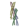
Mass: 65.409 Da / Num. of mol.: 2 / Source method: obtained synthetically / Formula: Zn
#3: Water ChemComp-HOH / water / Water


Mass: 18.015 Da / Num. of mol.: 3 / Source method: isolated from a natural source / Formula: H2O
Has ligand of interestN

-
Experimental details

-
Experiment

ExperimentMethod: X-RAY DIFFRACTION / Number of used crystals: 1

-
Sample preparation

CrystalDensity Matthews: 2.97 Å3/Da / Density % sol: 58.53 %
Crystal growTemperature: 293.15 K / Method: vapor diffusion, hanging drop / Details: 0.1 M MES, 0.6 M NaCl, 18% PEG4000 / PH range: 6.8

-
Data collection

DiffractionMean temperature: 100 K / Serial crystal experiment: N
Diffraction sourceSource: SYNCHROTRON / Site: PETRA III, EMBL c/o DESY / Beamline: P14 (MX2) / Wavelength: 1.2832 Å
DetectorType: DECTRIS EIGER X 16M / Detector: PIXEL / Date: Jul 5, 2021
RadiationProtocol: SINGLE WAVELENGTH / Monochromatic (M) / Laue (L): M / Scattering type: x-ray
Radiation wavelengthWavelength: 1.2832 Å / Relative weight: 1
ReflectionResolution: 2.51→104.88 Å / Num. obs: 63401 / % possible obs: 97.4 % / Redundancy: 3.55 % / CC1/2: 0.93 / Rmerge(I) obs: 0.078 / Net I/σ(I): 7.86
Reflection shellResolution: 2.51→2.54 Å / Num. unique obs: 1715 / CC1/2: 0.77

-
Processing

Software
NameVersionClassification
PHENIX(1.17.1_3660: ???)refinement
XDSdata reduction
XSCALEdata scaling
PHASERphasing
RefinementMethod to determine structure: MOLECULAR REPLACEMENT
Starting model: Alphafold2

Resolution: 2.51→104.88 Å / SU ML: 0.36 / Cross valid method: FREE R-VALUE / σ(F): 1.34 / Phase error: 37.03 / Stereochemistry target values: ML
RfactorNum. reflection% reflection
Rfree0.283 6487 10.23 %
Rwork0.2642 --
obs0.2661 63401 96.99 %
Solvent computationShrinkage radii: 0.9 Å / VDW probe radii: 1.11 Å / Solvent model: FLAT BULK SOLVENT MODEL
Refinement stepCycle: LAST / Resolution: 2.51→104.88 Å
ProteinNucleic acidLigandSolventTotal
Num. atoms5798 0 2 3 5803
Refine LS restraints
Refine-IDTypeDev idealNumber
X-RAY DIFFRACTIONf_bond_d0.0125854
X-RAY DIFFRACTIONf_angle_d1.7797850
X-RAY DIFFRACTIONf_dihedral_angle_d23.992362
X-RAY DIFFRACTIONf_chiral_restr0.12878
X-RAY DIFFRACTIONf_plane_restr0.0141034
LS refinement shell
Resolution (Å)Rfactor RfreeNum. reflection RfreeRfactor RworkNum. reflection RworkRefine-ID% reflection obs (%)
2.51-2.540.53361940.43291715X-RAY DIFFRACTION86
2.54-2.570.43282010.41272055X-RAY DIFFRACTION95
2.57-2.60.37831860.38751753X-RAY DIFFRACTION97
2.6-2.630.35721550.38282005X-RAY DIFFRACTION98
2.63-2.670.41072780.35521800X-RAY DIFFRACTION98
2.67-2.70.39962310.38291952X-RAY DIFFRACTION98
2.7-2.740.37662160.40161868X-RAY DIFFRACTION96
2.74-2.780.45492170.38611879X-RAY DIFFRACTION97
2.78-2.830.63561640.33841986X-RAY DIFFRACTION97
2.83-2.870.42422400.33981853X-RAY DIFFRACTION97
2.87-2.920.35831920.3361972X-RAY DIFFRACTION98
2.92-2.970.32672440.33151804X-RAY DIFFRACTION97
2.97-3.030.32262660.30851815X-RAY DIFFRACTION96
3.03-3.090.31352130.31451932X-RAY DIFFRACTION97
3.09-3.160.36582410.35441923X-RAY DIFFRACTION98
3.16-3.230.27961880.33481884X-RAY DIFFRACTION98
3.23-3.320.30782380.331937X-RAY DIFFRACTION98
3.32-3.410.31932490.30411863X-RAY DIFFRACTION99
3.41-3.510.35771790.28731965X-RAY DIFFRACTION98
3.51-3.620.29182200.27561986X-RAY DIFFRACTION98
3.62-3.750.2291840.23731918X-RAY DIFFRACTION98
3.75-3.90.27022220.25061925X-RAY DIFFRACTION98
3.9-4.080.22772300.23981955X-RAY DIFFRACTION98
4.08-4.290.26032020.22131877X-RAY DIFFRACTION98
4.29-4.560.20421880.19941880X-RAY DIFFRACTION97
4.56-4.910.20132590.20811932X-RAY DIFFRACTION98
4.91-5.410.24252630.22681830X-RAY DIFFRACTION97
5.41-6.190.24872010.2721975X-RAY DIFFRACTION99
6.19-7.790.31622420.26411877X-RAY DIFFRACTION97
7.8-104.880.2251840.17971798X-RAY DIFFRACTION92

+
About Yorodumi

-
News

-
Feb 9, 2022. New format data for meta-information of EMDB entries

New format data for meta-information of EMDB entries

  • Version 3 of the EMDB header file is now the official format.
  • The previous official version 1.9 will be removed from the archive.

Related info.:EMDB header

External links:wwPDB to switch to version 3 of the EMDB data model

-
Aug 12, 2020. Covid-19 info

Covid-19 info

URL: https://pdbj.org/emnavi/covid19.php

New page: Covid-19 featured information page in EM Navigator.

Related info.:Covid-19 info / Mar 5, 2020. Novel coronavirus structure data

+
Mar 5, 2020. Novel coronavirus structure data

Novel coronavirus structure data

Related info.:Yorodumi Speices / Aug 12, 2020. Covid-19 info

External links:COVID-19 featured content - PDBj / Molecule of the Month (242):Coronavirus Proteases

+
Jan 31, 2019. EMDB accession codes are about to change! (news from PDBe EMDB page)

EMDB accession codes are about to change! (news from PDBe EMDB page)

  • The allocation of 4 digits for EMDB accession codes will soon come to an end. Whilst these codes will remain in use, new EMDB accession codes will include an additional digit and will expand incrementally as the available range of codes is exhausted. The current 4-digit format prefixed with “EMD-” (i.e. EMD-XXXX) will advance to a 5-digit format (i.e. EMD-XXXXX), and so on. It is currently estimated that the 4-digit codes will be depleted around Spring 2019, at which point the 5-digit format will come into force.
  • The EM Navigator/Yorodumi systems omit the EMD- prefix.

Related info.:Q: What is EMD? / ID/Accession-code notation in Yorodumi/EM Navigator

External links:EMDB Accession Codes are Changing Soon! / Contact to PDBj

+
Jul 12, 2017. Major update of PDB

Major update of PDB

  • wwPDB released updated PDB data conforming to the new PDBx/mmCIF dictionary.
  • This is a major update changing the version number from 4 to 5, and with Remediation, in which all the entries are updated.
  • In this update, many items about electron microscopy experimental information are reorganized (e.g. em_software).
  • Now, EM Navigator and Yorodumi are based on the updated data.

External links:wwPDB Remediation / Enriched Model Files Conforming to OneDep Data Standards Now Available in the PDB FTP Archive

-
Yorodumi

Thousand views of thousand structures

  • Yorodumi is a browser for structure data from EMDB, PDB, SASBDB, etc.
  • This page is also the successor to EM Navigator detail page, and also detail information page/front-end page for Omokage search.
  • The word "yorodu" (or yorozu) is an old Japanese word meaning "ten thousand". "mi" (miru) is to see.

Related info.:EMDB / PDB / SASBDB / Comparison of 3 databanks / Yorodumi Search / Aug 31, 2016. New EM Navigator & Yorodumi / Yorodumi Papers / Jmol/JSmol / Function and homology information / Changes in new EM Navigator and Yorodumi

Read more