ジャーナル: Nucleic Acids Res / 年: 2023 タイトル: Cryo-EM reconstruction of the human 40S ribosomal subunit at 2.15 Å resolution. 著者: Simone Pellegrino / Kyle C Dent / Tobias Spikes / Alan J Warren / 要旨: The chemical modification of ribosomal RNA and proteins is critical for ribosome assembly, for protein synthesis and may drive ribosome specialisation in development and disease. However, the ...The chemical modification of ribosomal RNA and proteins is critical for ribosome assembly, for protein synthesis and may drive ribosome specialisation in development and disease. However, the inability to accurately visualise these modifications has limited mechanistic understanding of the role of these modifications in ribosome function. Here we report the 2.15 Å resolution cryo-EM reconstruction of the human 40S ribosomal subunit. We directly visualise post-transcriptional modifications within the 18S rRNA and four post-translational modifications of ribosomal proteins. Additionally, we interpret the solvation shells in the core regions of the 40S ribosomal subunit and reveal how potassium and magnesium ions establish both universally conserved and eukaryote-specific coordination to promote the stabilisation and folding of key ribosomal elements. This work provides unprecedented structural details for the human 40S ribosomal subunit that will serve as an important reference for unravelling the functional role of ribosomal RNA modifications.
 ムービー
ムービー コントローラー


 データを開く
 基本情報
基本情報
 マップデータ
 試料
試料 キーワード
キーワード Homo sapiens (ヒト)
 データ登録者
 英国, 4件
  引用
引用 構造の表示
構造の表示
 ダウンロードとリンク
 EMDBマップデータ形式
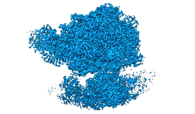 emd_14319.png
 http://ftp.pdbj.org/pub/emdb/structures/EMD-14319

 Z (Sec.)
 Y (Row.)
 X (Col.)


 試料の構成要素
 解析
解析 電子顕微鏡法
 FIELD EMISSION GUN

