 Movie
Movie Controller

[English] 日本語
 Yorodumi
- EMDB-13835: Subtomogram average of 80S ribosomes a cryo-FIB-lamella of Sum159... -
+ Open data

- Basic information
| Entry | Database: EMDB / ID: EMD-13835 | ||||||||||||
|---|---|---|---|---|---|---|---|---|---|---|---|---|---|
| Title | Subtomogram average of 80S ribosomes a cryo-FIB-lamella of Sum159 human cell line prepared after cryo-FIB-SEM volume imaging | ||||||||||||
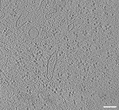
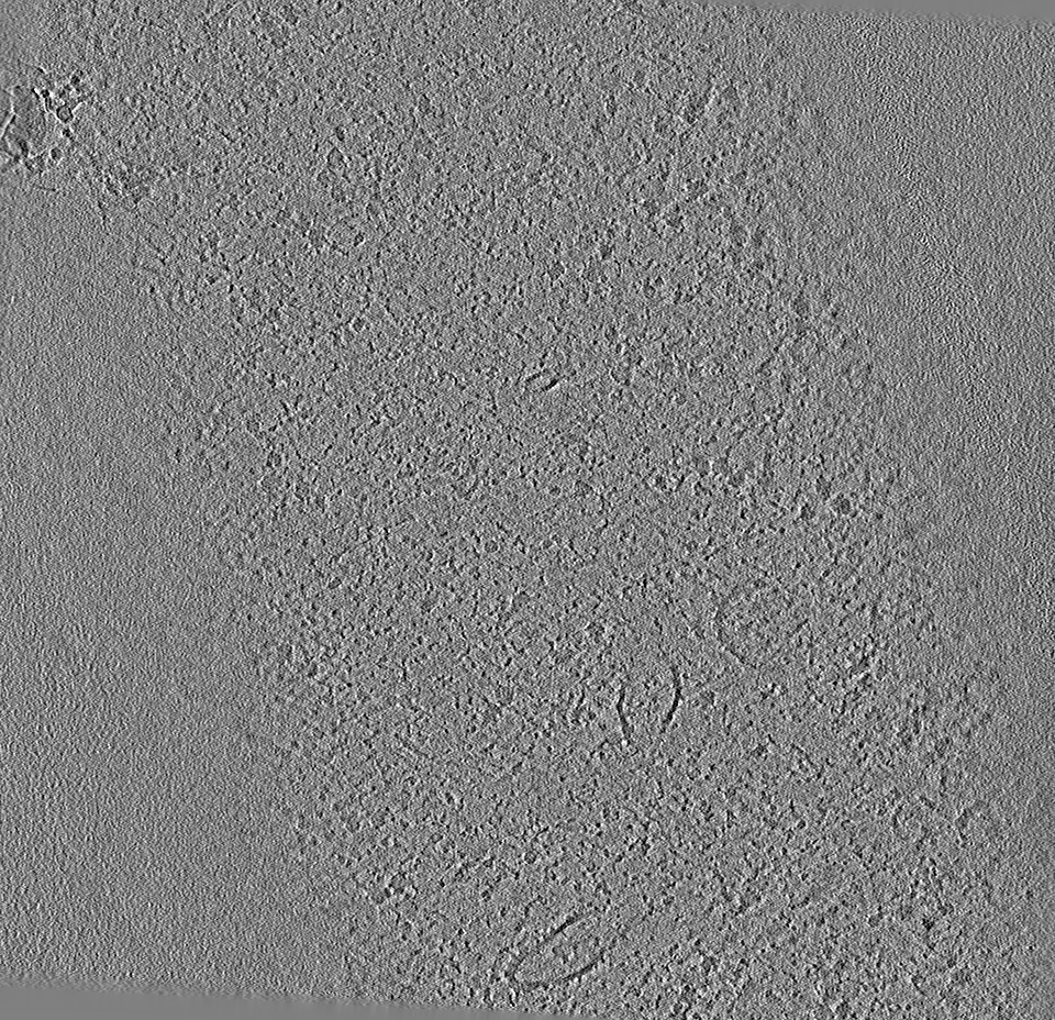
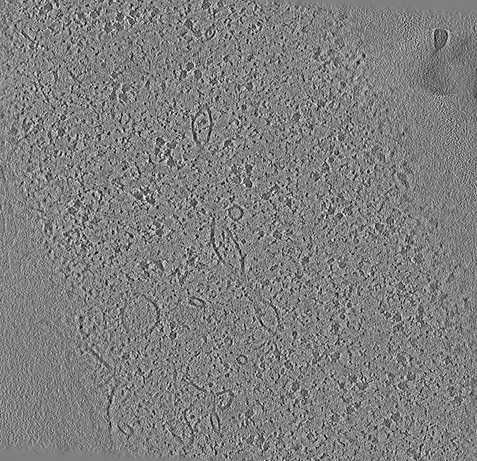
 Map data Map data | Tomogram used for subtomogram averaging, obtained from a cryo-FIB-milled Sum159 lamella after cryo-FIB SEM volume imaging. | ||||||||||||
 Sample Sample |
| ||||||||||||
| Biological species |  Homo sapiens (human) Homo sapiens (human) | ||||||||||||
| Method | electron tomography / cryo EM | ||||||||||||
 Authors Authors | Klumpe S / Fung HKH / Goetz SK / Plitzko JM / Mahamid J | ||||||||||||
| Funding support | European Union, 3 items
| ||||||||||||
 Citation Citation | Journal: Elife / Year: 2021 Title: A modular platform for automated cryo-FIB workflows. Authors: Sven Klumpe / Herman Kh Fung / Sara K Goetz / Ievgeniia Zagoriy / Bernhard Hampoelz / Xiaojie Zhang / Philipp S Erdmann / Janina Baumbach / Christoph W Müller / Martin Beck / Jürgen M ...Authors: Sven Klumpe / Herman Kh Fung / Sara K Goetz / Ievgeniia Zagoriy / Bernhard Hampoelz / Xiaojie Zhang / Philipp S Erdmann / Janina Baumbach / Christoph W Müller / Martin Beck / Jürgen M Plitzko / Julia Mahamid /  Abstract: Lamella micromachining by focused ion beam milling at cryogenic temperature (cryo-FIB) has matured into a preparation method widely used for cellular cryo-electron tomography. Due to the limited ...Lamella micromachining by focused ion beam milling at cryogenic temperature (cryo-FIB) has matured into a preparation method widely used for cellular cryo-electron tomography. Due to the limited ablation rates of low Ga ion beam currents required to maintain the structural integrity of vitreous specimens, common preparation protocols are time-consuming and labor intensive. The improved stability of new-generation cryo-FIB instruments now enables automated operations. Here, we present an open-source software tool, SerialFIB, for creating automated and customizable cryo-FIB preparation protocols. The software encompasses a graphical user interface for easy execution of routine lamellae preparations, a scripting module compatible with available Python packages, and interfaces with three-dimensional correlative light and electron microscopy (CLEM) tools. SerialFIB enables the streamlining of advanced cryo-FIB protocols such as multi-modal imaging, CLEM-guided lamella preparation and in situ lamella lift-out procedures. Our software therefore provides a foundation for further development of advanced cryogenic imaging and sample preparation protocols. | ||||||||||||
| History |
|
- Structure visualization
| Movie |
 Movie viewer Movie viewer |
|---|---|
| Supplemental images |
- Downloads & links
-EMDB archive
| Map data | emd_13835.map.gz | 1.5 GB | EMDB map data format | |
|---|---|---|---|---|
| Header (meta data) | emd-13835-v30.xmlemd-13835.xml | 11.7 KB 11.7 KB | Display Display | EMDB header |
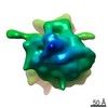
| Images |  emd_13835.png emd_13835.png | 248.9 KB | ||
| Others | emd_13835_additional_1.map.gz | 1.5 GB | ||
| Archive directory |  http://ftp.pdbj.org/pub/emdb/structures/EMD-13835ftp://ftp.pdbj.org/pub/emdb/structures/EMD-13835 http://ftp.pdbj.org/pub/emdb/structures/EMD-13835ftp://ftp.pdbj.org/pub/emdb/structures/EMD-13835 | HTTPS FTP |
-Validation report
| Summary document | emd_13835_validation.pdf.gz | 297.6 KB | Display | EMDB validaton report |
|---|---|---|---|---|
| Full document | emd_13835_full_validation.pdf.gz | 297.1 KB | Display | |
| Data in XML | emd_13835_validation.xml.gz | 4.8 KB | Display | |
| Data in CIF | emd_13835_validation.cif.gz | 5.3 KB | Display | |
| Arichive directory | https://ftp.pdbj.org/pub/emdb/validation_reports/EMD-13835ftp://ftp.pdbj.org/pub/emdb/validation_reports/EMD-13835 | HTTPS FTP |
-Related structure data


-Links
| EMDB pages | EMDB (EBI/PDBe) / EMDataResource |
|---|
-Map
| File | Download / File: emd_13835.map.gz / Format: CCP4 / Size: 1.7 GB / Type: IMAGE STORED AS FLOATING POINT NUMBER (4 BYTES) | ||||||||||||||||||||||||||||||||||||||||||||||||||||||||||||
|---|---|---|---|---|---|---|---|---|---|---|---|---|---|---|---|---|---|---|---|---|---|---|---|---|---|---|---|---|---|---|---|---|---|---|---|---|---|---|---|---|---|---|---|---|---|---|---|---|---|---|---|---|---|---|---|---|---|---|---|---|---|
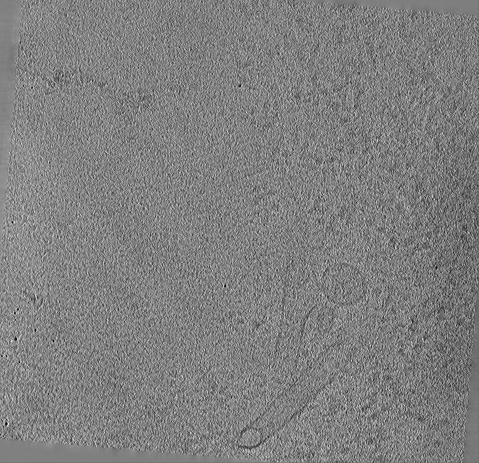
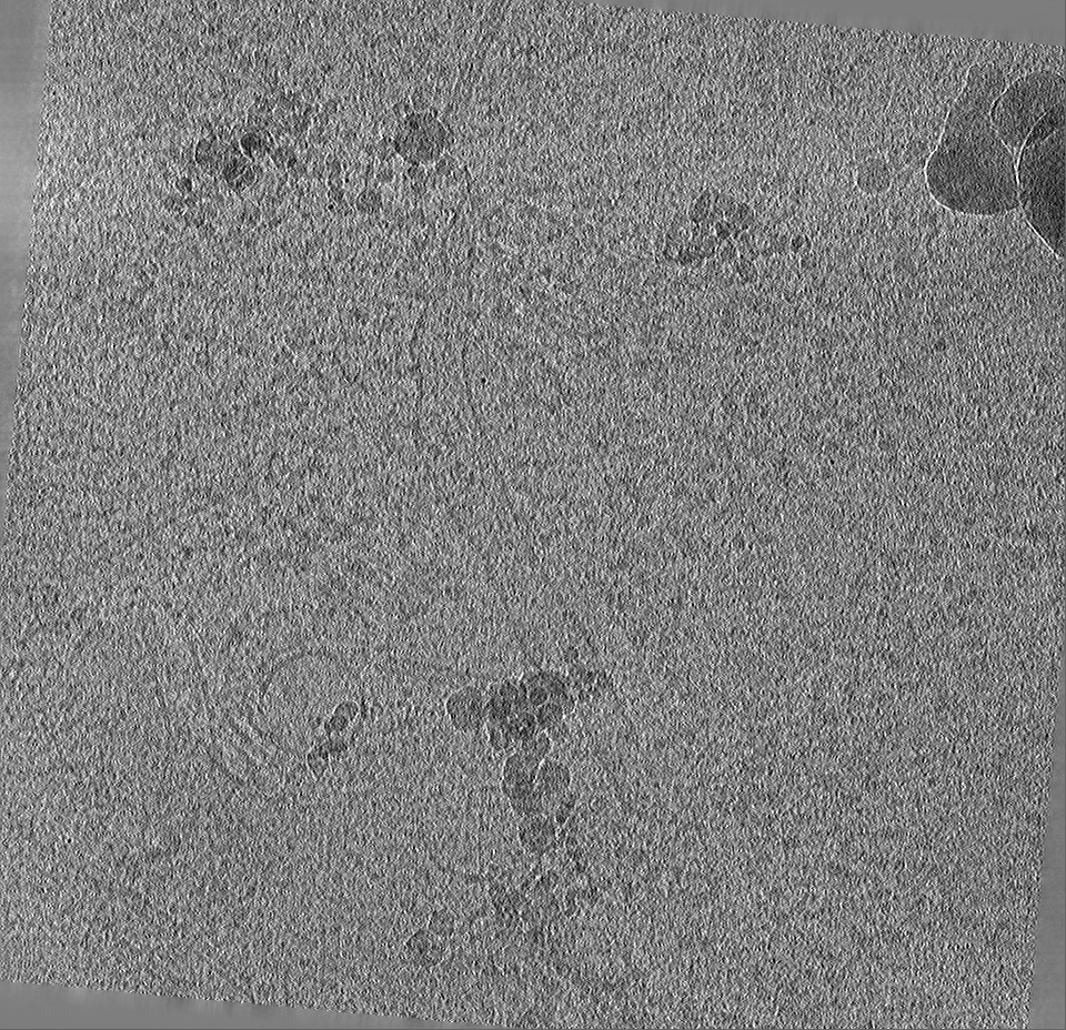
| Annotation | Tomogram used for subtomogram averaging, obtained from a cryo-FIB-milled Sum159 lamella after cryo-FIB SEM volume imaging. | ||||||||||||||||||||||||||||||||||||||||||||||||||||||||||||
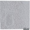
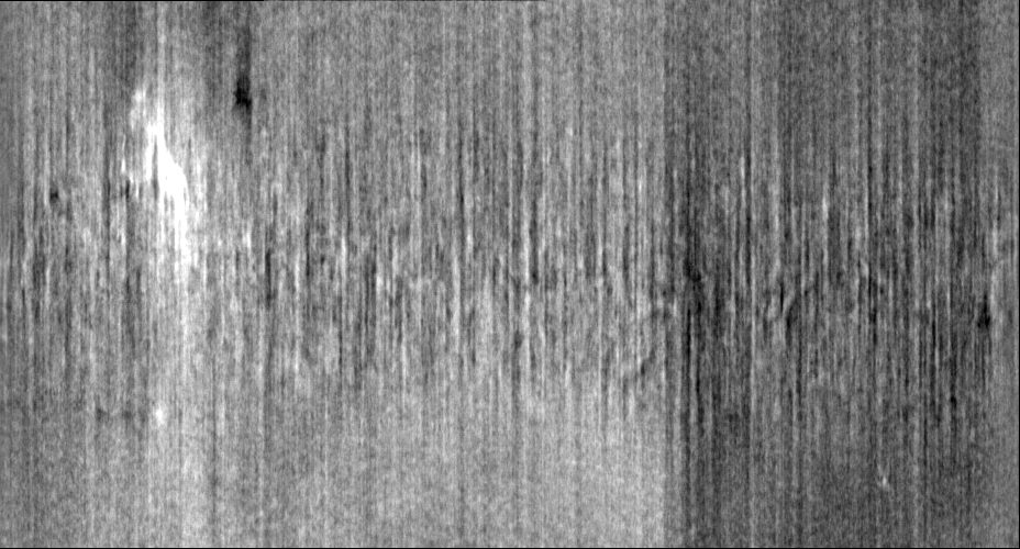
| Projections & slices | Image control
Images are generated by Spider. generated in cubic-lattice coordinate | ||||||||||||||||||||||||||||||||||||||||||||||||||||||||||||
| Voxel size | X=Y=Z: 13.4808 Å | ||||||||||||||||||||||||||||||||||||||||||||||||||||||||||||
| Density |
| ||||||||||||||||||||||||||||||||||||||||||||||||||||||||||||
| Symmetry | Space group: 1 | ||||||||||||||||||||||||||||||||||||||||||||||||||||||||||||
| Details | EMDB XML:
CCP4 map header:
| ||||||||||||||||||||||||||||||||||||||||||||||||||||||||||||
 Z (Sec.)
 Y (Row.)
 X (Col.)


-Supplemental data
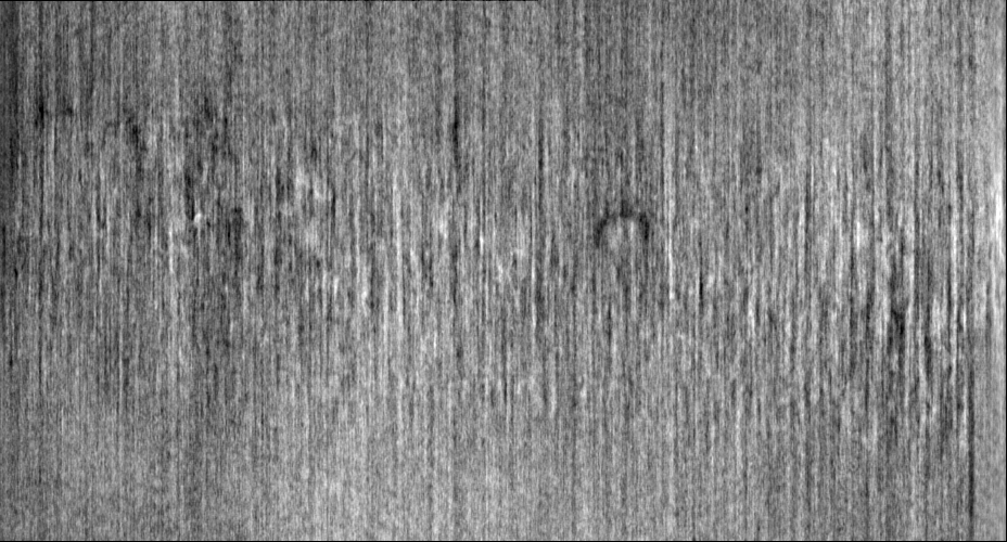
-Additional map: Tomogram used for subtomogram averaging, obtained from a...
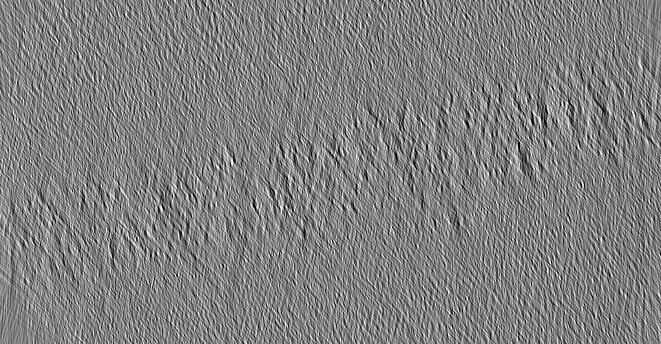
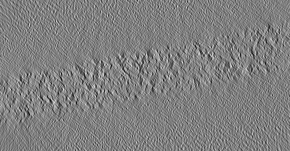
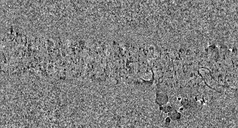
| File | emd_13835_additional_1.map | ||||||||||||
|---|---|---|---|---|---|---|---|---|---|---|---|---|---|
| Annotation | Tomogram used for subtomogram averaging, obtained from a cryo-FIB-milled Sum159 lamella after cryo-FIB SEM volume imaging. | ||||||||||||
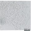
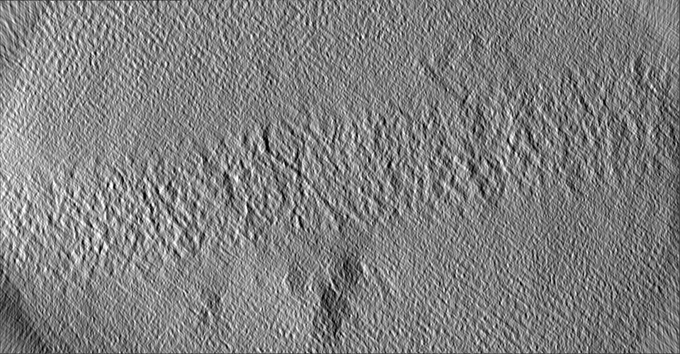
| Projections & Slices |
| ||||||||||||
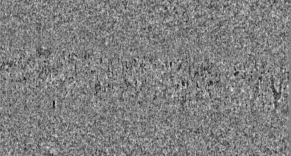
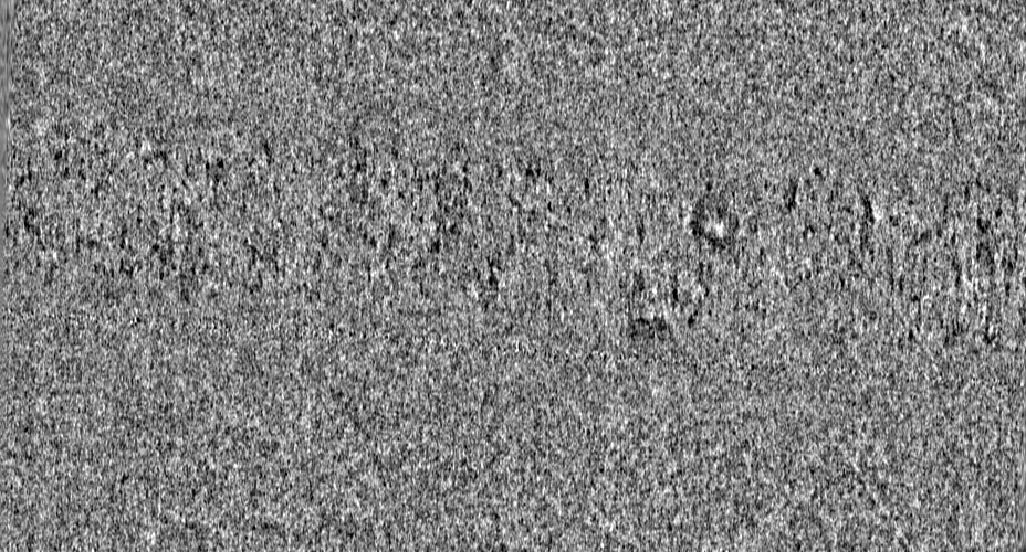
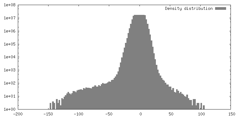
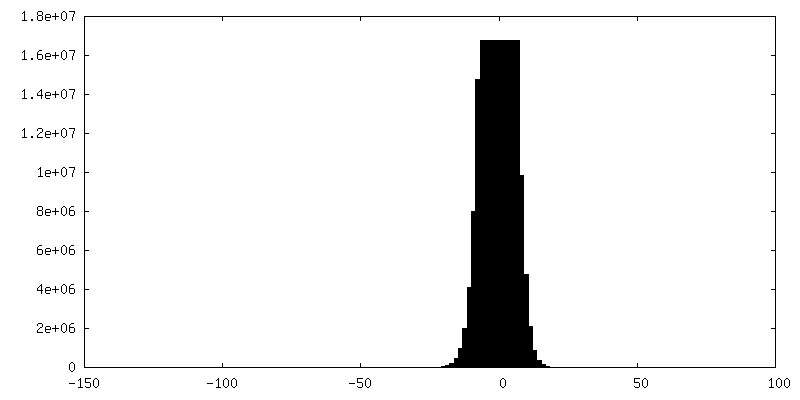
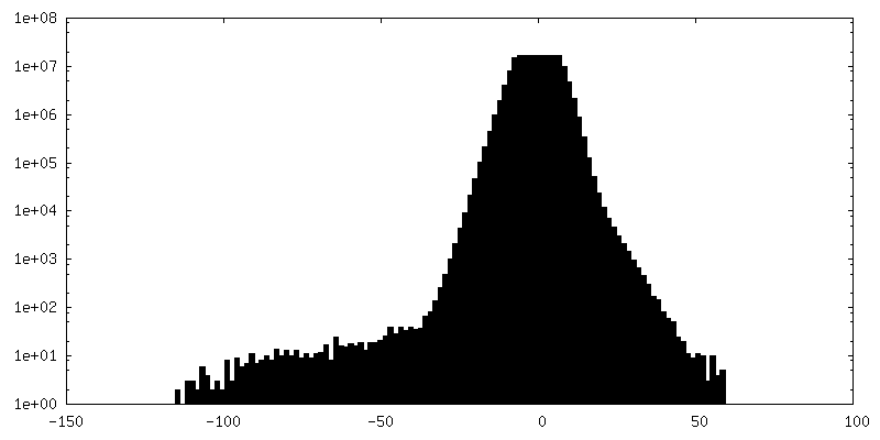
| Density Histograms |
- Sample components
-Entire : Sum159
| Entire | Name: Sum159 |
|---|---|
| Components |
|
-Supramolecule #1: Sum159
| Supramolecule | Name: Sum159 / type: cell / ID: 1 / Parent: 0 |
|---|---|
| Source (natural) | Organism: Homo sapiens (human) |
-Experimental details
-Structure determination
| Method | cryo EM |
|---|---|
 Processing Processing | electron tomography |
| Aggregation state | cell |
-Sample preparation
| Buffer | pH: 7 |
|---|---|
| Vitrification | Cryogen name: ETHANE |
| Sectioning | Focused ion beam - Instrument: OTHER / Focused ion beam - Ion: OTHER / Focused ion beam - Voltage: 30 kV / Focused ion beam - Current: 0.05 nA / Focused ion beam - Duration: 150 sec. / Focused ion beam - Temperature: 88 K / Focused ion beam - Initial thickness: 1000 nm / Focused ion beam - Final thickness: 200 nm Focused ion beam - Details: The value given for _emd_sectioning_focused_ion_beam.instrument is Thermo Fisher Aquilos. This is not in a list of allowed values {'OTHER', 'DB235'} so OTHER is written into the XML file. |
- Electron microscopy
| Microscope | FEI TITAN KRIOS |
|---|---|
| Specialist optics | Phase plate: VOLTA PHASE PLATE / Energy filter - Name: GIF Quantum LS / Energy filter - Slit width: 20 eV |
| Image recording | Film or detector model: GATAN K2 QUANTUM (4k x 4k) / Detector mode: COUNTING / Average electron dose: 2.27915 e/Å2 |
| Electron beam | Acceleration voltage: 300 kV / Electron source:  FIELD EMISSION GUN FIELD EMISSION GUN |
| Electron optics | Illumination mode: FLOOD BEAM / Imaging mode: BRIGHT FIELD / Cs: 2.7 mm |
| Experimental equipment |  Model: Titan Krios / Image courtesy: FEI Company |
-Image processing
| Final reconstruction | Software - Name: IMOD / Number images used: 57 |
|---|
