[English] 日本語
 Yorodumi
- EMDB-13819: CryoEM structure of electron bifurcating Fe-Fe hydrogenase HydABC... -
+ Open data
- Basic information
| Entry |  | |||||||||||||||||||||
|---|---|---|---|---|---|---|---|---|---|---|---|---|---|---|---|---|---|---|---|---|---|---|
| Title | CryoEM structure of electron bifurcating Fe-Fe hydrogenase HydABC complex A. woodii in the oxidised state | |||||||||||||||||||||
 Map data Map data | Single-Particle Cryo-EM Reconstructions of the Electron Bifurcating Hydrogenase HydABC symmetric(Processed on Cryosparc - non-sharpened map) | |||||||||||||||||||||
 Sample Sample |
| |||||||||||||||||||||
 Keywords Keywords | flavin-based electron bifurcating hydrogenase / FeFe hydrogenase / nicotine amide adenine dinucleotide / ferredoxin / ELECTRON TRANSPORT | |||||||||||||||||||||
| Function / homology |  Function and homology information Function and homology informationferredoxin hydrogenase / ferredoxin hydrogenase activity / NADH dehydrogenase (ubiquinone) activity / ATP synthesis coupled electron transport / FMN binding / 4 iron, 4 sulfur cluster binding / iron ion binding / membrane / metal ion binding Similarity search - Function | |||||||||||||||||||||
| Biological species |  Acetobacterium woodii DSM 1030 (bacteria) Acetobacterium woodii DSM 1030 (bacteria) | |||||||||||||||||||||
| Method | single particle reconstruction / cryo EM / Resolution: 3.78 Å | |||||||||||||||||||||
 Authors Authors | Kumar A / Saura P / Poeverlein MC / Gamiz-Hernandez AP / Kaila VRI / Mueller V / Schuller JM | |||||||||||||||||||||
| Funding support | 6 items
| |||||||||||||||||||||
 Citation Citation |  Journal: J Am Chem Soc / Year: 2023 Journal: J Am Chem Soc / Year: 2023Title: Molecular Basis of the Electron Bifurcation Mechanism in the [FeFe]-Hydrogenase Complex HydABC. Authors: Alexander Katsyv / Anuj Kumar / Patricia Saura / Maximilian C Pöverlein / Sven A Freibert / Sven T Stripp / Surbhi Jain / Ana P Gamiz-Hernandez / Ville R I Kaila / Volker Müller / Jan M Schuller /   Abstract: Electron bifurcation is a fundamental energy coupling mechanism widespread in microorganisms that thrive under anoxic conditions. These organisms employ hydrogen to reduce CO, but the molecular ...Electron bifurcation is a fundamental energy coupling mechanism widespread in microorganisms that thrive under anoxic conditions. These organisms employ hydrogen to reduce CO, but the molecular mechanisms have remained enigmatic. The key enzyme responsible for powering these thermodynamically challenging reactions is the electron-bifurcating [FeFe]-hydrogenase HydABC that reduces low-potential ferredoxins (Fd) by oxidizing hydrogen gas (H). By combining single-particle cryo-electron microscopy (cryoEM) under catalytic turnover conditions with site-directed mutagenesis experiments, functional studies, infrared spectroscopy, and molecular simulations, we show that HydABC from the acetogenic bacteria and employ a single flavin mononucleotide (FMN) cofactor to establish electron transfer pathways to the NAD(P) and Fd reduction sites by a mechanism that is fundamentally different from classical flavin-based electron bifurcation enzymes. By modulation of the NAD(P) binding affinity via reduction of a nearby iron-sulfur cluster, HydABC switches between the exergonic NAD(P) reduction and endergonic Fd reduction modes. Our combined findings suggest that the conformational dynamics establish a redox-driven kinetic gate that prevents the backflow of the electrons from the Fd reduction branch toward the FMN site, providing a basis for understanding general mechanistic principles of electron-bifurcating hydrogenases. | |||||||||||||||||||||
| History |
|
- Structure visualization
| Supplemental images |
|---|
- Downloads & links
-EMDB archive
| Map data |  emd_13819.map.gz emd_13819.map.gz | 15.7 MB |  EMDB map data format EMDB map data format | |
|---|---|---|---|---|
| Header (meta data) |  emd-13819-v30.xml emd-13819-v30.xml emd-13819.xml emd-13819.xml | 17 KB 17 KB | Display Display |  EMDB header EMDB header |
| Images |  emd_13819.png emd_13819.png | 69.7 KB | ||
| Filedesc metadata |  emd-13819.cif.gz emd-13819.cif.gz | 6.2 KB | ||
| Others |  emd_13819_additional_1.map.gz emd_13819_additional_1.map.gz | 15.8 MB | ||
| Archive directory |  http://ftp.pdbj.org/pub/emdb/structures/EMD-13819 http://ftp.pdbj.org/pub/emdb/structures/EMD-13819 ftp://ftp.pdbj.org/pub/emdb/structures/EMD-13819 ftp://ftp.pdbj.org/pub/emdb/structures/EMD-13819 | HTTPS FTP |
-Validation report
| Summary document |  emd_13819_validation.pdf.gz emd_13819_validation.pdf.gz | 413.7 KB | Display |  EMDB validaton report EMDB validaton report |
|---|---|---|---|---|
| Full document |  emd_13819_full_validation.pdf.gz emd_13819_full_validation.pdf.gz | 413.2 KB | Display | |
| Data in XML |  emd_13819_validation.xml.gz emd_13819_validation.xml.gz | 5.6 KB | Display | |
| Data in CIF |  emd_13819_validation.cif.gz emd_13819_validation.cif.gz | 6.4 KB | Display | |
| Arichive directory |  https://ftp.pdbj.org/pub/emdb/validation_reports/EMD-13819 https://ftp.pdbj.org/pub/emdb/validation_reports/EMD-13819 ftp://ftp.pdbj.org/pub/emdb/validation_reports/EMD-13819 ftp://ftp.pdbj.org/pub/emdb/validation_reports/EMD-13819 | HTTPS FTP |
-Related structure data
| Related structure data |  7q4wMC  7q4vC  8a5eC  8a6tC  8bewC M: atomic model generated by this map C: citing same article ( |
|---|---|
| Similar structure data | Similarity search - Function & homology  F&H Search F&H Search |
- Links
Links
| EMDB pages |  EMDB (EBI/PDBe) / EMDB (EBI/PDBe) /  EMDataResource EMDataResource |
|---|---|
| Related items in Molecule of the Month |
- Map
Map
| File |  Download / File: emd_13819.map.gz / Format: CCP4 / Size: 30.5 MB / Type: IMAGE STORED AS FLOATING POINT NUMBER (4 BYTES) Download / File: emd_13819.map.gz / Format: CCP4 / Size: 30.5 MB / Type: IMAGE STORED AS FLOATING POINT NUMBER (4 BYTES) | ||||||||||||||||||||||||||||||||||||
|---|---|---|---|---|---|---|---|---|---|---|---|---|---|---|---|---|---|---|---|---|---|---|---|---|---|---|---|---|---|---|---|---|---|---|---|---|---|
| Annotation | Single-Particle Cryo-EM Reconstructions of the Electron Bifurcating Hydrogenase HydABC symmetric(Processed on Cryosparc - non-sharpened map) | ||||||||||||||||||||||||||||||||||||
| Projections & slices | Image control
Images are generated by Spider. | ||||||||||||||||||||||||||||||||||||
| Voxel size | X=Y=Z: 1.09 Å | ||||||||||||||||||||||||||||||||||||
| Density |
| ||||||||||||||||||||||||||||||||||||
| Symmetry | Space group: 1 | ||||||||||||||||||||||||||||||||||||
| Details | EMDB XML:
|
-Supplemental data
-Additional map: Single-Particle Cryo-EM Reconstructions of the Electron Bifurcating Hydrogenase...
| File | emd_13819_additional_1.map | ||||||||||||
|---|---|---|---|---|---|---|---|---|---|---|---|---|---|
| Annotation | Single-Particle Cryo-EM Reconstructions of the Electron Bifurcating Hydrogenase HydABC symmetric(Processed on Cryosparc sharpened map) | ||||||||||||
| Projections & Slices |
| ||||||||||||
| Density Histograms |
- Sample components
-Entire : Homodimer complex of HydABC trimer
| Entire | Name: Homodimer complex of HydABC trimer |
|---|---|
| Components |
|
-Supramolecule #1: Homodimer complex of HydABC trimer
| Supramolecule | Name: Homodimer complex of HydABC trimer / type: complex / ID: 1 / Parent: 0 / Macromolecule list: #1-#3 |
|---|---|
| Source (natural) | Organism:  Acetobacterium woodii DSM 1030 (bacteria) Acetobacterium woodii DSM 1030 (bacteria) |
| Molecular weight | Theoretical: 350 kDa/nm |
-Macromolecule #1: Iron hydrogenase HydA1
| Macromolecule | Name: Iron hydrogenase HydA1 / type: protein_or_peptide / ID: 1 / Number of copies: 2 / Enantiomer: LEVO / EC number: ferredoxin hydrogenase |
|---|---|
| Source (natural) | Organism:  Acetobacterium woodii DSM 1030 (bacteria) Acetobacterium woodii DSM 1030 (bacteria) |
| Molecular weight | Theoretical: 63.692887 KDa |
| Sequence | String: MKEITFKING QEMIVPEGTT ILEAARMNNI DIPTLCYLKD INEIGACRMC LVEIAGARAL QAACVYPVAN GIEVLTNSPK VREARRVNL ELILSNHNRE CTTCIRSENC ELQTLATDLG VSDIPFEGEK SGKLIDDLST SVVRDESKCI LCKRCVSVCR D VQSVAVLG ...String: MKEITFKING QEMIVPEGTT ILEAARMNNI DIPTLCYLKD INEIGACRMC LVEIAGARAL QAACVYPVAN GIEVLTNSPK VREARRVNL ELILSNHNRE CTTCIRSENC ELQTLATDLG VSDIPFEGEK SGKLIDDLST SVVRDESKCI LCKRCVSVCR D VQSVAVLG TVGRGFTSQV QPVFNKSLAD VGCINCGQCI INCPVGALKE KSDIQRVWDA IADPSKTVIV QTAPAVRAAL GE EFGYPMG TSVTGKMAAA LRRLGFDKVF DTDFGADVCI MEEGTELIGR VTNGGVLPMI TSCSPGWIKF IETYYPEAIP HLS SCKSPQ NITGALLKNH YAQTNNIDPK DMVVVSIMPC TAKKYEVQRE ELCTDGNADV DISITTRELA RMIKEARILF NKLP DEDFD DYYGESTGAA VIFGATGGVM EAAVRTVADV LNKKDIQEID YQIVRGVDGI KKASVEVTPD LTVNLVVAHG GANIR EVME QLKAGELADT HFIELMACPG GCVNGGGQPI VSAKDKMDID IRTERAKALY DEDANVLTYR KSHQNPSVIR LYEEYL EEP NSPKAHHILH TKYSAKPKLV UniProtKB: Iron hydrogenase HydA1 |
-Macromolecule #2: Iron hydrogenase HydB
| Macromolecule | Name: Iron hydrogenase HydB / type: protein_or_peptide / ID: 2 / Number of copies: 2 / Enantiomer: LEVO / EC number: ferredoxin hydrogenase |
|---|---|
| Source (natural) | Organism:  Acetobacterium woodii DSM 1030 (bacteria) / Strain: ATCC 29683 / DSM 1030 / JCM 2381 / KCTC 1655 / WB1 Acetobacterium woodii DSM 1030 (bacteria) / Strain: ATCC 29683 / DSM 1030 / JCM 2381 / KCTC 1655 / WB1 |
| Molecular weight | Theoretical: 50.264406 KDa |
| Sequence | String: NIDEYIGFDG YLALEKVLLT MSPVDVINEV KASGLRGRGG GGFPTGLKWQ FAHDAVSEDG IKYVACNADE GDPGAFMDRS VLEGDPHAV IEAMAIAGYA VGASKGYVYV RAEYPIAVNR LQIAIDQAKE YGILGENIFE TDFSFDLEIR LGAGAFVCGE E TALMNSIE ...String: NIDEYIGFDG YLALEKVLLT MSPVDVINEV KASGLRGRGG GGFPTGLKWQ FAHDAVSEDG IKYVACNADE GDPGAFMDRS VLEGDPHAV IEAMAIAGYA VGASKGYVYV RAEYPIAVNR LQIAIDQAKE YGILGENIFE TDFSFDLEIR LGAGAFVCGE E TALMNSIE GKRGEPRPRP PFPANKGLFG KPTVLNNVET YANIPKIILN GAEWFASVGT EKSKGTKVFA LGGKINNTGL LE IPMGTTL REIIYEIGGG IPNGKAFKAA QTGGPSGGCL PESLLDTEID YDNLIAAGSM MGSGGLIVMD EDNCMVDVAR FFL DFTQDE SCGKCPPCRI GTKRMLEILE RICDGKGVEG DIERLEELAV GIKSSALCGL GQTAPNPVLS TIRFFRDEYE AHIR DKKCP AGVCKHLLDF KINADTCKGC GICAKKCPAD AISGEKKKPY NIDTSKCIKC GACIEACPFG SISKA UniProtKB: Iron hydrogenase HydB |
-Macromolecule #3: Iron hydrogenase HydC
| Macromolecule | Name: Iron hydrogenase HydC / type: protein_or_peptide / ID: 3 / Number of copies: 2 / Enantiomer: LEVO / EC number: ferredoxin hydrogenase |
|---|---|
| Source (natural) | Organism:  Acetobacterium woodii DSM 1030 (bacteria) Acetobacterium woodii DSM 1030 (bacteria) |
| Molecular weight | Theoretical: 16.999689 KDa |
| Sequence | String: MAELIPVENL DVVKAIVAEH REVPGCLMQI LQETQLKYGY LPLELQGTIA DELGIPLTEV YGVATFYSQF TLKPKGKYKI GICLGTACY VRGSQAIIDK VNSVLGTQVG DTTEDGKWSV DATRCVGACG LAPVMMINEE VFGRLTVDEI PGILEKY |
-Macromolecule #4: IRON/SULFUR CLUSTER
| Macromolecule | Name: IRON/SULFUR CLUSTER / type: ligand / ID: 4 / Number of copies: 14 / Formula: SF4 |
|---|---|
| Molecular weight | Theoretical: 351.64 Da |
| Chemical component information |  ChemComp-FS1: |
-Macromolecule #5: FE2/S2 (INORGANIC) CLUSTER
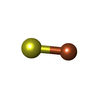
| Macromolecule | Name: FE2/S2 (INORGANIC) CLUSTER / type: ligand / ID: 5 / Number of copies: 4 / Formula: FES |
|---|---|
| Molecular weight | Theoretical: 175.82 Da |
| Chemical component information |  ChemComp-FES: |
-Macromolecule #6: FLAVIN MONONUCLEOTIDE
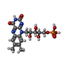
| Macromolecule | Name: FLAVIN MONONUCLEOTIDE / type: ligand / ID: 6 / Number of copies: 2 / Formula: FMN |
|---|---|
| Molecular weight | Theoretical: 456.344 Da |
| Chemical component information |  ChemComp-FMN: |
-Macromolecule #7: ZINC ION
| Macromolecule | Name: ZINC ION / type: ligand / ID: 7 / Number of copies: 2 / Formula: ZN |
|---|---|
| Molecular weight | Theoretical: 65.409 Da |
-Experimental details
-Structure determination
| Method | cryo EM |
|---|---|
 Processing Processing | single particle reconstruction |
| Aggregation state | particle |
- Sample preparation
| Buffer | pH: 7.6 |
|---|---|
| Vitrification | Cryogen name: ETHANE-PROPANE |
- Electron microscopy
| Microscope | FEI TITAN KRIOS |
|---|---|
| Image recording | Film or detector model: GATAN K3 (6k x 4k) / Average electron dose: 55.0 e/Å2 |
| Electron beam | Acceleration voltage: 300 kV / Electron source:  FIELD EMISSION GUN FIELD EMISSION GUN |
| Electron optics | Illumination mode: FLOOD BEAM / Imaging mode: BRIGHT FIELD / Nominal defocus max: 2.0 µm / Nominal defocus min: 0.8 µm |
| Experimental equipment |  Model: Titan Krios / Image courtesy: FEI Company |
- Image processing
| Startup model | Type of model: INSILICO MODEL |
|---|---|
| Final reconstruction | Resolution.type: BY AUTHOR / Resolution: 3.78 Å / Resolution method: FSC 0.143 CUT-OFF / Number images used: 250709 |
| Initial angle assignment | Type: PROJECTION MATCHING |
| Final angle assignment | Type: PROJECTION MATCHING |
 Movie
Movie Controller


 Z (Sec.)
 Y (Row.)
 X (Col.)


