 Movie
Movie Controller


+ Open data

- Basic information
| Entry | Database: EMDB / ID: EMD-12321 | |||||||||
|---|---|---|---|---|---|---|---|---|---|---|
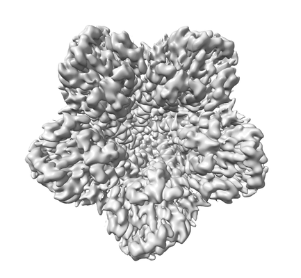
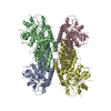
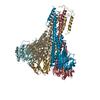
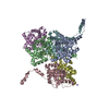
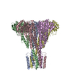
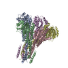
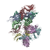
| Title | structure of the full-length CmaX protein | |||||||||
 Map data Map data | ||||||||||
 Sample Sample |
| |||||||||
 Keywords Keywords | divalent transport / zinc transporter / CorA family / membrane protein / TRANSPORT PROTEIN | |||||||||
| Function / homology | Mg2+ transporter protein, CorA-like/Zinc transport protein ZntB / CorA-like Mg2+ transporter protein / cobalt ion transmembrane transporter activity / magnesium ion transmembrane transporter activity / cobalt ion binding / plasma membrane => GO:0005886 / magnesium ion binding / CmaX protein Function and homology information Function and homology information | |||||||||
| Biological species |   Pseudomonas aeruginosa (bacteria) / Pseudomonas aeruginosa (strain ATCC 15692 / DSM 22644 / CIP 104116 / JCM 14847 / LMG 12228 / 1C / PRS 101 / PAO1) (bacteria) Pseudomonas aeruginosa (bacteria) / Pseudomonas aeruginosa (strain ATCC 15692 / DSM 22644 / CIP 104116 / JCM 14847 / LMG 12228 / 1C / PRS 101 / PAO1) (bacteria) | |||||||||
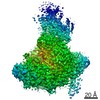
| Method | single particle reconstruction / cryo EM / Resolution: 3.03 Å | |||||||||
 Authors Authors | Stetsenko A / Stehantsev P | |||||||||
| Funding support | 1 items
| |||||||||
 Citation Citation | Journal: Int J Biol Macromol / Year: 2021 Title: Structural and biochemical characterization of a novel ZntB (CmaX) transporter protein from Pseudomonas aeruginosa. Authors: Artem Stetsenko / Pavlo Stehantsev / Natalia O Dranenko / Mikhail S Gelfand / Albert Guskov /   Abstract: The 2-TM-GxN family of membrane proteins is widespread in prokaryotes and plays an important role in transport of divalent cations. The canonical signature motif, which is also a selectivity filter, ...The 2-TM-GxN family of membrane proteins is widespread in prokaryotes and plays an important role in transport of divalent cations. The canonical signature motif, which is also a selectivity filter, has a composition of Gly-Met-Asn. Some members though deviate from this composition, however no data are available as to whether this has any functional implications. Here we report the functional and structural analysis of CmaX protein from a pathogenic Pseudomonas aeruginosa bacterium, which has a Gly-Ile-Asn signature motif. CmaX readily transports Zn, Mg, Cd, Ni and Co ions, but it does not utilize proton-symport as does ZntB from Escherichia coli. Together with the bioinformatics analysis, our data suggest that deviations from the canonical signature motif do not reveal any changes in substrate selectivity or transport and easily alter in course of evolution. | |||||||||
| History |
|
- Structure visualization
| Movie |
Movie viewer |
|---|---|
| Structure viewer | EM map: SurfViewMolmilJmol/JSmol |
| Supplemental images |
- Downloads & links
-EMDB archive
| Map data | emd_12321.map.gz | 96.1 MB | EMDB map data format | |
|---|---|---|---|---|
| Header (meta data) | emd-12321-v30.xmlemd-12321.xml | 9.3 KB 9.3 KB | Display Display | EMDB header |
| Images |  emd_12321.png emd_12321.png | 75.8 KB | ||
| Filedesc metadata | emd-12321.cif.gz | 5 KB | ||
| Archive directory |  http://ftp.pdbj.org/pub/emdb/structures/EMD-12321ftp://ftp.pdbj.org/pub/emdb/structures/EMD-12321 http://ftp.pdbj.org/pub/emdb/structures/EMD-12321ftp://ftp.pdbj.org/pub/emdb/structures/EMD-12321 | HTTPS FTP |
-Validation report
| Summary document | emd_12321_validation.pdf.gz | 456.6 KB | Display | EMDB validaton report |
|---|---|---|---|---|
| Full document | emd_12321_full_validation.pdf.gz | 456.2 KB | Display | |
| Data in XML | emd_12321_validation.xml.gz | 7 KB | Display | |
| Data in CIF | emd_12321_validation.cif.gz | 7.9 KB | Display | |
| Arichive directory | https://ftp.pdbj.org/pub/emdb/validation_reports/EMD-12321ftp://ftp.pdbj.org/pub/emdb/validation_reports/EMD-12321 | HTTPS FTP |
-Related structure data
| Related structure data | 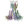 7nh9MC M: atomic model generated by this map C: citing same article ( |
|---|---|
| Similar structure data |

-Links
| EMDB pages | EMDB (EBI/PDBe) / EMDataResource |
|---|
-Map
| File | Download / File: emd_12321.map.gz / Format: CCP4 / Size: 193.2 MB / Type: IMAGE STORED AS FLOATING POINT NUMBER (4 BYTES) | ||||||||||||||||||||||||||||||||||||||||||||||||||||||||||||
|---|---|---|---|---|---|---|---|---|---|---|---|---|---|---|---|---|---|---|---|---|---|---|---|---|---|---|---|---|---|---|---|---|---|---|---|---|---|---|---|---|---|---|---|---|---|---|---|---|---|---|---|---|---|---|---|---|---|---|---|---|---|
| Projections & slices | Image control
Images are generated by Spider. | ||||||||||||||||||||||||||||||||||||||||||||||||||||||||||||
| Voxel size | X=Y=Z: 0.656 Å | ||||||||||||||||||||||||||||||||||||||||||||||||||||||||||||
| Density |
| ||||||||||||||||||||||||||||||||||||||||||||||||||||||||||||
| Symmetry | Space group: 1 | ||||||||||||||||||||||||||||||||||||||||||||||||||||||||||||
| Details | EMDB XML:
CCP4 map header:
| ||||||||||||||||||||||||||||||||||||||||||||||||||||||||||||
 Z (Sec.)
 Y (Row.)
 X (Col.)


-Supplemental data
- Sample components
-Entire : CmaX protein pentamer
| Entire | Name: CmaX protein pentamer |
|---|---|
| Components |
|
-Supramolecule #1: CmaX protein pentamer
| Supramolecule | Name: CmaX protein pentamer / type: complex / ID: 1 / Parent: 0 / Macromolecule list: all |
|---|---|
| Source (natural) | Organism: Pseudomonas aeruginosa (bacteria) |
-Macromolecule #1: CmaX protein
| Macromolecule | Name: CmaX protein / type: protein_or_peptide / ID: 1 / Details: N-terminal His tag and thrombin cleavage site / Number of copies: 5 / Enantiomer: LEVO |
|---|---|
| Source (natural) | Organism: Pseudomonas aeruginosa (strain ATCC 15692 / DSM 22644 / CIP 104116 / JCM 14847 / LMG 12228 / 1C / PRS 101 / PAO1) (bacteria) Strain: ATCC 15692 / DSM 22644 / CIP 104116 / JCM 14847 / LMG 12228 / 1C / PRS 101 / PAO1 |
| Molecular weight | Theoretical: 41.36502 KDa |
| Recombinant expression | Organism: |
| Sequence | String: MGSSHHHHHH SSGLVPRGSH MASMTGGQQM GRGSMQAYES GDERGLIYGY VLNGRGGGRR VGRNQIAVLD LLPEESLWLH WDRGVPEAQ AWLRDSAGLS EFACDLLLEE ATRPRLLDLG AESLLVFLRG VNLNPGAEPE DMVSLRVFAD ARRVISLRLR P LKAVADLL ...String: MGSSHHHHHH SSGLVPRGSH MASMTGGQQM GRGSMQAYES GDERGLIYGY VLNGRGGGRR VGRNQIAVLD LLPEESLWLH WDRGVPEAQ AWLRDSAGLS EFACDLLLEE ATRPRLLDLG AESLLVFLRG VNLNPGAEPE DMVSLRVFAD ARRVISLRLR P LKAVADLL EDLEAGKGPK TASEVVYYLA HYLTDRVDTL ISGIADQLDA VEELVEADER ASPDQHQLRT LRRRSAGLRR YL APQRDIY SQLARYKLSW FVEDDADYWN ELNNRLTRNL EELELIRERI SVLQEAESRR ITERMNRTMY LLGIITGFFL PMS FVTGLL GINVGGIPGA DAPHGFWLAC LLIGGVATFQ WWVFRRLRWL UniProtKB: CmaX protein |
-Experimental details
-Structure determination
| Method | cryo EM |
|---|---|
 Processing Processing | single particle reconstruction |
| Aggregation state | particle |
-Sample preparation
| Buffer | pH: 8 |
|---|---|
| Vitrification | Cryogen name: ETHANE |
- Electron microscopy
| Microscope | FEI TITAN KRIOS |
|---|---|
| Image recording | Film or detector model: GATAN K3 BIOQUANTUM (6k x 4k) / Average electron dose: 60.0 e/Å2 |
| Electron beam | Acceleration voltage: 300 kV / Electron source:  FIELD EMISSION GUN FIELD EMISSION GUN |
| Electron optics | Illumination mode: FLOOD BEAM / Imaging mode: BRIGHT FIELD |
| Experimental equipment |  Model: Titan Krios / Image courtesy: FEI Company |
-Image processing
| Startup model | Type of model: PDB ENTRY / Details: 5N9Y |
|---|---|
| Final reconstruction | Resolution.type: BY AUTHOR / Resolution: 3.03 Å / Resolution method: FSC 0.143 CUT-OFF / Number images used: 243386 |
| Initial angle assignment | Type: NOT APPLICABLE |
| Final angle assignment | Type: NOT APPLICABLE |
