Movie
Movie Controller

[English] 日本語
 Yorodumi
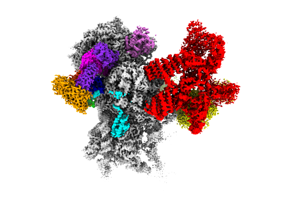
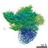
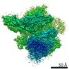
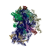
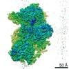
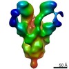
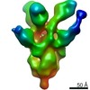
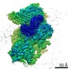
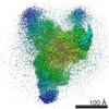
- EMDB-10775: Structure of a human 48S translational initiation complex - 40S body -
+ Open data

- Basic information
| Entry | Database: EMDB / ID: EMD-10775 | ||||||||||||
|---|---|---|---|---|---|---|---|---|---|---|---|---|---|
| Title | Structure of a human 48S translational initiation complex - 40S body | ||||||||||||
 Map data Map data | |||||||||||||
 Sample Sample |
| ||||||||||||
 Keywords Keywords | eIF3 / ribosome / translation / initiation complex | ||||||||||||
| Function / homology |  Function and homology information Function and homology informationformation of cytoplasmic translation initiation complex / multi-eIF complex / eukaryotic translation initiation factor 3 complex / eukaryotic 43S preinitiation complex / translation factor activity, RNA binding / eukaryotic 48S preinitiation complex / positive regulation of ubiquitin-protein transferase activity / regulation of translational initiation / negative regulation of RNA splicing / neural crest cell differentiation ...formation of cytoplasmic translation initiation complex / multi-eIF complex / eukaryotic translation initiation factor 3 complex / eukaryotic 43S preinitiation complex / translation factor activity, RNA binding / eukaryotic 48S preinitiation complex / positive regulation of ubiquitin-protein transferase activity / regulation of translational initiation / negative regulation of RNA splicing / neural crest cell differentiation / rRNA modification in the nucleus and cytosol / negative regulation of bicellular tight junction assembly / erythrocyte homeostasis / cytoplasmic side of rough endoplasmic reticulum membrane / Formation of the ternary complex, and subsequently, the 43S complex / laminin receptor activity / Ribosomal scanning and start codon recognition / Translation initiation complex formation / fibroblast growth factor binding / Protein hydroxylation / TOR signaling / ribosomal small subunit binding / SARS-CoV-1 modulates host translation machinery / mTORC1-mediated signalling / Peptide chain elongation / cellular response to ethanol / Selenocysteine synthesis / positive regulation of intrinsic apoptotic signaling pathway by p53 class mediator / Formation of a pool of free 40S subunits / Eukaryotic Translation Termination / ubiquitin ligase inhibitor activity / SRP-dependent cotranslational protein targeting to membrane / Response of EIF2AK4 (GCN2) to amino acid deficiency / endonucleolytic cleavage to generate mature 3'-end of SSU-rRNA from (SSU-rRNA, 5.8S rRNA, LSU-rRNA) / Viral mRNA Translation / negative regulation of ubiquitin-dependent protein catabolic process / Nonsense Mediated Decay (NMD) independent of the Exon Junction Complex (EJC) / GTP hydrolysis and joining of the 60S ribosomal subunit / L13a-mediated translational silencing of Ceruloplasmin expression / Major pathway of rRNA processing in the nucleolus and cytosol / Nonsense Mediated Decay (NMD) enhanced by the Exon Junction Complex (EJC) / Protein methylation / Nuclear events stimulated by ALK signaling in cancer / endonucleolytic cleavage in ITS1 to separate SSU-rRNA from 5.8S rRNA and LSU-rRNA from tricistronic rRNA transcript (SSU-rRNA, 5.8S rRNA, LSU-rRNA) / rough endoplasmic reticulum / positive regulation of cell cycle / laminin binding / translation initiation factor binding / Amplification of signal from unattached kinetochores via a MAD2 inhibitory signal / translation initiation factor activity / Mitotic Prometaphase / EML4 and NUDC in mitotic spindle formation / cytosolic ribosome / antiviral innate immune response / Resolution of Sister Chromatid Cohesion / stress granule assembly / ribosome assembly / erythrocyte differentiation / positive regulation of translation / maturation of SSU-rRNA from tricistronic rRNA transcript (SSU-rRNA, 5.8S rRNA, LSU-rRNA) / innate immune response in mucosa / maturation of SSU-rRNA / mRNA 3'-UTR binding / neural tube closure / translational initiation / small-subunit processome / RHO GTPases Activate Formins / response to insulin / maintenance of translational fidelity / modification-dependent protein catabolic process / GABA-ergic synapse / mRNA 5'-UTR binding / response to virus / RMTs methylate histone arginines / Regulation of expression of SLITs and ROBOs / protein tag activity / cytoplasmic ribonucleoprotein granule / rRNA processing / Separation of Sister Chromatids / antimicrobial humoral immune response mediated by antimicrobial peptide / glucose homeostasis / antibacterial humoral response / ribosomal small subunit assembly / virus receptor activity / presynapse / ribosome binding / ribosomal small subunit biogenesis / cell body / small ribosomal subunit / small ribosomal subunit rRNA binding / cytosolic small ribosomal subunit / cytosolic large ribosomal subunit / SARS-CoV-2 modulates host translation machinery / cell differentiation / cytoplasmic translation / tRNA binding / postsynaptic density / rRNA binding / defense response to Gram-positive bacterium / structural constituent of ribosome Similarity search - Function | ||||||||||||
| Biological species |  Homo sapiens (human) Homo sapiens (human) | ||||||||||||
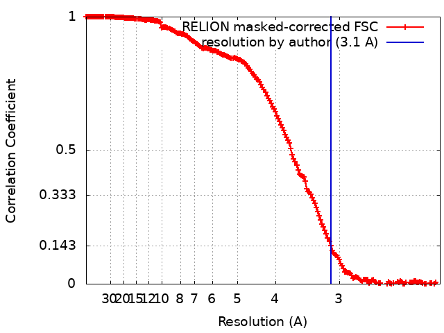
| Method | single particle reconstruction / cryo EM / Resolution: 3.1 Å | ||||||||||||
 Authors Authors | Brito Querido J / Sokabe M | ||||||||||||
| Funding support |  United Kingdom, United Kingdom,  United States, 3 items United States, 3 items
| ||||||||||||
 Citation Citation | Journal: Science / Year: 2020 Title: Structure of a human 48 translational initiation complex. Authors: Jailson Brito Querido / Masaaki Sokabe / Sebastian Kraatz / Yuliya Gordiyenko / J Mark Skehel / Christopher S Fraser / V Ramakrishnan / Abstract: A key step in translational initiation is the recruitment of the 43 preinitiation complex by the cap-binding complex [eukaryotic initiation factor 4F (eIF4F)] at the 5' end of messenger RNA (mRNA) to ...A key step in translational initiation is the recruitment of the 43 preinitiation complex by the cap-binding complex [eukaryotic initiation factor 4F (eIF4F)] at the 5' end of messenger RNA (mRNA) to form the 48 initiation complex (i.e., the 48). The 48 then scans along the mRNA to locate a start codon. To understand the mechanisms involved, we used cryo-electron microscopy to determine the structure of a reconstituted human 48 The structure reveals insights into early events of translation initiation complex assembly, as well as how eIF4F interacts with subunits of eIF3 near the mRNA exit channel in the 43 The location of eIF4F is consistent with a slotting model of mRNA recruitment and suggests that downstream mRNA is unwound at least in part by being "pulled" through the 40 subunit during scanning. | ||||||||||||
| History |
|
- Structure visualization
| Movie |
Movie viewer |
|---|---|
| Structure viewer | EM map: SurfViewMolmilJmol/JSmol |
| Supplemental images |
- Downloads & links
-EMDB archive
| Map data | emd_10775.map.gz | 36.6 MB | EMDB map data format | |
|---|---|---|---|---|
| Header (meta data) | emd-10775-v30.xmlemd-10775.xml | 50.7 KB 50.7 KB | Display Display | EMDB header |
| FSC (resolution estimation) | emd_10775_fsc.xml | 17.7 KB | Display | FSC data file |
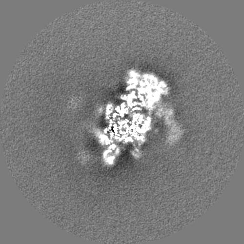
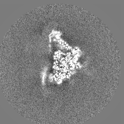
| Images |  emd_10775.png emd_10775.png | 168.4 KB | ||
| Filedesc metadata | emd-10775.cif.gz | 11 KB | ||
| Others | emd_10775_additional_1.map.gzemd_10775_additional_2.map.gzemd_10775_half_map_1.map.gzemd_10775_half_map_2.map.gz | 382 MB 391.4 MB 394 MB 393.9 MB | ||
| Archive directory |  http://ftp.pdbj.org/pub/emdb/structures/EMD-10775ftp://ftp.pdbj.org/pub/emdb/structures/EMD-10775 http://ftp.pdbj.org/pub/emdb/structures/EMD-10775ftp://ftp.pdbj.org/pub/emdb/structures/EMD-10775 | HTTPS FTP |
-Related structure data
| Related structure data | 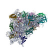 6ybwMC  6ybdC 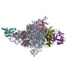 6ybsC  6ybtC  6ybvC  6zmwC M: atomic model generated by this map C: citing same article ( |
|---|---|
| Similar structure data |


-Links
| EMDB pages | EMDB (EBI/PDBe) / EMDataResource |
|---|---|
| Related items in Molecule of the Month |


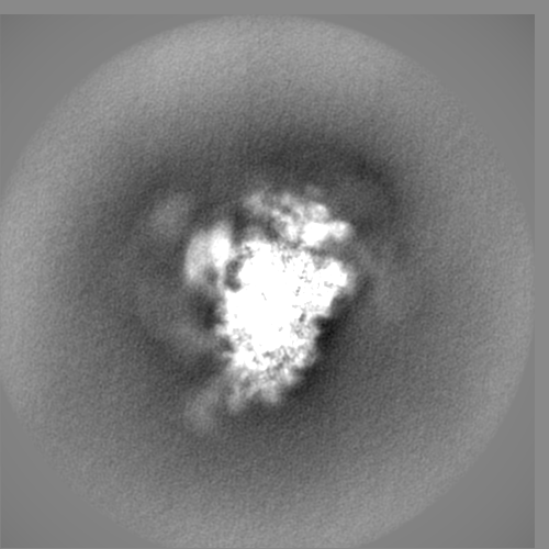
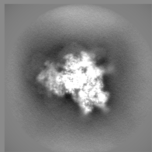
-Map
| File | Download / File: emd_10775.map.gz / Format: CCP4 / Size: 476.8 MB / Type: IMAGE STORED AS FLOATING POINT NUMBER (4 BYTES) | ||||||||||||||||||||||||||||||||||||||||||||||||||||||||||||
|---|---|---|---|---|---|---|---|---|---|---|---|---|---|---|---|---|---|---|---|---|---|---|---|---|---|---|---|---|---|---|---|---|---|---|---|---|---|---|---|---|---|---|---|---|---|---|---|---|---|---|---|---|---|---|---|---|---|---|---|---|---|
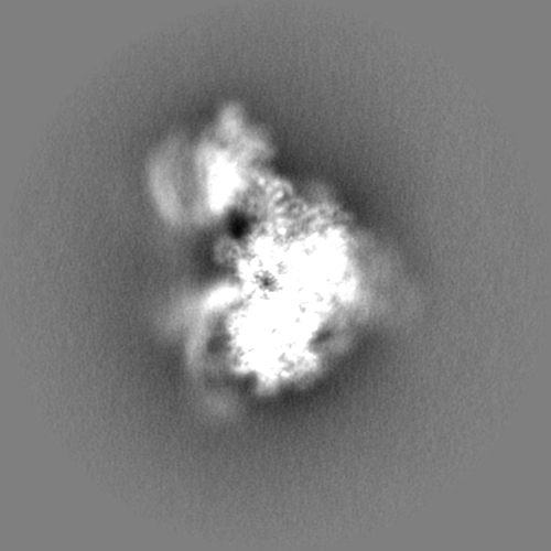
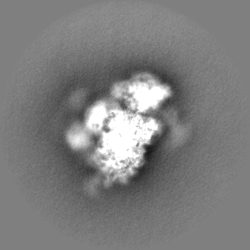
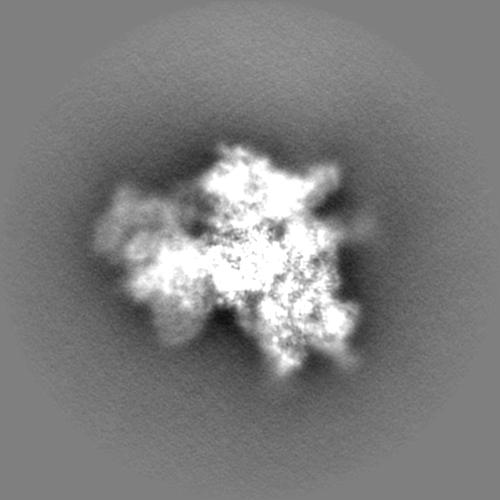
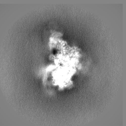
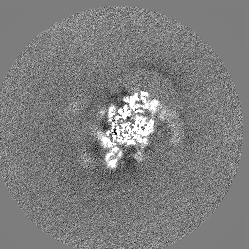
| Projections & slices | Image control
Images are generated by Spider. | ||||||||||||||||||||||||||||||||||||||||||||||||||||||||||||
| Voxel size | X=Y=Z: 1.074 Å | ||||||||||||||||||||||||||||||||||||||||||||||||||||||||||||
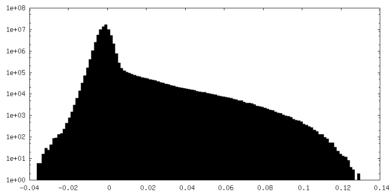
| Density |
| ||||||||||||||||||||||||||||||||||||||||||||||||||||||||||||
| Symmetry | Space group: 1 | ||||||||||||||||||||||||||||||||||||||||||||||||||||||||||||
| Details | EMDB XML:
CCP4 map header:
| ||||||||||||||||||||||||||||||||||||||||||||||||||||||||||||
 Z (Sec.)
 Y (Row.)
 X (Col.)


-Supplemental data
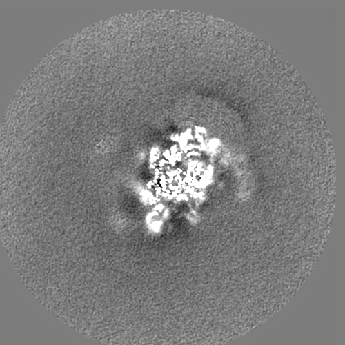
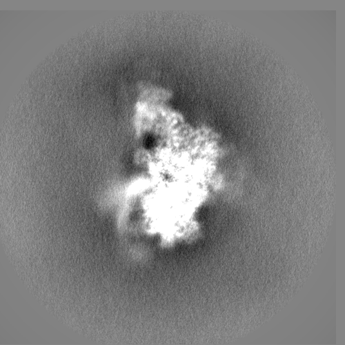
-Additional map: #1
| File | emd_10775_additional_1.map | ||||||||||||
|---|---|---|---|---|---|---|---|---|---|---|---|---|---|
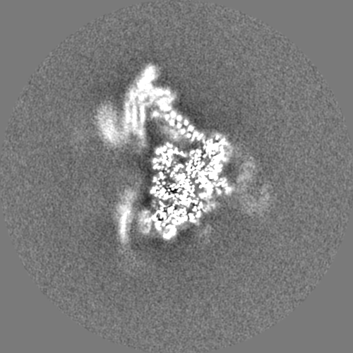
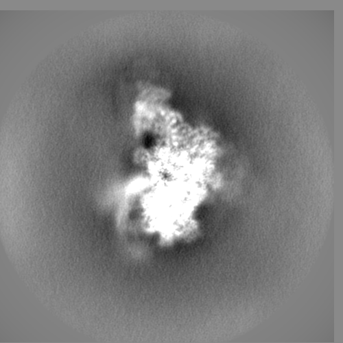
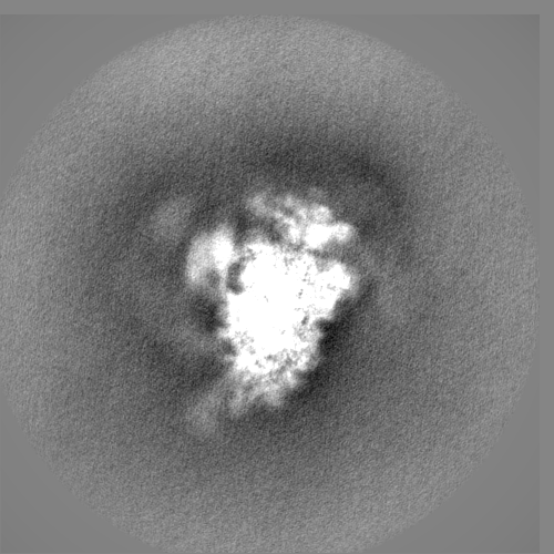
| Projections & Slices |
| ||||||||||||
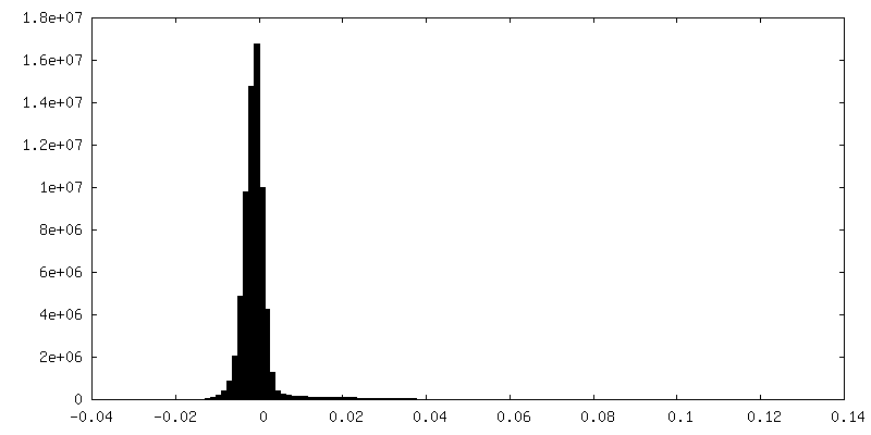
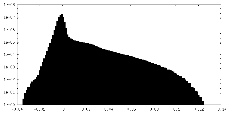
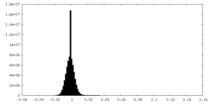
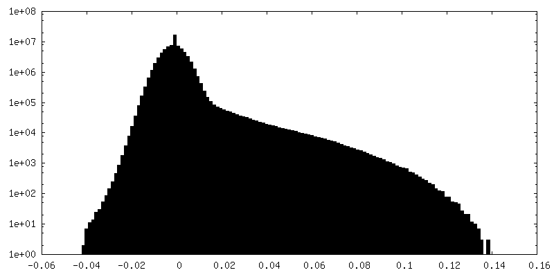
| Density Histograms |

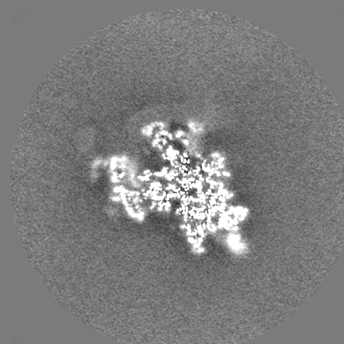
-Additional map: #2
| File | emd_10775_additional_2.map | ||||||||||||
|---|---|---|---|---|---|---|---|---|---|---|---|---|---|
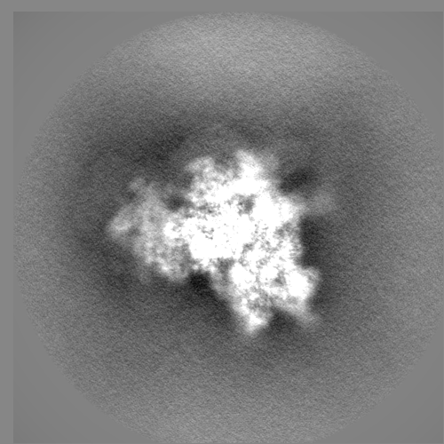
| Projections & Slices |
| ||||||||||||
| Density Histograms |


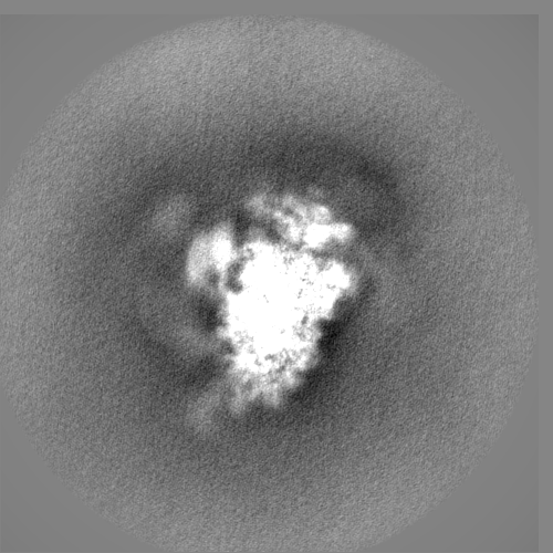
-Half map: #1
| File | emd_10775_half_map_1.map | ||||||||||||
|---|---|---|---|---|---|---|---|---|---|---|---|---|---|
| Projections & Slices |
| ||||||||||||
| Density Histograms |


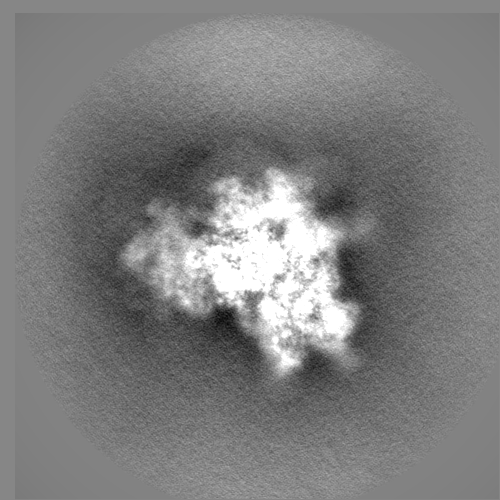
-Half map: #2
| File | emd_10775_half_map_2.map | ||||||||||||
|---|---|---|---|---|---|---|---|---|---|---|---|---|---|
| Projections & Slices |
| ||||||||||||
| Density Histograms |


- Sample components
+Entire : Human 48S initiation complex
+Supramolecule #1: Human 48S initiation complex
+Supramolecule #2: Human 48S initiation complex
+Supramolecule #3: Human 48S initiation complex
+Supramolecule #4: Human 48S initiation complex
+Macromolecule #1: 40S ribosomal protein S4, X isoform
+Macromolecule #2: 40S ribosomal protein S11
+Macromolecule #3: 40S ribosomal protein S23
+Macromolecule #4: 40S ribosomal protein S9
+Macromolecule #5: 40S ribosomal protein S7
+Macromolecule #6: 40S ribosomal protein S30
+Macromolecule #7: 40S ribosomal protein S27
+Macromolecule #8: 40S ribosomal protein S21
+Macromolecule #9: 40S ribosomal protein S15a
+Macromolecule #10: 40S ribosomal protein S17
+Macromolecule #11: 40S ribosomal protein S2
+Macromolecule #12: 40S ribosomal protein S3a
+Macromolecule #13: 40S ribosomal protein SA
+Macromolecule #14: 40S ribosomal protein S26
+Macromolecule #15: 40S ribosomal protein S14
+Macromolecule #16: 40S ribosomal protein S6
+Macromolecule #17: 40S ribosomal protein S8
+Macromolecule #18: 40S ribosomal protein S24
+Macromolecule #19: Eukaryotic translation initiation factor 1A, X-chromosomal

+Macromolecule #20: Eukaryotic translation initiation factor 1
+Macromolecule #21: 40S ribosomal protein S13
+Macromolecule #22: Eukaryotic translation initiation factor 3 subunit J
+Macromolecule #23: Eukaryotic translation initiation factor 3 subunit C
+Macromolecule #24: 60S ribosomal protein L41
+Macromolecule #25: 18S rRNA
+Macromolecule #26: mRNA
+Macromolecule #27: MAGNESIUM ION
+Macromolecule #28: ZINC ION
-Experimental details
-Structure determination
| Method | cryo EM |
|---|---|
 Processing Processing | single particle reconstruction |
| Aggregation state | particle |
-Sample preparation
| Buffer | pH: 7.4 |
|---|---|
| Vitrification | Cryogen name: ETHANE / Chamber humidity: 100 % |
- Electron microscopy
| Microscope | FEI TITAN KRIOS |
|---|---|
| Image recording | Film or detector model: FEI FALCON III (4k x 4k) / Average exposure time: 1.0 sec. / Average electron dose: 107.0 e/Å2 |
| Electron beam | Acceleration voltage: 300 kV / Electron source:  FIELD EMISSION GUN FIELD EMISSION GUN |
| Electron optics | Illumination mode: FLOOD BEAM / Imaging mode: BRIGHT FIELD |
| Experimental equipment |  Model: Titan Krios / Image courtesy: FEI Company |