[English] 日本語
 Yorodumi
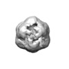
- EMDB-10008: Native encapsulin with bound dye decolourising peroxidase on thre... -
+ Open data
- Basic information
| Entry | Database: EMDB / ID: EMD-10008 | |||||||||
|---|---|---|---|---|---|---|---|---|---|---|
| Title | Native encapsulin with bound dye decolourising peroxidase on three fold axis | |||||||||
 Map data Map data | Encapsulin with Dyp on three fold axis | |||||||||
 Sample Sample |
| |||||||||
| Biological species |  Mycobacterium smegmatis str. MC2 155 (bacteria) Mycobacterium smegmatis str. MC2 155 (bacteria) | |||||||||
| Method | single particle reconstruction / negative staining / Resolution: 30.0 Å | |||||||||
 Authors Authors | Kirykowicz AM / Woodward JD | |||||||||
| Funding support |  South Africa, 1 items South Africa, 1 items
| |||||||||
 Citation Citation |  Journal: To Be Published Journal: To Be PublishedTitle: Structural pipeline for the investigation of Mycobacterial protein complexes involved in the stationary phase stress response Authors: Kirykowicz AM / Woodward JD | |||||||||
| History |
|
- Structure visualization
| Movie |
 Movie viewer Movie viewer |
|---|---|
| Structure viewer | EM map:  SurfView SurfView Molmil Molmil Jmol/JSmol Jmol/JSmol |
| Supplemental images |
- Downloads & links
-EMDB archive
| Map data |  emd_10008.map.gz emd_10008.map.gz | 3.9 MB |  EMDB map data format EMDB map data format | |
|---|---|---|---|---|
| Header (meta data) |  emd-10008-v30.xml emd-10008-v30.xml emd-10008.xml emd-10008.xml | 10.3 KB 10.3 KB | Display Display |  EMDB header EMDB header |
| Images |  emd_10008.png emd_10008.png | 31.8 KB | ||
| Archive directory |  http://ftp.pdbj.org/pub/emdb/structures/EMD-10008 http://ftp.pdbj.org/pub/emdb/structures/EMD-10008 ftp://ftp.pdbj.org/pub/emdb/structures/EMD-10008 ftp://ftp.pdbj.org/pub/emdb/structures/EMD-10008 | HTTPS FTP |
-Validation report
| Summary document |  emd_10008_validation.pdf.gz emd_10008_validation.pdf.gz | 224.9 KB | Display |  EMDB validaton report EMDB validaton report |
|---|---|---|---|---|
| Full document |  emd_10008_full_validation.pdf.gz emd_10008_full_validation.pdf.gz | 224 KB | Display | |
| Data in XML |  emd_10008_validation.xml.gz emd_10008_validation.xml.gz | 5.5 KB | Display | |
| Arichive directory |  https://ftp.pdbj.org/pub/emdb/validation_reports/EMD-10008 https://ftp.pdbj.org/pub/emdb/validation_reports/EMD-10008 ftp://ftp.pdbj.org/pub/emdb/validation_reports/EMD-10008 ftp://ftp.pdbj.org/pub/emdb/validation_reports/EMD-10008 | HTTPS FTP |
-Related structure data
| Related structure data | C: citing same article ( |
|---|---|
| Similar structure data |
- Links
Links
| EMDB pages |  EMDB (EBI/PDBe) / EMDB (EBI/PDBe) /  EMDataResource EMDataResource |
|---|
- Map
Map
| File |  Download / File: emd_10008.map.gz / Format: CCP4 / Size: 8 MB / Type: IMAGE STORED AS FLOATING POINT NUMBER (4 BYTES) Download / File: emd_10008.map.gz / Format: CCP4 / Size: 8 MB / Type: IMAGE STORED AS FLOATING POINT NUMBER (4 BYTES) | ||||||||||||||||||||||||||||||||||||||||||||||||||||||||||||
|---|---|---|---|---|---|---|---|---|---|---|---|---|---|---|---|---|---|---|---|---|---|---|---|---|---|---|---|---|---|---|---|---|---|---|---|---|---|---|---|---|---|---|---|---|---|---|---|---|---|---|---|---|---|---|---|---|---|---|---|---|---|
| Annotation | Encapsulin with Dyp on three fold axis | ||||||||||||||||||||||||||||||||||||||||||||||||||||||||||||
| Projections & slices | Image control
Images are generated by Spider. | ||||||||||||||||||||||||||||||||||||||||||||||||||||||||||||
| Voxel size | X=Y=Z: 3.84 Å | ||||||||||||||||||||||||||||||||||||||||||||||||||||||||||||
| Density |
| ||||||||||||||||||||||||||||||||||||||||||||||||||||||||||||
| Symmetry | Space group: 1 | ||||||||||||||||||||||||||||||||||||||||||||||||||||||||||||
| Details | EMDB XML:
CCP4 map header:
| ||||||||||||||||||||||||||||||||||||||||||||||||||||||||||||
-Supplemental data
- Sample components
-Entire : Encapsulin with bound DyP on three fold axis
| Entire | Name: Encapsulin with bound DyP on three fold axis |
|---|---|
| Components |
|
-Supramolecule #1: Encapsulin with bound DyP on three fold axis
| Supramolecule | Name: Encapsulin with bound DyP on three fold axis / type: complex / ID: 1 / Parent: 0 / Macromolecule list: #1 / Details: C3 symmetry applied to native encapsulin particles |
|---|---|
| Source (natural) | Organism:  Mycobacterium smegmatis str. MC2 155 (bacteria) / Location in cell: Cytoplasm Mycobacterium smegmatis str. MC2 155 (bacteria) / Location in cell: Cytoplasm |
| Molecular weight | Experimental: 1.962 MDa |
-Experimental details
-Structure determination
| Method | negative staining |
|---|---|
 Processing Processing | single particle reconstruction |
| Aggregation state | particle |
- Sample preparation
| Concentration | 0.3 mg/mL |
|---|---|
| Buffer | pH: 8 / Details: 200 mM NaCl, 50 mM Tris-HCl |
| Staining | Type: NEGATIVE / Material: Uranyl acetate / Details: Sample washed/blotted with 2% UA and air-dried |
| Details | Negative stain |
- Electron microscopy
| Microscope | FEI TECNAI F20 |
|---|---|
| Image recording | Film or detector model: GATAN ULTRASCAN 4000 (4k x 4k) / Digitization - Dimensions - Width: 4000 pixel / Digitization - Dimensions - Height: 4000 pixel / Average exposure time: 0.5 sec. / Average electron dose: 20.0 e/Å2 |
| Electron beam | Acceleration voltage: 200 kV / Electron source:  FIELD EMISSION GUN FIELD EMISSION GUN |
| Electron optics | C2 aperture diameter: 70.0 µm / Illumination mode: OTHER / Imaging mode: OTHER / Cs: 1.2 mm / Nominal magnification: 29000 |
| Experimental equipment |  Model: Tecnai F20 / Image courtesy: FEI Company |
 Movie
Movie Controller


 Z (Sec.)
 Y (Row.)
 X (Col.)


