+ Open data
- Basic information
| Entry | Database: EMDB / ID: EMD-0633 | |||||||||
|---|---|---|---|---|---|---|---|---|---|---|
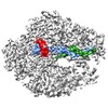
| Title | RNA polymerase II elongation complex arrested at a CPD lesion | |||||||||
 Map data Map data | RNA polymerase II elongation complex stalled at a CPD lesion | |||||||||
 Sample Sample |
| |||||||||
 Keywords Keywords | RNA polymerase / CPD / elongation complex / streptavidin grids / transcription / transferase-DNA-RNA complex | |||||||||
| Function / homology |  Function and homology information Function and homology informationRNA Polymerase I Transcription Initiation / Processing of Capped Intron-Containing Pre-mRNA / RNA Polymerase III Transcription Initiation From Type 2 Promoter / RNA Pol II CTD phosphorylation and interaction with CE / Formation of the Early Elongation Complex / mRNA Capping / RNA polymerase II transcribes snRNA genes / TP53 Regulates Transcription of DNA Repair Genes / termination of RNA polymerase II transcription / RNA Polymerase II Promoter Escape ...RNA Polymerase I Transcription Initiation / Processing of Capped Intron-Containing Pre-mRNA / RNA Polymerase III Transcription Initiation From Type 2 Promoter / RNA Pol II CTD phosphorylation and interaction with CE / Formation of the Early Elongation Complex / mRNA Capping / RNA polymerase II transcribes snRNA genes / TP53 Regulates Transcription of DNA Repair Genes / termination of RNA polymerase II transcription / RNA Polymerase II Promoter Escape / RNA Polymerase II Transcription Pre-Initiation And Promoter Opening / RNA Polymerase II Transcription Initiation / RNA Polymerase II Transcription Initiation And Promoter Clearance / RNA Polymerase II Pre-transcription Events / RNA-templated transcription / termination of RNA polymerase III transcription / Formation of TC-NER Pre-Incision Complex / transcription initiation at RNA polymerase III promoter / maintenance of transcriptional fidelity during transcription elongation by RNA polymerase II / termination of RNA polymerase I transcription / RNA Polymerase I Promoter Escape / nucleolar large rRNA transcription by RNA polymerase I / Gap-filling DNA repair synthesis and ligation in TC-NER / transcription by RNA polymerase I / transcription initiation at RNA polymerase I promoter / Estrogen-dependent gene expression / transcription by RNA polymerase III / RNA polymerase II activity / Dual incision in TC-NER / transcription elongation by RNA polymerase I / transcription-coupled nucleotide-excision repair / tRNA transcription by RNA polymerase III / RNA polymerase I complex / RNA polymerase I activity / RNA polymerase III complex / translesion synthesis / RNA polymerase II, core complex / transcription initiation at RNA polymerase II promoter / transcription elongation by RNA polymerase II / ribonucleoside binding / DNA-directed 5'-3' RNA polymerase activity / DNA-directed RNA polymerase / cytoplasmic stress granule / peroxisome / ribosome biogenesis / transcription by RNA polymerase II / nucleic acid binding / protein dimerization activity / mRNA binding / nucleolus / mitochondrion / DNA binding / zinc ion binding / nucleoplasm / nucleus / metal ion binding / cytoplasm Similarity search - Function | |||||||||
| Biological species |  | |||||||||
| Method | single particle reconstruction / cryo EM / Resolution: 3.1 Å | |||||||||
 Authors Authors | Lahiri I / Leshziner AE | |||||||||
| Funding support |  United States, 2 items United States, 2 items
| |||||||||
 Citation Citation |  Journal: J Struct Biol / Year: 2019 Journal: J Struct Biol / Year: 2019Title: 3.1 Å structure of yeast RNA polymerase II elongation complex stalled at a cyclobutane pyrimidine dimer lesion solved using streptavidin affinity grids. Authors: Indrajit Lahiri / Jun Xu / Bong Gyoon Han / Juntaek Oh / Dong Wang / Frank DiMaio / Andres E Leschziner /  Abstract: Despite significant advances in all aspects of single particle cryo-electron microscopy (cryo-EM), specimen preparation still remains a challenge. During sample preparation, macromolecules interact ...Despite significant advances in all aspects of single particle cryo-electron microscopy (cryo-EM), specimen preparation still remains a challenge. During sample preparation, macromolecules interact with the air-water interface, which often leads to detrimental effects such as denaturation or adoption of preferred orientations, ultimately hindering structure determination. Randomly biotinylating the protein of interest (for example, at its primary amines) and then tethering it to a cryo-EM grid coated with two-dimensional crystals of streptavidin (acting as an affinity surface) can prevent the protein from interacting with the air-water interface. Recently, this approach was successfully used to solve a high-resolution structure of a test sample, a bacterial ribosome. However, whether this method can be used for samples where interaction with the air-water interface has been shown to be problematic remains to be determined. Here we report a 3.1 Å structure of an RNA polymerase II elongation complex stalled at a cyclobutane pyrimidine dimer lesion (Pol II EC(CPD)) solved using streptavidin grids. Our previous attempt to solve this structure using conventional sample preparation methods resulted in a poor quality cryo-EM map due to Pol II EC(CPD)'s adopting a strong preferred orientation. Imaging the same sample on streptavidin grids improved the angular distribution of its view, resulting in a high-resolution structure. This work shows that streptavidin affinity grids can be used to address known challenges posed by the interaction with the air-water interface. | |||||||||
| History |
|
- Structure visualization
| Movie |
 Movie viewer Movie viewer |
|---|---|
| Structure viewer | EM map:  SurfView SurfView Molmil Molmil Jmol/JSmol Jmol/JSmol |
| Supplemental images |
- Downloads & links
-EMDB archive
| Map data |  emd_0633.map.gz emd_0633.map.gz | 124.6 MB |  EMDB map data format EMDB map data format | |
|---|---|---|---|---|
| Header (meta data) |  emd-0633-v30.xml emd-0633-v30.xml emd-0633.xml emd-0633.xml | 26.3 KB 26.3 KB | Display Display |  EMDB header EMDB header |
| Images |  emd_0633.png emd_0633.png | 201.6 KB | ||
| Filedesc metadata |  emd-0633.cif.gz emd-0633.cif.gz | 9.3 KB | ||
| Archive directory |  http://ftp.pdbj.org/pub/emdb/structures/EMD-0633 http://ftp.pdbj.org/pub/emdb/structures/EMD-0633 ftp://ftp.pdbj.org/pub/emdb/structures/EMD-0633 ftp://ftp.pdbj.org/pub/emdb/structures/EMD-0633 | HTTPS FTP |
-Validation report
| Summary document |  emd_0633_validation.pdf.gz emd_0633_validation.pdf.gz | 404.1 KB | Display |  EMDB validaton report EMDB validaton report |
|---|---|---|---|---|
| Full document |  emd_0633_full_validation.pdf.gz emd_0633_full_validation.pdf.gz | 403.7 KB | Display | |
| Data in XML |  emd_0633_validation.xml.gz emd_0633_validation.xml.gz | 7.1 KB | Display | |
| Data in CIF |  emd_0633_validation.cif.gz emd_0633_validation.cif.gz | 8 KB | Display | |
| Arichive directory |  https://ftp.pdbj.org/pub/emdb/validation_reports/EMD-0633 https://ftp.pdbj.org/pub/emdb/validation_reports/EMD-0633 ftp://ftp.pdbj.org/pub/emdb/validation_reports/EMD-0633 ftp://ftp.pdbj.org/pub/emdb/validation_reports/EMD-0633 | HTTPS FTP |
-Related structure data
| Related structure data |  6o6cMC M: atomic model generated by this map C: citing same article ( |
|---|---|
| Similar structure data |
- Links
Links
| EMDB pages |  EMDB (EBI/PDBe) / EMDB (EBI/PDBe) /  EMDataResource EMDataResource |
|---|---|
| Related items in Molecule of the Month |
- Map
Map
| File |  Download / File: emd_0633.map.gz / Format: CCP4 / Size: 216 MB / Type: IMAGE STORED AS FLOATING POINT NUMBER (4 BYTES) Download / File: emd_0633.map.gz / Format: CCP4 / Size: 216 MB / Type: IMAGE STORED AS FLOATING POINT NUMBER (4 BYTES) | ||||||||||||||||||||||||||||||||||||||||||||||||||||||||||||||||||||
|---|---|---|---|---|---|---|---|---|---|---|---|---|---|---|---|---|---|---|---|---|---|---|---|---|---|---|---|---|---|---|---|---|---|---|---|---|---|---|---|---|---|---|---|---|---|---|---|---|---|---|---|---|---|---|---|---|---|---|---|---|---|---|---|---|---|---|---|---|---|
| Annotation | RNA polymerase II elongation complex stalled at a CPD lesion | ||||||||||||||||||||||||||||||||||||||||||||||||||||||||||||||||||||
| Projections & slices | Image control
Images are generated by Spider. | ||||||||||||||||||||||||||||||||||||||||||||||||||||||||||||||||||||
| Voxel size | X=Y=Z: 1.16 Å | ||||||||||||||||||||||||||||||||||||||||||||||||||||||||||||||||||||
| Density |
| ||||||||||||||||||||||||||||||||||||||||||||||||||||||||||||||||||||
| Symmetry | Space group: 1 | ||||||||||||||||||||||||||||||||||||||||||||||||||||||||||||||||||||
| Details | EMDB XML:
CCP4 map header:
| ||||||||||||||||||||||||||||||||||||||||||||||||||||||||||||||||||||
-Supplemental data
- Sample components
+Entire : RNA polymerase II elongation complex stalled at a CPD lesion
+Supramolecule #1: RNA polymerase II elongation complex stalled at a CPD lesion
+Macromolecule #1: DNA-directed RNA polymerase II subunit RPB1
+Macromolecule #2: DNA-directed RNA polymerase II subunit RPB2
+Macromolecule #3: DNA-directed RNA polymerase II subunit RPB3
+Macromolecule #4: DNA-directed RNA polymerases I, II, and III subunit RPABC1
+Macromolecule #5: DNA-directed RNA polymerases I, II, and III subunit RPABC2
+Macromolecule #6: DNA-directed RNA polymerases I, II, and III subunit RPABC3
+Macromolecule #7: DNA-directed RNA polymerase II subunit RPB9
+Macromolecule #8: DNA-directed RNA polymerases I, II, and III subunit RPABC5
+Macromolecule #9: DNA-directed RNA polymerase II subunit RPB11
+Macromolecule #10: DNA-directed RNA polymerases I, II, and III subunit RPABC4
+Macromolecule #11: RNA (5'-R(P*AP*UP*CP*GP*AP*GP*AP*GP*G)-3')
+Macromolecule #12: DNA (5'-D(P*GP*GP*AP*GP*AP*AP*GP*GP*AP*GP*CP*AP*GP*AP*GP*C)-3')
+Macromolecule #13: DNA (27-MER)
+Macromolecule #14: ZINC ION
+Macromolecule #15: MAGNESIUM ION
-Experimental details
-Structure determination
| Method | cryo EM |
|---|---|
 Processing Processing | single particle reconstruction |
| Aggregation state | particle |
- Sample preparation
| Buffer | pH: 7.5 |
|---|---|
| Grid | Model: Quantifoil R2/2 / Material: GOLD / Mesh: 300 Details: The grids had a monolayer of streptavidin crystals on them. |
| Vitrification | Cryogen name: ETHANE / Chamber humidity: 100 % / Chamber temperature: 293.15 K / Instrument: FEI VITROBOT MARK IV Details: The sample was manually wicked from the grid. 1.2 uL sample buffer was then applied to the streptavidin side. The grid was then blotted.. |
- Electron microscopy
| Microscope | FEI TALOS ARCTICA |
|---|---|
| Image recording | Film or detector model: GATAN K2 SUMMIT (4k x 4k) / Detector mode: COUNTING / Number grids imaged: 1 / Average exposure time: 6.0 sec. / Average electron dose: 51.7 e/Å2 |
| Electron beam | Acceleration voltage: 200 kV / Electron source:  FIELD EMISSION GUN FIELD EMISSION GUN |
| Electron optics | Illumination mode: OTHER / Imaging mode: BRIGHT FIELD / Cs: 2.7 mm |
| Experimental equipment |  Model: Talos Arctica / Image courtesy: FEI Company |
- Image processing
| Startup model | Type of model: PDB ENTRY PDB model - PDB ID: |
|---|---|
| Final reconstruction | Applied symmetry - Point group: C1 (asymmetric) / Algorithm: FOURIER SPACE / Resolution.type: BY AUTHOR / Resolution: 3.1 Å / Resolution method: FSC 0.143 CUT-OFF / Number images used: 61654 |
| Initial angle assignment | Type: PROJECTION MATCHING / Software - Name: RELION (ver. 3.0) |
| Final angle assignment | Type: PROJECTION MATCHING / Software - Name: RELION (ver. 3.0) |
-Atomic model buiding 1
| Refinement | Space: REAL / Protocol: FLEXIBLE FIT |
|---|---|
| Output model |  PDB-6o6c: |
 Movie
Movie Controller


 Z (Sec.)
 Y (Row.)
 X (Col.)


