 Movie
Movie Controller


+ Open data

- Basic information
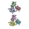
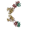
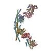
| Entry | Database: EMDB / ID: EMD-0347 | |||||||||
|---|---|---|---|---|---|---|---|---|---|---|
| Title | Apo form metabotropic glutamate receptor 5 with Nanobody 43 | |||||||||
 Map data Map data | Apo form metabotropic glutamate receptor 5 with Nanobody 43, primary map | |||||||||
 Sample Sample |
| |||||||||
| Function / homology |  Function and homology information Function and homology informationA2A adenosine receptor binding / sensory perception of hot stimulus / : / negative regulation of dendritic spine morphogenesis / G protein-coupled receptor activity involved in regulation of postsynaptic membrane potential / adenylate cyclase inhibiting G protein-coupled glutamate receptor activity / phospholipase C-activating G protein-coupled glutamate receptor signaling pathway / operant conditioning / protein localization to nuclear inner membrane / positive regulation of cellular response to hypoxia ...A2A adenosine receptor binding / sensory perception of hot stimulus / : / negative regulation of dendritic spine morphogenesis / G protein-coupled receptor activity involved in regulation of postsynaptic membrane potential / adenylate cyclase inhibiting G protein-coupled glutamate receptor activity / phospholipase C-activating G protein-coupled glutamate receptor signaling pathway / operant conditioning / protein localization to nuclear inner membrane / positive regulation of cellular response to hypoxia / positive regulation of long-term neuronal synaptic plasticity / positive regulation of sensory perception of pain / negative regulation of excitatory postsynaptic potential / positive regulation of dopamine secretion / desensitization of G protein-coupled receptor signaling pathway / G protein-coupled glutamate receptor signaling pathway / positive regulation of neural precursor cell proliferation / Class C/3 (Metabotropic glutamate/pheromone receptors) / glutamate receptor activity / nuclear inner membrane / Neurexins and neuroligins / astrocyte projection / response to morphine / response to corticosterone / temperature homeostasis / protein tyrosine kinase activator activity / conditioned place preference / regulation of synaptic transmission, glutamatergic / regulation of long-term synaptic depression / positive regulation of calcium-mediated signaling / protein tyrosine kinase binding / dendritic shaft / response to amphetamine / locomotory behavior / synapse organization / postsynaptic density membrane / G protein-coupled receptor activity / Schaffer collateral - CA1 synapse / cognition / cellular response to amyloid-beta / positive regulation of cytosolic calcium ion concentration / G alpha (q) signalling events / dendritic spine / chemical synaptic transmission / response to ethanol / learning or memory / positive regulation of MAPK cascade / response to antibiotic / neuronal cell body / regulation of DNA-templated transcription / dendrite / glutamatergic synapse / identical protein binding / plasma membrane / cytoplasm Similarity search - Function | |||||||||
| Biological species |  Homo sapiens (human) / Homo sapiens (human) /  | |||||||||
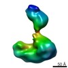
| Method | single particle reconstruction / cryo EM / Resolution: 7.9 Å | |||||||||
 Authors Authors | Koehl A / Hu H / Feng D / Zhang Y / Sun B / Kobilka TS / Pardon E / Steyaert J / Mathiesen JM / Skiniotis G / Kobilka BK | |||||||||
 Citation Citation | Journal: Nature / Year: 2019 Title: Structural insights into the activation of metabotropic glutamate receptors. Authors: Antoine Koehl / Hongli Hu / Dan Feng / Bingfa Sun / Yan Zhang / Michael J Robertson / Matthew Chu / Tong Sun Kobilka / Toon Laeremans / Jan Steyaert / Jeffrey Tarrasch / Somnath Dutta / ...Authors: Antoine Koehl / Hongli Hu / Dan Feng / Bingfa Sun / Yan Zhang / Michael J Robertson / Matthew Chu / Tong Sun Kobilka / Toon Laeremans / Jan Steyaert / Jeffrey Tarrasch / Somnath Dutta / Rasmus Fonseca / William I Weis / Jesper M Mathiesen / Georgios Skiniotis / Brian K Kobilka /     Abstract: Metabotropic glutamate receptors are family C G-protein-coupled receptors. They form obligate dimers and possess extracellular ligand-binding Venus flytrap domains, which are linked by cysteine-rich ...Metabotropic glutamate receptors are family C G-protein-coupled receptors. They form obligate dimers and possess extracellular ligand-binding Venus flytrap domains, which are linked by cysteine-rich domains to their 7-transmembrane domains. Spectroscopic studies show that signalling is a dynamic process, in which large-scale conformational changes underlie the transmission of signals from the extracellular Venus flytraps to the G protein-coupling domains-the 7-transmembrane domains-in the membrane. Here, using a combination of X-ray crystallography, cryo-electron microscopy and signalling studies, we present a structural framework for the activation mechanism of metabotropic glutamate receptor subtype 5. Our results show that agonist binding at the Venus flytraps leads to a compaction of the intersubunit dimer interface, thereby bringing the cysteine-rich domains into close proximity. Interactions between the cysteine-rich domains and the second extracellular loops of the receptor enable the rigid-body repositioning of the 7-transmembrane domains, which come into contact with each other to initiate signalling. | |||||||||
| History |
|
- Structure visualization
| Movie |
Movie viewer |
|---|---|
| Structure viewer | EM map: SurfViewMolmilJmol/JSmol |
| Supplemental images |

- Downloads & links
-EMDB archive
| Map data | emd_0347.map.gz | 2.6 MB | EMDB map data format | |
|---|---|---|---|---|
| Header (meta data) | emd-0347-v30.xmlemd-0347.xml | 14 KB 14 KB | Display Display | EMDB header |
| Images | 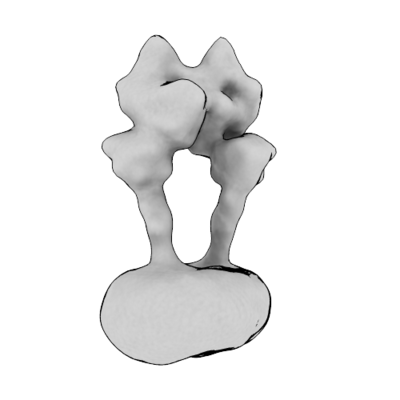 emd_0347.png emd_0347.png | 30.8 KB | ||
| Archive directory |  http://ftp.pdbj.org/pub/emdb/structures/EMD-0347ftp://ftp.pdbj.org/pub/emdb/structures/EMD-0347 http://ftp.pdbj.org/pub/emdb/structures/EMD-0347ftp://ftp.pdbj.org/pub/emdb/structures/EMD-0347 | HTTPS FTP |
-Related structure data
| Related structure data |  0345C 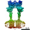 0346C 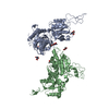 6n4xC 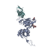 6n4yC 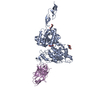 6n50C 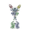 6n51C 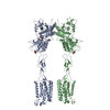 6n52C C: citing same article ( |
|---|---|
| Similar structure data |

-Links
| EMDB pages | EMDB (EBI/PDBe) / EMDataResource |
|---|---|
| Related items in Molecule of the Month |


-Map
| File | Download / File: emd_0347.map.gz / Format: CCP4 / Size: 6.6 MB / Type: IMAGE STORED AS FLOATING POINT NUMBER (4 BYTES) | ||||||||||||||||||||||||||||||||||||||||||||||||||||||||||||
|---|---|---|---|---|---|---|---|---|---|---|---|---|---|---|---|---|---|---|---|---|---|---|---|---|---|---|---|---|---|---|---|---|---|---|---|---|---|---|---|---|---|---|---|---|---|---|---|---|---|---|---|---|---|---|---|---|---|---|---|---|---|
| Annotation | Apo form metabotropic glutamate receptor 5 with Nanobody 43, primary map | ||||||||||||||||||||||||||||||||||||||||||||||||||||||||||||
| Projections & slices | Image control
Images are generated by Spider. | ||||||||||||||||||||||||||||||||||||||||||||||||||||||||||||
| Voxel size | X=Y=Z: 2.58 Å | ||||||||||||||||||||||||||||||||||||||||||||||||||||||||||||
| Density |
| ||||||||||||||||||||||||||||||||||||||||||||||||||||||||||||
| Symmetry | Space group: 1 | ||||||||||||||||||||||||||||||||||||||||||||||||||||||||||||
| Details | EMDB XML:
CCP4 map header:
| ||||||||||||||||||||||||||||||||||||||||||||||||||||||||||||
 Z (Sec.)
 Y (Row.)
 X (Col.)


-Supplemental data
- Sample components
-Entire : Metabotropic Glutamate Receptor 5 - Nb43 Complex
| Entire | Name: Metabotropic Glutamate Receptor 5 - Nb43 Complex |
|---|---|
| Components |
|
-Supramolecule #1: Metabotropic Glutamate Receptor 5 - Nb43 Complex
| Supramolecule | Name: Metabotropic Glutamate Receptor 5 - Nb43 Complex / type: complex / ID: 1 / Parent: 0 / Macromolecule list: all Details: Complex between metabotropic glutamate receptor 5 and Nb43 |
|---|---|
| Source (natural) | Organism: Homo sapiens (human) |
| Recombinant expression | Organism:   Spodoptera frugiperda (fall armyworm) / Recombinant strain: SF9 Spodoptera frugiperda (fall armyworm) / Recombinant strain: SF9 |
| Molecular weight | Theoretical: 220 KDa |
-Macromolecule #1: Metabotropic Glutamate Receptor 5
| Macromolecule | Name: Metabotropic Glutamate Receptor 5 / type: protein_or_peptide / ID: 1 Details: Polypeptide encoding metabotropic glutamate receptor 5 Enantiomer: LEVO |
|---|---|
| Source (natural) | Organism: Homo sapiens (human) |
| Recombinant expression | Organism: Spodoptera frugiperda (fall armyworm) |
| Sequence | String: QSSERRVVAH MPGDIIIGAL FSVHHQPTVD KVHERKCGAV REQYGIQRV EAMLHTLERI NSDPTLLPNI TLGCEIRDSC WHSAVALEQS IEFIRDSLIS S EEEEGLVR CVDGSSSSFR SKKPIVGVIG PGSSSVAIQV QNLLQLFNIP QIAYSATSMD LS DKTLFKY ...String: QSSERRVVAH MPGDIIIGAL FSVHHQPTVD KVHERKCGAV REQYGIQRV EAMLHTLERI NSDPTLLPNI TLGCEIRDSC WHSAVALEQS IEFIRDSLIS S EEEEGLVR CVDGSSSSFR SKKPIVGVIG PGSSSVAIQV QNLLQLFNIP QIAYSATSMD LS DKTLFKY FMRVVPSDAQ QARAMVDIVK RYNWTYVSAV HTEGNYGESG MEAFKDMSAK EGI CIAHSY KIYSNAGEQS FDKLLKKLTS HLPKARVVAC FCEGMTVRGL LMAMRRLGLA GEFL LLGSD GWADRYDVTD GYQREAVGGI TIKLQSPDVK WFDDYYLKLR PETNHRNPWF QEFWQ HRFQ CRLEGFPQEN SKYNKTCNSS LTLKTHHVQD SKMGFVINAI YSMAYGLHNM QMSLCP GYA GLCDAMKPID GRKLLESLMK TNFTGVSGDT ILFDENGDSP GRYEIMNFKE MGKDYFD YI NVGSWDNGEL KMDDDEVWSK KSNIIRSVCS EPCEKGQIKV IRKGEVSCCW TCTPCKEN E YVFDEYTCKA CQLGSWPTDD LTGCDLIPVQ YLRWGDPEPI AAVVFACLGL LATLFVTVV FIIYRDTPVV KSSSRELCYI ILAGICLGYL CTFCLIAKPK QIYCYLQRIG IGLSPAMSYS ALVTKTNRI ARILAGSKKK ICTKKPRFMS ACAQLVIAFI LICIQLGIIV ALFIMEPPDI M HDYPSIRE VYLICNTTNL GVVTPLGYNG LLILSCTFYA FKTRNVPANF NEAKYIAFTM YT TCIIWLA FVPIYFGSNY KIITMCFSVS LSATVALGCM FVPKVYIILA KPERNVRSAF TTS TVVRMH VGDGKSSSAA SRSSSLVNL |
-Macromolecule #2: Nanobody 43
| Macromolecule | Name: Nanobody 43 / type: protein_or_peptide / ID: 2 / Details: Nanobody 43 / Enantiomer: LEVO |
|---|---|
| Source (natural) | Organism: |
| Recombinant expression | Organism:  |
| Sequence | String: QVQLVESGGG LVQAGGSLRL SCAASGRTFT SYAMGWFRQA PGKERESVAA ISSSGGSTHY ADSVKGRFTI SRDNSKNTVY LQMNSLKPED TAVYYCAAAM YGSRWPDWEY DYWGQGTQVT VSSAA |
-Experimental details
-Structure determination
| Method | cryo EM |
|---|---|
 Processing Processing | single particle reconstruction |
| Aggregation state | particle |
-Sample preparation
| Concentration | 10 mg/mL | |||||||||||||||
|---|---|---|---|---|---|---|---|---|---|---|---|---|---|---|---|---|
| Buffer | pH: 7.5 Component:
| |||||||||||||||
| Grid | Model: Quantifoil R1.2/1.3 / Material: GOLD / Mesh: 200 / Pretreatment - Type: GLOW DISCHARGE | |||||||||||||||
| Vitrification | Cryogen name: ETHANE | |||||||||||||||
| Details | Sample was mono disperse as assayed by gel filtration and SDS PAGE. |
- Electron microscopy
| Microscope | FEI TITAN KRIOS |
|---|---|
| Image recording | Film or detector model: GATAN K2 SUMMIT (4k x 4k) / Detector mode: COUNTING / Average electron dose: 50.0 e/Å2 |
| Electron beam | Acceleration voltage: 300 kV / Electron source:  FIELD EMISSION GUN FIELD EMISSION GUN |
| Electron optics | C2 aperture diameter: 50.0 µm / Calibrated magnification: 58139 / Illumination mode: FLOOD BEAM / Imaging mode: BRIGHT FIELD / Nominal magnification: 29000 |
| Experimental equipment |  Model: Titan Krios / Image courtesy: FEI Company |
-Image processing
| Startup model | Type of model: OTHER / Details: The apo structure was used as starting model. |
|---|---|
| Final reconstruction | Resolution.type: BY AUTHOR / Resolution: 7.9 Å / Resolution method: FSC 0.143 CUT-OFF / Software - Name: RELION (ver. 2.1) / Number images used: 44831 |
| Initial angle assignment | Type: RANDOM ASSIGNMENT |
| Final angle assignment | Type: MAXIMUM LIKELIHOOD |
