Movie
Movie Controller

[English] 日本語
 Yorodumi
- EMDB-0112: Hinge region of the Membrane Attack Complex in the open conformation -
+ Open data

- Basic information
| Entry | Database: EMDB / ID: EMD-0112 | |||||||||
|---|---|---|---|---|---|---|---|---|---|---|
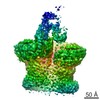
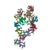
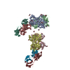
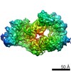
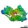
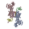
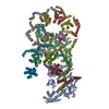
| Title | Hinge region of the Membrane Attack Complex in the open conformation | |||||||||
 Map data Map data | ||||||||||
 Sample Sample |
| |||||||||
| Biological species |  Homo sapiens (human) Homo sapiens (human) | |||||||||
| Method | single particle reconstruction / cryo EM / Resolution: 4.9 Å | |||||||||
 Authors Authors | Menny A / Serna M / Boyd CB / Gardener S / Joseph AP / Topf M / Bubeck D | |||||||||
 Citation Citation | Journal: Nat Commun / Year: 2018 Title: CryoEM reveals how the complement membrane attack complex ruptures lipid bilayers. Authors: Anaïs Menny / Marina Serna / Courtney M Boyd / Scott Gardner / Agnel Praveen Joseph / B Paul Morgan / Maya Topf / Nicholas J Brooks / Doryen Bubeck /   Abstract: The membrane attack complex (MAC) is one of the immune system's first responders. Complement proteins assemble on target membranes to form pores that lyse pathogens and impact tissue homeostasis of ...The membrane attack complex (MAC) is one of the immune system's first responders. Complement proteins assemble on target membranes to form pores that lyse pathogens and impact tissue homeostasis of self-cells. How MAC disrupts the membrane barrier remains unclear. Here we use electron cryo-microscopy and flicker spectroscopy to show that MAC interacts with lipid bilayers in two distinct ways. Whereas C6 and C7 associate with the outer leaflet and reduce the energy for membrane bending, C8 and C9 traverse the bilayer increasing membrane rigidity. CryoEM reconstructions reveal plasticity of the MAC pore and demonstrate how C5b6 acts as a platform, directing assembly of a giant β-barrel whose structure is supported by a glycan scaffold. Our work provides a structural basis for understanding how β-pore forming proteins breach the membrane and reveals a mechanism for how MAC kills pathogens and regulates cell functions. | |||||||||
| History |
|
- Structure visualization
| Movie |
 Movie viewer Movie viewer |
|---|---|
| Structure viewer | EM map: SurfViewMolmilJmol/JSmol |
| Supplemental images |

- Downloads & links
-EMDB archive
| Map data | emd_0112.map.gz | 102.3 MB | EMDB map data format | |
|---|---|---|---|---|
| Header (meta data) | emd-0112-v30.xmlemd-0112.xml | 16.1 KB 16.1 KB | Display Display | EMDB header |
| Images | 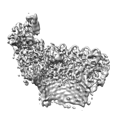 emd_0112.png emd_0112.png | 57 KB | ||
| Archive directory |  http://ftp.pdbj.org/pub/emdb/structures/EMD-0112ftp://ftp.pdbj.org/pub/emdb/structures/EMD-0112 http://ftp.pdbj.org/pub/emdb/structures/EMD-0112ftp://ftp.pdbj.org/pub/emdb/structures/EMD-0112 | HTTPS FTP |
-Related structure data
| Related structure data |  0106C 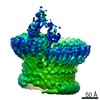 0107C 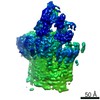 0109C 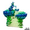 0110C  0111C  0113C  6h03C 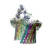 6h04C C: citing same article ( |
|---|---|
| Similar structure data |


-Links
| EMDB pages | EMDB (EBI/PDBe) / EMDataResource |
|---|
-Map
| File | Download / File: emd_0112.map.gz / Format: CCP4 / Size: 178 MB / Type: IMAGE STORED AS FLOATING POINT NUMBER (4 BYTES) | ||||||||||||||||||||||||||||||||||||||||||||||||||||||||||||
|---|---|---|---|---|---|---|---|---|---|---|---|---|---|---|---|---|---|---|---|---|---|---|---|---|---|---|---|---|---|---|---|---|---|---|---|---|---|---|---|---|---|---|---|---|---|---|---|---|---|---|---|---|---|---|---|---|---|---|---|---|---|
| Projections & slices | Image control
Images are generated by Spider. | ||||||||||||||||||||||||||||||||||||||||||||||||||||||||||||
| Voxel size | X=Y=Z: 1.384 Å | ||||||||||||||||||||||||||||||||||||||||||||||||||||||||||||
| Density |
| ||||||||||||||||||||||||||||||||||||||||||||||||||||||||||||
| Symmetry | Space group: 1 | ||||||||||||||||||||||||||||||||||||||||||||||||||||||||||||
| Details | EMDB XML:
CCP4 map header:
| ||||||||||||||||||||||||||||||||||||||||||||||||||||||||||||
 Z (Sec.)
 Y (Row.)
 X (Col.)


-Supplemental data
- Sample components
-Entire : Membrane Attack Complex
| Entire | Name: Membrane Attack Complex |
|---|---|
| Components |
|
-Supramolecule #1: Membrane Attack Complex
| Supramolecule | Name: Membrane Attack Complex / type: complex / ID: 1 / Parent: 0 / Macromolecule list: #1-#7 Details: Protein complex was assembled on liposomes and detergent solubilized |
|---|---|
| Source (natural) | Organism: Homo sapiens (human) |
| Molecular weight | Theoretical: 69 KDa |
-Supramolecule #2: C5b
| Supramolecule | Name: C5b / type: complex / ID: 2 / Parent: 1 / Macromolecule list: #1 / Details: Component of the Membrane Attack Complex |
|---|---|
| Source (natural) | Organism: Homo sapiens (human) |
-Supramolecule #3: C6
| Supramolecule | Name: C6 / type: complex / ID: 3 / Parent: 1 / Macromolecule list: #2 / Details: Component of the Membrane Attack Complex |
|---|---|
| Source (natural) | Organism: Homo sapiens (human) |
-Supramolecule #4: C7
| Supramolecule | Name: C7 / type: complex / ID: 4 / Parent: 1 / Macromolecule list: #4 / Details: Component of the Membrane Attack Complex |
|---|---|
| Source (natural) | Organism: Homo sapiens (human) |
-Supramolecule #5: C8 alpha
| Supramolecule | Name: C8 alpha / type: complex / ID: 5 / Parent: 1 / Macromolecule list: #6 / Details: Component of the Membrane Attack Complex |
|---|---|
| Source (natural) | Organism: Homo sapiens (human) |
-Supramolecule #6: C8 beta
| Supramolecule | Name: C8 beta / type: complex / ID: 6 / Parent: 1 / Macromolecule list: #3 / Details: Component of the Membrane Attack Complex |
|---|---|
| Source (natural) | Organism: Homo sapiens (human) |
-Supramolecule #7: C8 gamma
| Supramolecule | Name: C8 gamma / type: complex / ID: 7 / Parent: 1 / Macromolecule list: #5 / Details: Component of the Membrane Attack Complex |
|---|---|
| Source (natural) | Organism: Homo sapiens (human) |
-Supramolecule #8: C9
| Supramolecule | Name: C9 / type: complex / ID: 8 / Parent: 1 / Macromolecule list: #7 / Details: Component of the Membrane Attack Complex |
|---|---|
| Source (natural) | Organism: Homo sapiens (human) |
-Experimental details
-Structure determination
| Method | cryo EM |
|---|---|
 Processing Processing | single particle reconstruction |
| Aggregation state | particle |
-Sample preparation
| Buffer | pH: 7.4 |
|---|---|
| Vitrification | Cryogen name: ETHANE |
- Electron microscopy
| Microscope | FEI TITAN KRIOS |
|---|---|
| Image recording | Film or detector model: FEI FALCON II (4k x 4k) / Detector mode: COUNTING / Number grids imaged: 8 / Number real images: 13009 / Average exposure time: 2.0 sec. / Average electron dose: 50.0 e/Å2 |
| Electron beam | Acceleration voltage: 300 kV / Electron source:  FIELD EMISSION GUN FIELD EMISSION GUN |
| Electron optics | Illumination mode: SPOT SCAN / Imaging mode: BRIGHT FIELD / Nominal magnification: 59000 |
| Sample stage | Cooling holder cryogen: NITROGEN |
| Experimental equipment |  Model: Titan Krios / Image courtesy: FEI Company |