 Movie
Movie Controller


+ Open data

- Basic information
| Entry | Database: PDB / ID: 8trs | |||||||||
|---|---|---|---|---|---|---|---|---|---|---|
| Title | Structure of the EphA2 CRD bound to FabS1CE_C1, trigonal form | |||||||||
 Components Components |
| |||||||||
 Keywords Keywords |  SIGNALING PROTEIN/IMMUNE SYSTEM / Cell signalling / antibody / agonist / high affinity / clustering / activation / SIGNALING PROTEIN-IMMUNE SYSTEM complex SIGNALING PROTEIN/IMMUNE SYSTEM / Cell signalling / antibody / agonist / high affinity / clustering / activation / SIGNALING PROTEIN-IMMUNE SYSTEM complex | |||||||||
| Function / homology |  Function and homology information Function and homology informationnotochord cell development / notochord formation / lens fiber cell morphogenesis / blood vessel endothelial cell proliferation involved in sprouting angiogenesis / negative regulation of lymphangiogenesis / axial mesoderm formation / pericyte cell differentiation / cAMP metabolic process / positive regulation of bicellular tight junction assembly / regulation of blood vessel endothelial cell migration ...notochord cell development / notochord formation / lens fiber cell morphogenesis / blood vessel endothelial cell proliferation involved in sprouting angiogenesis / negative regulation of lymphangiogenesis / axial mesoderm formation / pericyte cell differentiation / cAMP metabolic process / positive regulation of bicellular tight junction assembly / regulation of blood vessel endothelial cell migration / leading edge membrane / negative regulation of chemokine production / bone remodeling / transmembrane-ephrin receptor activity / post-anal tail morphogenesis / response to growth factor / activation of GTPase activity / regulation of lamellipodium assembly / tight junction / branching involved in mammary gland duct morphogenesis / EPH-Ephrin signaling / neural tube development / RND1 GTPase cycle / RND2 GTPase cycle / RND3 GTPase cycle / mammary gland epithelial cell proliferation / RHOV GTPase cycle / EPHA-mediated growth cone collapse / growth factor binding / regulation of cell adhesion mediated by integrin / lamellipodium membrane / RHOU GTPase cycle / RHOG GTPase cycle / EPH-ephrin mediated repulsion of cells / RAC2 GTPase cycle / ephrin receptor signaling pathway / RAC3 GTPase cycle / negative regulation of phosphatidylinositol 3-kinase/protein kinase B signal transduction / vasculogenesis / regulation of angiogenesis / keratinocyte differentiation / RAC1 GTPase cycle / transmembrane receptor protein tyrosine kinase activity / cell chemotaxis / negative regulation of angiogenesis / osteoclast differentiation / regulation of ERK1 and ERK2 cascade / phosphatidylinositol 3-kinase/protein kinase B signal transduction / skeletal system development / molecular function activator activity / protein localization to plasma membrane / cell motility / positive regulation of protein localization to plasma membrane / axon guidance / receptor protein-tyrosine kinase / ruffle membrane / osteoblast differentiation / intrinsic apoptotic signaling pathway in response to DNA damage / cell migration / virus receptor activity / lamellipodium / receptor complex / cell adhesion / positive regulation of cell migration / defense response to Gram-positive bacterium / cadherin binding / inflammatory response / phosphorylation / focal adhesion / dendrite / cell surface / ATP binding / plasma membraneSimilarity search - Function | |||||||||
| Biological species |  Homo sapiens (human) Homo sapiens (human) | |||||||||
| Method | X-RAY DIFFRACTION / SYNCHROTRON / MOLECULAR REPLACEMENT / Resolution: 1.9 Å | |||||||||
 Authors Authors | Singer, A.U. / Bruce, H.A. / Blazer, L. / Adams, J.J. / Sicheri, F. / Sidhu, S.S. | |||||||||
| Funding support |  United States, 2items United States, 2items
| |||||||||
 Citation Citation | Journal: Protein Sci. / Year: 2024 Title: Engineered antigen-binding fragments for enhanced crystallization of antibody:antigen complexes. Authors: Bruce, H.A. / Singer, A.U. / Filippova, E.V. / Blazer, L.L. / Adams, J.J. / Enderle, L. / Ben-David, M. / Radley, E.H. / Mao, D.Y.L. / Pau, V. / Orlicky, S. / Sicheri, F. / Kurinov, I. / ...Authors: Bruce, H.A. / Singer, A.U. / Filippova, E.V. / Blazer, L.L. / Adams, J.J. / Enderle, L. / Ben-David, M. / Radley, E.H. / Mao, D.Y.L. / Pau, V. / Orlicky, S. / Sicheri, F. / Kurinov, I. / Atwell, S. / Kossiakoff, A.A. / Sidhu, S.S. | |||||||||
| History |
|
- Structure visualization
| Structure viewer | Molecule: MolmilJmol/JSmol |
|---|
- Downloads & links
-Download
| PDBx/mmCIF format | 8trs.cif.gz | 238.5 KB | Display | PDBx/mmCIF format |
|---|---|---|---|---|
| PDB format | pdb8trs.ent.gz | 190.2 KB | Display | PDB format |
| PDBx/mmJSON format | 8trs.json.gz | Tree view | PDBx/mmJSON format | |
| Others |  Other downloads Other downloads |
-Validation report
| Arichive directory | https://data.pdbj.org/pub/pdb/validation_reports/tr/8trsftp://data.pdbj.org/pub/pdb/validation_reports/tr/8trs | HTTPS FTP |
|---|
-Related structure data
| Related structure data |  8t58C  8t6iC  8t7fC  8t7gC  8t7iC 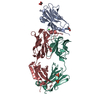 8t8iC 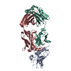 8t9yC  8trtC 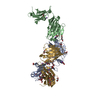 8ts5C  3fl7S  8trv C: citing same article ( S: Starting model for refinement |
|---|---|
| Similar structure data |
-Links
 PDBj
PDBj


- Assembly
| Deposited unit | 
| ||||||||||
|---|---|---|---|---|---|---|---|---|---|---|---|
| 1 |
| ||||||||||
| Unit cell |
|
-Components
-Protein , 1 types, 1 molecules D
| #3: Protein | Mass: 13961.746 Da / Num. of mol.: 1 Source method: isolated from a genetically manipulated source Details: residues 203-330 / Source: (gene. exp.) Homo sapiens (human) / Gene: EPHA2, ECK / Plasmid: pSCSTaDetails (production host): expressed with a C-terminal thrombin cleavage site and 6-his sequence. The residues LTPR at the C-terminus are derived from the thrombin cleavage site Cell line (production host): Expi293 / Production host: Homo sapiens (human)References: UniProt: P29317, receptor protein-tyrosine kinase |
|---|
-Antibody , 2 types, 2 molecules AG
| #1: Antibody | Mass: 23883.762 Da / Num. of mol.: 1 / Mutation: SSASTK replaced by FNQIK Source method: isolated from a genetically manipulated source Details: Fab produced by randomization of CDR regions and selected by phage display. Fabs utilize IMGT (LeClerc et al., Dev Comp Immunol. 2003 Jan;27(1):55-77) numbering. Source: (gene. exp.) Homo sapiens (human) / Plasmid: pSCSTa / Cell line (production host): Expi293 / Production host: Homo sapiens (human) |
|---|---|
| #2: Antibody | Mass: 23432.895 Da / Num. of mol.: 1 / Mutation: SPHAGLSSP replaced by QGTTS; Q165S, K167Y Source method: isolated from a genetically manipulated source Details: Fab produced by randomization of CDR regions and selected by phage display. Fabs utilize IMGT (LeClerc et al., Dev Comp Immunol. 2003 Jan;27(1):55-77) numbering. Source: (gene. exp.) Homo sapiens (human) / Plasmid: pSCSTa / Cell line (production host): Expi293 / Production host: Homo sapiens (human) |
-Non-polymers , 5 types, 281 molecules 


| #4: Chemical | ChemComp-EDO / Ethylene glycol Mass: 62.068 Da / Num. of mol.: 19 / Source method: obtained synthetically / Formula: C2H6O2 Mass: 62.068 Da / Num. of mol.: 19 / Source method: obtained synthetically / Formula: C2H6O2#5: Chemical | ChemComp-PEG / Diethylene glycol Mass: 106.120 Da / Num. of mol.: 7 / Source method: obtained synthetically / Formula: C4H10O3 Mass: 106.120 Da / Num. of mol.: 7 / Source method: obtained synthetically / Formula: C4H10O3#6: Chemical | ChemComp-CL / Chloride Mass: 35.453 Da / Num. of mol.: 17 / Source method: obtained synthetically / Formula: Cl Mass: 35.453 Da / Num. of mol.: 17 / Source method: obtained synthetically / Formula: Cl#7: Chemical | ChemComp-NA / |  Mass: 22.990 Da / Num. of mol.: 1 / Source method: obtained synthetically / Formula: Na Mass: 22.990 Da / Num. of mol.: 1 / Source method: obtained synthetically / Formula: Na#8: Water | ChemComp-HOH / | WaterMass: 18.015 Da / Num. of mol.: 237 / Source method: isolated from a natural source / Formula: H2O |
|---|
-Details
| Has ligand of interest | Y |
|---|
-Experimental details
-Experiment
| Experiment | Method: X-RAY DIFFRACTION / Number of used crystals: 1 |
|---|
- Sample preparation
| Crystal | Density Matthews: 2.82 Å3/Da / Density % sol: 56.39 % |
|---|---|
| Crystal grow | Temperature: 298 K / Method: vapor diffusion, sitting drop / pH: 5.5 Details: 200 mM ammonium chloride, 30% PEG600 (GRAS1 condition A8) Temp details: room temperature |
-Data collection
| Diffraction | Mean temperature: 100 K / Serial crystal experiment: N |
|---|---|
| Diffraction source | Source: SYNCHROTRON / Site: APS / Beamline: 24-ID-E / Wavelength: 0.97918 Å |
| Detector | Type: DECTRIS EIGER X 16M / Detector: PIXEL / Date: Apr 5, 2022 / Details: Mirrors |
| Radiation | Protocol: SINGLE WAVELENGTH / Monochromatic (M) / Laue (L): M / Scattering type: x-ray |
| Radiation wavelength | Wavelength: 0.97918 Å / Relative weight: 1 |
| Reflection | Resolution: 1.85→117.51 Å / Num. obs: 60648 / % possible obs: 100 % / Redundancy: 11.5 % / Biso Wilson estimate: 41.8 Å2 / CC1/2: 1 / Rmerge(I) obs: 0.05 / Rpim(I) all: 0.015 / Rrim(I) all: 0.052 / Net I/σ(I): 28.7 |
| Reflection shell | Resolution: 1.85→1.89 Å / Rmerge(I) obs: 1.516 / Mean I/σ(I) obs: 1.6 / Num. unique obs: 3633 / CC1/2: 0.692 / Rpim(I) all: 0.458 / Rrim(I) all: 1.585 / % possible all: 99.3 |
- Processing
| Software |
| |||||||||||||||||||||||||||||||||||||||||||||||||||||||||||||||||||||||||||||||||||||||||||||||||||||||||||||||||||||||||||||||||||||||||||||||||||||||||||||||||||||||||||||||||||||||||||||||||||||||||||||||||||||||||||||||||||||||||||||||||||||||||||||||||||||||||||||||||||||||||||||||||||||||||||||||||||||||||||||||||||||||||||||||||||||||||||||||||||||||||||||||||||||||||||||||||||||||||||||||||||||||||||||||||||||||||
|---|---|---|---|---|---|---|---|---|---|---|---|---|---|---|---|---|---|---|---|---|---|---|---|---|---|---|---|---|---|---|---|---|---|---|---|---|---|---|---|---|---|---|---|---|---|---|---|---|---|---|---|---|---|---|---|---|---|---|---|---|---|---|---|---|---|---|---|---|---|---|---|---|---|---|---|---|---|---|---|---|---|---|---|---|---|---|---|---|---|---|---|---|---|---|---|---|---|---|---|---|---|---|---|---|---|---|---|---|---|---|---|---|---|---|---|---|---|---|---|---|---|---|---|---|---|---|---|---|---|---|---|---|---|---|---|---|---|---|---|---|---|---|---|---|---|---|---|---|---|---|---|---|---|---|---|---|---|---|---|---|---|---|---|---|---|---|---|---|---|---|---|---|---|---|---|---|---|---|---|---|---|---|---|---|---|---|---|---|---|---|---|---|---|---|---|---|---|---|---|---|---|---|---|---|---|---|---|---|---|---|---|---|---|---|---|---|---|---|---|---|---|---|---|---|---|---|---|---|---|---|---|---|---|---|---|---|---|---|---|---|---|---|---|---|---|---|---|---|---|---|---|---|---|---|---|---|---|---|---|---|---|---|---|---|---|---|---|---|---|---|---|---|---|---|---|---|---|---|---|---|---|---|---|---|---|---|---|---|---|---|---|---|---|---|---|---|---|---|---|---|---|---|---|---|---|---|---|---|---|---|---|---|---|---|---|---|---|---|---|---|---|---|---|---|---|---|---|---|---|---|---|---|---|---|---|---|---|---|---|---|---|---|---|---|---|---|---|---|---|---|---|---|---|---|---|---|---|---|---|---|---|---|---|---|---|---|---|---|---|---|---|---|---|---|---|---|---|---|---|---|---|---|---|---|---|---|---|---|---|---|---|---|---|---|---|---|---|---|---|---|---|---|---|---|---|---|---|---|---|---|---|---|---|---|---|---|---|---|---|---|---|---|---|---|---|---|
| Refinement | Method to determine structure: MOLECULAR REPLACEMENT Starting model: PDB entry 3FL7 Resolution: 1.9→61.81 Å / SU ML: 0.229 / Cross valid method: FREE R-VALUE / σ(F): 1.34 / Phase error: 22.437 Stereochemistry target values: GEOSTD + MONOMER LIBRARY + CDL V1.2
| |||||||||||||||||||||||||||||||||||||||||||||||||||||||||||||||||||||||||||||||||||||||||||||||||||||||||||||||||||||||||||||||||||||||||||||||||||||||||||||||||||||||||||||||||||||||||||||||||||||||||||||||||||||||||||||||||||||||||||||||||||||||||||||||||||||||||||||||||||||||||||||||||||||||||||||||||||||||||||||||||||||||||||||||||||||||||||||||||||||||||||||||||||||||||||||||||||||||||||||||||||||||||||||||||||||||||
| Solvent computation | Shrinkage radii: 0.9 Å / VDW probe radii: 1.11 Å / Solvent model: FLAT BULK SOLVENT MODEL | |||||||||||||||||||||||||||||||||||||||||||||||||||||||||||||||||||||||||||||||||||||||||||||||||||||||||||||||||||||||||||||||||||||||||||||||||||||||||||||||||||||||||||||||||||||||||||||||||||||||||||||||||||||||||||||||||||||||||||||||||||||||||||||||||||||||||||||||||||||||||||||||||||||||||||||||||||||||||||||||||||||||||||||||||||||||||||||||||||||||||||||||||||||||||||||||||||||||||||||||||||||||||||||||||||||||||
| Displacement parameters | Biso mean: 50.17 Å2 | |||||||||||||||||||||||||||||||||||||||||||||||||||||||||||||||||||||||||||||||||||||||||||||||||||||||||||||||||||||||||||||||||||||||||||||||||||||||||||||||||||||||||||||||||||||||||||||||||||||||||||||||||||||||||||||||||||||||||||||||||||||||||||||||||||||||||||||||||||||||||||||||||||||||||||||||||||||||||||||||||||||||||||||||||||||||||||||||||||||||||||||||||||||||||||||||||||||||||||||||||||||||||||||||||||||||||
| Refinement step | Cycle: LAST / Resolution: 1.9→61.81 Å
| |||||||||||||||||||||||||||||||||||||||||||||||||||||||||||||||||||||||||||||||||||||||||||||||||||||||||||||||||||||||||||||||||||||||||||||||||||||||||||||||||||||||||||||||||||||||||||||||||||||||||||||||||||||||||||||||||||||||||||||||||||||||||||||||||||||||||||||||||||||||||||||||||||||||||||||||||||||||||||||||||||||||||||||||||||||||||||||||||||||||||||||||||||||||||||||||||||||||||||||||||||||||||||||||||||||||||
| Refine LS restraints |
| |||||||||||||||||||||||||||||||||||||||||||||||||||||||||||||||||||||||||||||||||||||||||||||||||||||||||||||||||||||||||||||||||||||||||||||||||||||||||||||||||||||||||||||||||||||||||||||||||||||||||||||||||||||||||||||||||||||||||||||||||||||||||||||||||||||||||||||||||||||||||||||||||||||||||||||||||||||||||||||||||||||||||||||||||||||||||||||||||||||||||||||||||||||||||||||||||||||||||||||||||||||||||||||||||||||||||
| LS refinement shell |
| |||||||||||||||||||||||||||||||||||||||||||||||||||||||||||||||||||||||||||||||||||||||||||||||||||||||||||||||||||||||||||||||||||||||||||||||||||||||||||||||||||||||||||||||||||||||||||||||||||||||||||||||||||||||||||||||||||||||||||||||||||||||||||||||||||||||||||||||||||||||||||||||||||||||||||||||||||||||||||||||||||||||||||||||||||||||||||||||||||||||||||||||||||||||||||||||||||||||||||||||||||||||||||||||||||||||||
| Refinement TLS params. | Method: refined / Refine-ID: X-RAY DIFFRACTION
| |||||||||||||||||||||||||||||||||||||||||||||||||||||||||||||||||||||||||||||||||||||||||||||||||||||||||||||||||||||||||||||||||||||||||||||||||||||||||||||||||||||||||||||||||||||||||||||||||||||||||||||||||||||||||||||||||||||||||||||||||||||||||||||||||||||||||||||||||||||||||||||||||||||||||||||||||||||||||||||||||||||||||||||||||||||||||||||||||||||||||||||||||||||||||||||||||||||||||||||||||||||||||||||||||||||||||
| Refinement TLS group |
|