 Movie
Movie Controller


+ Open data

- Basic information
| Entry | Database: PDB / ID: 3fl7 | ||||||
|---|---|---|---|---|---|---|---|
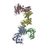
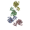
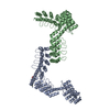
| Title | Crystal structure of the human ephrin A2 ectodomain | ||||||
 Components Components | Ephrin receptor | ||||||
 Keywords Keywords | TRANSFERASE / SIGNALING PROTEIN / ATP-BINDING / KINASE / NUCLEOTIDE-BINDING / RECEPTOR / PHOSPHORYLATION / TRANSMEMBRANE / TYROSINE-PROTEIN KINASE / GLYCOPROTEIN / LIGAND BINDING DOMAIN / CYSTEINE-RICH DOMAIN / SUSHI DOMAIN / EGF-LIKE MOTIF / FIBRONECTIN DOMAIN / STRUCTURAL GENOMICS CONSORTIUM / SGC / Membrane / Phosphoprotein | ||||||
| Function / homology |  Function and homology information Function and homology informationtransmembrane-ephrin receptor activity / notochord cell development / notochord formation / lens fiber cell morphogenesis / blood vessel endothelial cell proliferation involved in sprouting angiogenesis / negative regulation of lymphangiogenesis / axial mesoderm formation / multicellular organism development / pericyte cell differentiation / cAMP metabolic process ...transmembrane-ephrin receptor activity / notochord cell development / notochord formation / lens fiber cell morphogenesis / blood vessel endothelial cell proliferation involved in sprouting angiogenesis / negative regulation of lymphangiogenesis / axial mesoderm formation / multicellular organism development / pericyte cell differentiation / cAMP metabolic process / positive regulation of bicellular tight junction assembly / regulation of blood vessel endothelial cell migration / negative regulation of chemokine production / ephrin receptor activity / leading edge membrane / bone remodeling / post-anal tail morphogenesis / response to growth factor / activation of GTPase activity / positive regulation of kinase activity / regulation of lamellipodium assembly / tight junction / branching involved in mammary gland duct morphogenesis / EPH-Ephrin signaling / neural tube development / RND1 GTPase cycle / RND2 GTPase cycle / RND3 GTPase cycle / mammary gland epithelial cell proliferation / RHOV GTPase cycle / EPHA-mediated growth cone collapse / growth factor binding / plasma membrane => GO:0005886 / regulation of cell adhesion mediated by integrin / lamellipodium membrane / RHOU GTPase cycle / RHOG GTPase cycle / EPH-ephrin mediated repulsion of cells / negative regulation of phosphatidylinositol 3-kinase/protein kinase B signal transduction / RAC3 GTPase cycle / RAC2 GTPase cycle / ephrin receptor signaling pathway / vasculogenesis / regulation of angiogenesis / keratinocyte differentiation / RAC1 GTPase cycle / transmembrane receptor protein tyrosine kinase activity / cell chemotaxis / negative regulation of angiogenesis / osteoclast differentiation / regulation of ERK1 and ERK2 cascade / phosphatidylinositol 3-kinase/protein kinase B signal transduction / skeletal system development / molecular function activator activity / cell motility / axon guidance / protein localization to plasma membrane / positive regulation of protein localization to plasma membrane / receptor protein-tyrosine kinase / neuron differentiation / ruffle membrane / osteoblast differentiation / cell surface receptor protein tyrosine kinase signaling pathway / intrinsic apoptotic signaling pathway in response to DNA damage / cell migration / virus receptor activity / lamellipodium / receptor complex / cell adhesion / neuron projection / positive regulation of cell migration / defense response to Gram-positive bacterium / cadherin binding / inflammatory response / phosphorylation / focal adhesion / cell surface / ATP binding / plasma membraneSimilarity search - Function | ||||||
| Biological species |  Homo sapiens (human) Homo sapiens (human) | ||||||
| Method | X-RAY DIFFRACTION / SYNCHROTRON / MOLECULAR REPLACEMENT / Resolution: 2.5 Å | ||||||
 Authors Authors | Walker, J.R. / Yermekbayeva, L. / Seitova, A. / Butler-Cole, C. / Bountra, C. / Weigelt, J. / Arrowsmith, C.H. / Edwards, A.M. / Bochkarev, A. / Dhe-Paganon, S. / Structural Genomics Consortium (SGC) | ||||||
 Citation Citation | Journal: Proc.Natl.Acad.Sci.USA / Year: 2010 Title: Architecture of Eph receptor clusters. Authors: Himanen, J.P. / Yermekbayeva, L. / Janes, P.W. / Walker, J.R. / Xu, K. / Atapattu, L. / Rajashankar, K.R. / Mensinga, A. / Lackmann, M. / Nikolov, D.B. / Dhe-Paganon, S. #1: Journal: J.Cell Biol. / Year: 2004 Title: Recruitment of Eph receptors into signaling clusters does not require ephrin contact. Authors: Wimmer-Kleikamp, S.H. / Janes, P.W. / Squire, A. / Bastiaens, P.I. / Lackmann, M. #2: Journal: J.Biol.Chem. / Year: 1998 Title: Distinct subdomains of the EphA3 receptor mediate ligand binding and receptor dimerization. Authors: Lackmann, M. / Oates, A.C. / Dottori, M. / Smith, F.M. / Do, C. / Power, M. / Kravets, L. / Boyd, A.W. #3: Journal: Nature / Year: 2001Title: Crystal structure of an Eph receptor-ephrin complex. Authors: Himanen, J.P. / Rajashankar, K.R. / Lackmann, M. / Cowan, C.A. / Henkemeyer, M. / Nikolov, D.B. | ||||||
| History |
|
- Structure visualization
| Structure viewer | Molecule: MolmilJmol/JSmol |
|---|
- Downloads & links
-Download
| PDBx/mmCIF format | 3fl7.cif.gz | 202.4 KB | Display | PDBx/mmCIF format |
|---|---|---|---|---|
| PDB format | pdb3fl7.ent.gz | 161.5 KB | Display | PDB format |
| PDBx/mmJSON format | 3fl7.json.gz | Tree view | PDBx/mmJSON format | |
| Others |  Other downloads Other downloads |
-Validation report
| Arichive directory | https://data.pdbj.org/pub/pdb/validation_reports/fl/3fl7ftp://data.pdbj.org/pub/pdb/validation_reports/fl/3fl7 | HTTPS FTP |
|---|
-Related structure data
| Related structure data |  3c8xSC  3czuC  3mbwC  3mx0C  2e7hS S: Starting model for refinement C: citing same article ( |
|---|---|
| Similar structure data |

-Links
 PDBj
PDBj


- Assembly
| Deposited unit | 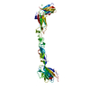
| ||||||||
|---|---|---|---|---|---|---|---|---|---|
| 1 | 
| ||||||||
| 2 |
| ||||||||
| Unit cell |
|
-Components
| #1: Protein | Mass: 59218.930 Da / Num. of mol.: 1 / Fragment: Ectodomain, UNP residues 23-531 Source method: isolated from a genetically manipulated source Source: (gene. exp.) Homo sapiens (human) / Gene: ECK, EPHA2, hCG_24712, RP11-276H7.1-001 / Plasmid: pFHMSP-LIC-N / Production host:   Spodoptera frugiperda (fall armyworm) / Strain (production host): SF9 Spodoptera frugiperda (fall armyworm) / Strain (production host): SF9References: UniProt: Q8N3Z2, UniProt: P29317*PLUS, receptor protein-tyrosine kinase | ||||
|---|---|---|---|---|---|
| #2: Chemical | ChemComp-NA /   Mass: 22.990 Da / Num. of mol.: 1 / Source method: obtained synthetically / Formula: Na Mass: 22.990 Da / Num. of mol.: 1 / Source method: obtained synthetically / Formula: Na | ||||
| #3: Chemical | Chloride  Mass: 35.453 Da / Num. of mol.: 3 / Source method: obtained synthetically / Formula: Cl Mass: 35.453 Da / Num. of mol.: 3 / Source method: obtained synthetically / Formula: Cl#4: Sugar | ChemComp-NAG / | N-Acetylglucosamine  Type: D-saccharide, beta linking / Mass: 221.208 Da / Num. of mol.: 1 Type: D-saccharide, beta linking / Mass: 221.208 Da / Num. of mol.: 1Source method: isolated from a genetically manipulated source Formula: C8H15NO6 #5: Water | ChemComp-HOH / | Water Mass: 18.015 Da / Num. of mol.: 46 / Source method: isolated from a natural source / Formula: H2O Mass: 18.015 Da / Num. of mol.: 46 / Source method: isolated from a natural source / Formula: H2O |
-Experimental details
-Experiment
| Experiment | Method: X-RAY DIFFRACTION / Number of used crystals: 1 |
|---|
- Sample preparation
| Crystal | Density Matthews: 3.08 Å3/Da / Density % sol: 60.03 % |
|---|---|
| Crystal grow | Temperature: 290.9 K / Method: vapor diffusion, hanging drop / pH: 5.5 Details: 3.0% PEG 4000, 0.1M Sodium acetate, 0.1 M Cacodylate pH 5.5, 0.5 M NDSB 256, VAPOR DIFFUSION, HANGING DROP, temperature 290.9K |
-Data collection
| Diffraction | Mean temperature: 100 K |
|---|---|
| Diffraction source | Source: SYNCHROTRON / Site: APS  / Beamline: 23-ID-B / Wavelength: 0.97948 Å / Beamline: 23-ID-B / Wavelength: 0.97948 Å |
| Detector | Type: MARMOSAIC 300 mm CCD / Detector: CCD / Date: Nov 20, 2008 / Details: Mirrors |
| Radiation | Monochromator: DOUBLE CRYSTAL / Protocol: SINGLE WAVELENGTH / Monochromatic (M) / Laue (L): M / Scattering type: x-ray |
| Radiation wavelength | Wavelength: 0.97948 Å / Relative weight: 1 |
| Reflection | Resolution: 2.5→41 Å / Num. obs: 25966 / % possible obs: 99.7 % / Observed criterion σ(I): -3 / Redundancy: 4.1 % / Rsym value: 0.07 / Net I/σ(I): 22.43 |
| Reflection shell | Resolution: 2.5→2.59 Å / Redundancy: 4 % / Mean I/σ(I) obs: 2.94 / Num. unique all: 2510 / Rsym value: 0.375 / % possible all: 98.1 |
- Processing
| Software |
| ||||||||||||||||||||||||||||||||||||||||||||||||||||||||||||||||||||||||||||||||||||||||||||||||||||||||||||||||||||||||||||||||||||||||||||||||||||||||||||||||||||||||||||||||||||||||||||||||||||||||
|---|---|---|---|---|---|---|---|---|---|---|---|---|---|---|---|---|---|---|---|---|---|---|---|---|---|---|---|---|---|---|---|---|---|---|---|---|---|---|---|---|---|---|---|---|---|---|---|---|---|---|---|---|---|---|---|---|---|---|---|---|---|---|---|---|---|---|---|---|---|---|---|---|---|---|---|---|---|---|---|---|---|---|---|---|---|---|---|---|---|---|---|---|---|---|---|---|---|---|---|---|---|---|---|---|---|---|---|---|---|---|---|---|---|---|---|---|---|---|---|---|---|---|---|---|---|---|---|---|---|---|---|---|---|---|---|---|---|---|---|---|---|---|---|---|---|---|---|---|---|---|---|---|---|---|---|---|---|---|---|---|---|---|---|---|---|---|---|---|---|---|---|---|---|---|---|---|---|---|---|---|---|---|---|---|---|---|---|---|---|---|---|---|---|---|---|---|---|---|---|---|---|
| Refinement | Method to determine structure: MOLECULAR REPLACEMENT Starting model: PDB entries 3C8X, 2E7H Resolution: 2.5→40.59 Å / Cor.coef. Fo:Fc: 0.927 / Cor.coef. Fo:Fc free: 0.895 / SU B: 25.979 / SU ML: 0.259 / Cross valid method: THROUGHOUT / ESU R: 0.398 / ESU R Free: 0.3 / Stereochemistry target values: MAXIMUM LIKELIHOOD Details: HYDROGENS HAVE BEEN ADDED IN THE RIDING POSITIONS. ATOM RECORD CONTAINS SUM OF TLS AND RESIDUAL B FACTORS. ANISOU RECORD CONTAINS SUM OF TLS AND RESIDUAL U FACTORS
| ||||||||||||||||||||||||||||||||||||||||||||||||||||||||||||||||||||||||||||||||||||||||||||||||||||||||||||||||||||||||||||||||||||||||||||||||||||||||||||||||||||||||||||||||||||||||||||||||||||||||
| Solvent computation | Ion probe radii: 0.8 Å / Shrinkage radii: 0.8 Å / VDW probe radii: 1.2 Å / Solvent model: BABINET MODEL WITH MASK | ||||||||||||||||||||||||||||||||||||||||||||||||||||||||||||||||||||||||||||||||||||||||||||||||||||||||||||||||||||||||||||||||||||||||||||||||||||||||||||||||||||||||||||||||||||||||||||||||||||||||
| Displacement parameters | Biso mean: 35.468 Å2
| ||||||||||||||||||||||||||||||||||||||||||||||||||||||||||||||||||||||||||||||||||||||||||||||||||||||||||||||||||||||||||||||||||||||||||||||||||||||||||||||||||||||||||||||||||||||||||||||||||||||||
| Refinement step | Cycle: LAST / Resolution: 2.5→40.59 Å
| ||||||||||||||||||||||||||||||||||||||||||||||||||||||||||||||||||||||||||||||||||||||||||||||||||||||||||||||||||||||||||||||||||||||||||||||||||||||||||||||||||||||||||||||||||||||||||||||||||||||||
| Refine LS restraints |
| ||||||||||||||||||||||||||||||||||||||||||||||||||||||||||||||||||||||||||||||||||||||||||||||||||||||||||||||||||||||||||||||||||||||||||||||||||||||||||||||||||||||||||||||||||||||||||||||||||||||||
| LS refinement shell | Resolution: 2.5→2.565 Å / Total num. of bins used: 20
| ||||||||||||||||||||||||||||||||||||||||||||||||||||||||||||||||||||||||||||||||||||||||||||||||||||||||||||||||||||||||||||||||||||||||||||||||||||||||||||||||||||||||||||||||||||||||||||||||||||||||
| Refinement TLS params. | Method: refined / Refine-ID: X-RAY DIFFRACTION
| ||||||||||||||||||||||||||||||||||||||||||||||||||||||||||||||||||||||||||||||||||||||||||||||||||||||||||||||||||||||||||||||||||||||||||||||||||||||||||||||||||||||||||||||||||||||||||||||||||||||||
| Refinement TLS group |
|
