 Movie
Movie Controller

[English] 日本語
 Yorodumi
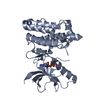
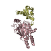
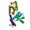
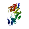
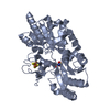
- PDB-3c8x: Crystal structure of the ligand binding domain of human Ephrin A2... -
+ Open data

- Basic information
| Entry | Database: PDB / ID: 3c8x | ||||||
|---|---|---|---|---|---|---|---|
| Title | Crystal structure of the ligand binding domain of human Ephrin A2 (Epha2) receptor protein kinase | ||||||
 Components Components | Ephrin type-A receptor 2 | ||||||
 Keywords Keywords |  TRANSFERASE / ATP-BINDING / KINASE / NUCLEOTIDE-BINDING / RECEPTOR / PHOSPHORYLATION / TRANSMEMBRANE / TYROSINE-PROTEIN KINASE / GLYCOPROTEIN / STRUCTURAL GENOMICS CONSORTIUM / SGC / Phosphoprotein TRANSFERASE / ATP-BINDING / KINASE / NUCLEOTIDE-BINDING / RECEPTOR / PHOSPHORYLATION / TRANSMEMBRANE / TYROSINE-PROTEIN KINASE / GLYCOPROTEIN / STRUCTURAL GENOMICS CONSORTIUM / SGC / Phosphoprotein | ||||||
| Function / homology |  Function and homology information Function and homology informationnotochord cell development / notochord formation / lens fiber cell morphogenesis / blood vessel endothelial cell proliferation involved in sprouting angiogenesis / negative regulation of lymphangiogenesis / axial mesoderm formation / pericyte cell differentiation / cAMP metabolic process / positive regulation of bicellular tight junction assembly / regulation of blood vessel endothelial cell migration ...notochord cell development / notochord formation / lens fiber cell morphogenesis / blood vessel endothelial cell proliferation involved in sprouting angiogenesis / negative regulation of lymphangiogenesis / axial mesoderm formation / pericyte cell differentiation / cAMP metabolic process / positive regulation of bicellular tight junction assembly / regulation of blood vessel endothelial cell migration / negative regulation of chemokine production / ephrin receptor activity / leading edge membrane / bone remodeling / post-anal tail morphogenesis / response to growth factor / activation of GTPase activity / regulation of lamellipodium assembly / tight junction / branching involved in mammary gland duct morphogenesis / EPH-Ephrin signaling / neural tube development / RND1 GTPase cycle / RND2 GTPase cycle / RND3 GTPase cycle / mammary gland epithelial cell proliferation / RHOV GTPase cycle / EPHA-mediated growth cone collapse / growth factor binding / regulation of cell adhesion mediated by integrin / lamellipodium membrane / RHOU GTPase cycle / RHOG GTPase cycle / EPH-ephrin mediated repulsion of cells / negative regulation of phosphatidylinositol 3-kinase/protein kinase B signal transduction / ephrin receptor signaling pathway / RAC2 GTPase cycle / RAC3 GTPase cycle / vasculogenesis / regulation of angiogenesis / keratinocyte differentiation / RAC1 GTPase cycle / transmembrane receptor protein tyrosine kinase activity / cell chemotaxis / negative regulation of angiogenesis / osteoclast differentiation / regulation of ERK1 and ERK2 cascade / phosphatidylinositol 3-kinase/protein kinase B signal transduction / skeletal system development / molecular function activator activity / cell motility / protein localization to plasma membrane / positive regulation of protein localization to plasma membrane / receptor protein-tyrosine kinase / neuron differentiation / ruffle membrane / osteoblast differentiation / cell surface receptor protein tyrosine kinase signaling pathway / intrinsic apoptotic signaling pathway in response to DNA damage / cell migration / virus receptor activity / lamellipodium / receptor complex / cell adhesion / positive regulation of cell migration / defense response to Gram-positive bacterium / cadherin binding / inflammatory response / phosphorylation / focal adhesion / cell surface / ATP binding / plasma membraneSimilarity search - Function | ||||||
| Biological species |  Homo sapiens (human) Homo sapiens (human) | ||||||
| Method | X-RAY DIFFRACTION / MOLECULAR REPLACEMENT / Resolution: 1.95 Å | ||||||
 Authors Authors | Walker, J.R. / Yermekbayeva, L. / Seitova, A. / Butler-Cole, C. / Bountra, C. / Weigelt, J. / Arrowsmith, C.H. / Edwards, A.M. / Bochkarev, A. / Dhe-Paganon, S. / Structural Genomics Consortium (SGC) | ||||||
 Citation Citation | Journal: Proc.Natl.Acad.Sci.USA / Year: 2010 Title: Architecture of Eph receptor clusters. Authors: Himanen, J.P. / Yermekbayeva, L. / Janes, P.W. / Walker, J.R. / Xu, K. / Atapattu, L. / Rajashankar, K.R. / Mensinga, A. / Lackmann, M. / Nikolov, D.B. / Dhe-Paganon, S. | ||||||
| History |
|
- Structure visualization
| Structure viewer | Molecule: MolmilJmol/JSmol |
|---|
- Downloads & links
-Download
| PDBx/mmCIF format | 3c8x.cif.gz | 84.5 KB | Display | PDBx/mmCIF format |
|---|---|---|---|---|
| PDB format | pdb3c8x.ent.gz | 63.4 KB | Display | PDB format |
| PDBx/mmJSON format | 3c8x.json.gz | Tree view | PDBx/mmJSON format | |
| Others |  Other downloads Other downloads |
-Validation report
| Arichive directory | https://data.pdbj.org/pub/pdb/validation_reports/c8/3c8xftp://data.pdbj.org/pub/pdb/validation_reports/c8/3c8x | HTTPS FTP |
|---|
-Related structure data
| Related structure data |  3czuC  3fl7C  3mbwC  3mx0C 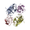 1kgyS S: Starting model for refinement C: citing same article ( |
|---|---|
| Similar structure data |


-Links
 PDBj
PDBj


- Assembly
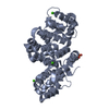
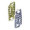
| Deposited unit | 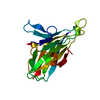
| ||||||||
|---|---|---|---|---|---|---|---|---|---|
| 1 | 
| ||||||||
| Unit cell |
|
-Components
| #1: Protein | Mass: 23568.516 Da / Num. of mol.: 1 / Fragment: Ligand binding domain: Residues 23-202 Source method: isolated from a genetically manipulated source Source: (gene. exp.) Homo sapiens (human) / Gene: EPHA2, ECKPlasmid details: Plasmid transfer vector pFHMSP-LIC-N containing the gene was transformed into DH10Bac E.coli cells (Invitrogen) to obtain recombinant viral DNA. SF9 cells were transfected with ...Plasmid details: Plasmid transfer vector pFHMSP-LIC-N containing the gene was transformed into DH10Bac E.coli cells (Invitrogen) to obtain recombinant viral DNA. SF9 cells were transfected with Bacmid DNA using Cellfectin reagent (Invitrogen), and recombinant baculovirus was generated. Viral stock was amplified from P1 to P3. Plasmid: pFHMSP-LIC-N / Production host:   Spodoptera frugiperda (fall armyworm) / Strain (production host): SF9 Spodoptera frugiperda (fall armyworm) / Strain (production host): SF9References: UniProt: P29317, receptor protein-tyrosine kinase |
|---|---|
| #2: Water | ChemComp-HOH / Water Mass: 18.015 Da / Num. of mol.: 110 / Source method: isolated from a natural source / Formula: H2O Mass: 18.015 Da / Num. of mol.: 110 / Source method: isolated from a natural source / Formula: H2O |
| Sequence details | AUTHORS STATE THAT THE CORRECT SEQUENCE IS PROVIDED IN GENBANK ENTRY NP_004422. |
-Experimental details
-Experiment
| Experiment | Method: X-RAY DIFFRACTION / Number of used crystals: 1 |
|---|
- Sample preparation
| Crystal | Density Matthews: 2.17 Å3/Da / Density % sol: 43.27 % |
|---|---|
| Crystal grow | Temperature: 291.2 K / Method: vapor diffusion, sitting drop / pH: 5.5 Details: AN IN-SITU PROTEOLYSIS STRATEGY WAS USED TO GENERATE HIGH QUALITY CRYSTALS. TRYPSIN WAS ADDED FROM A 1.5 GRAM/L STOCK TO A PROTEIN SAMPLE (AT 6.2 GRAM/L) TO A FINAL TRYPSIN CONC OF 6.2 ...Details: AN IN-SITU PROTEOLYSIS STRATEGY WAS USED TO GENERATE HIGH QUALITY CRYSTALS. TRYPSIN WAS ADDED FROM A 1.5 GRAM/L STOCK TO A PROTEIN SAMPLE (AT 6.2 GRAM/L) TO A FINAL TRYPSIN CONC OF 6.2 MICROGRAM/L BEFORE CRYSTAL PLATES WERE SET AT 291.2K. 25% PEG 3350, 0.1 M AMMONIUM SULFATE, 0.1 M BIS-TRIS PH 5.5. PARATONE-N WAS USED AS THE CRYOPROTECTANT, VAPOR DIFFUSION, SITTING DROP |
-Data collection
| Diffraction | Mean temperature: 100 K |
|---|---|
| Diffraction source | Source: ROTATING ANODE / Type: RIGAKU FR-E+ SUPERBRIGHT / Wavelength: 1.5418 |
| Detector | Type: RIGAKU RAXIS IV / Detector: IMAGE PLATE / Date: Jan 11, 2008 |
| Radiation | Monochromator: GRAPHITE / Protocol: SINGLE WAVELENGTH / Monochromatic (M) / Laue (L): M / Scattering type: x-ray |
| Radiation wavelength | Wavelength: 1.5418 Å / Relative weight: 1 |
| Reflection | Resolution: 1.95→35 Å / Num. all: 14975 / Num. obs: 14975 / % possible obs: 99.9 % / Observed criterion σ(F): 0 / Observed criterion σ(I): -3 / Redundancy: 6.8 % / Rsym value: 0.151 / Net I/σ(I): 15.44 |
| Reflection shell | Resolution: 1.95→2.02 Å / Redundancy: 5 % / Mean I/σ(I) obs: 1.8281 / Num. unique all: 1453 / Rsym value: 0.849 / % possible all: 98.6 |
- Processing
| Software |
| ||||||||||||||||||||||||||||||||||||||||||||||||||||||||||||||||||||||||||||||||||||||||||||||||||||||||||||||||||||||||||||||||||||||||||||||||||||||||||||||||||||||||||||||||||||||||||||||||||||||||||||||||||||||||||||||||||||||||||||||||||||||||||||||||||||||||||||||||||||||||||||||||||||||||||||||||||||||||||||||||||||||||||||||||||||||||||||||||||||||||||||||||||||||||||||||||||||||||||||||||||||||||||||||||||||||||||||||||||||||||||||||||||
|---|---|---|---|---|---|---|---|---|---|---|---|---|---|---|---|---|---|---|---|---|---|---|---|---|---|---|---|---|---|---|---|---|---|---|---|---|---|---|---|---|---|---|---|---|---|---|---|---|---|---|---|---|---|---|---|---|---|---|---|---|---|---|---|---|---|---|---|---|---|---|---|---|---|---|---|---|---|---|---|---|---|---|---|---|---|---|---|---|---|---|---|---|---|---|---|---|---|---|---|---|---|---|---|---|---|---|---|---|---|---|---|---|---|---|---|---|---|---|---|---|---|---|---|---|---|---|---|---|---|---|---|---|---|---|---|---|---|---|---|---|---|---|---|---|---|---|---|---|---|---|---|---|---|---|---|---|---|---|---|---|---|---|---|---|---|---|---|---|---|---|---|---|---|---|---|---|---|---|---|---|---|---|---|---|---|---|---|---|---|---|---|---|---|---|---|---|---|---|---|---|---|---|---|---|---|---|---|---|---|---|---|---|---|---|---|---|---|---|---|---|---|---|---|---|---|---|---|---|---|---|---|---|---|---|---|---|---|---|---|---|---|---|---|---|---|---|---|---|---|---|---|---|---|---|---|---|---|---|---|---|---|---|---|---|---|---|---|---|---|---|---|---|---|---|---|---|---|---|---|---|---|---|---|---|---|---|---|---|---|---|---|---|---|---|---|---|---|---|---|---|---|---|---|---|---|---|---|---|---|---|---|---|---|---|---|---|---|---|---|---|---|---|---|---|---|---|---|---|---|---|---|---|---|---|---|---|---|---|---|---|---|---|---|---|---|---|---|---|---|---|---|---|---|---|---|---|---|---|---|---|---|---|---|---|---|---|---|---|---|---|---|---|---|---|---|---|---|---|---|---|---|---|---|---|---|---|---|---|---|---|---|---|---|---|---|---|---|---|---|---|---|---|---|---|---|---|---|---|---|---|---|---|---|---|---|---|---|---|---|---|---|---|---|---|---|---|---|---|---|---|---|---|---|---|---|---|---|---|---|---|---|---|---|---|---|---|---|---|---|---|---|
| Refinement | Method to determine structure: MOLECULAR REPLACEMENT Starting model: PDB entry 1KGY Resolution: 1.95→35 Å / Cor.coef. Fo:Fc: 0.967 / Cor.coef. Fo:Fc free: 0.928 / SU B: 6.795 / SU ML: 0.1 / Cross valid method: THROUGHOUT / σ(F): 0 / ESU R: 0.13 / ESU R Free: 0.141 / Stereochemistry target values: MAXIMUM LIKELIHOOD Details: HYDROGENS HAVE BEEN ADDED IN THE RIDING POSITIONS. ATOM RECORD CONTAINS SUM OF TLS AND RESIDUAL B FACTORS. ANISOU RECORD CONTAINS SUM OF TLS AND RESIDUAL U FACTORS
| ||||||||||||||||||||||||||||||||||||||||||||||||||||||||||||||||||||||||||||||||||||||||||||||||||||||||||||||||||||||||||||||||||||||||||||||||||||||||||||||||||||||||||||||||||||||||||||||||||||||||||||||||||||||||||||||||||||||||||||||||||||||||||||||||||||||||||||||||||||||||||||||||||||||||||||||||||||||||||||||||||||||||||||||||||||||||||||||||||||||||||||||||||||||||||||||||||||||||||||||||||||||||||||||||||||||||||||||||||||||||||||||||||
| Solvent computation | Ion probe radii: 0.8 Å / Shrinkage radii: 0.8 Å / VDW probe radii: 1.2 Å / Solvent model: BABINET MODEL WITH MASK | ||||||||||||||||||||||||||||||||||||||||||||||||||||||||||||||||||||||||||||||||||||||||||||||||||||||||||||||||||||||||||||||||||||||||||||||||||||||||||||||||||||||||||||||||||||||||||||||||||||||||||||||||||||||||||||||||||||||||||||||||||||||||||||||||||||||||||||||||||||||||||||||||||||||||||||||||||||||||||||||||||||||||||||||||||||||||||||||||||||||||||||||||||||||||||||||||||||||||||||||||||||||||||||||||||||||||||||||||||||||||||||||||||
| Displacement parameters | Biso mean: 24.222 Å2
| ||||||||||||||||||||||||||||||||||||||||||||||||||||||||||||||||||||||||||||||||||||||||||||||||||||||||||||||||||||||||||||||||||||||||||||||||||||||||||||||||||||||||||||||||||||||||||||||||||||||||||||||||||||||||||||||||||||||||||||||||||||||||||||||||||||||||||||||||||||||||||||||||||||||||||||||||||||||||||||||||||||||||||||||||||||||||||||||||||||||||||||||||||||||||||||||||||||||||||||||||||||||||||||||||||||||||||||||||||||||||||||||||||
| Refinement step | Cycle: LAST / Resolution: 1.95→35 Å
| ||||||||||||||||||||||||||||||||||||||||||||||||||||||||||||||||||||||||||||||||||||||||||||||||||||||||||||||||||||||||||||||||||||||||||||||||||||||||||||||||||||||||||||||||||||||||||||||||||||||||||||||||||||||||||||||||||||||||||||||||||||||||||||||||||||||||||||||||||||||||||||||||||||||||||||||||||||||||||||||||||||||||||||||||||||||||||||||||||||||||||||||||||||||||||||||||||||||||||||||||||||||||||||||||||||||||||||||||||||||||||||||||||
| Refine LS restraints |
| ||||||||||||||||||||||||||||||||||||||||||||||||||||||||||||||||||||||||||||||||||||||||||||||||||||||||||||||||||||||||||||||||||||||||||||||||||||||||||||||||||||||||||||||||||||||||||||||||||||||||||||||||||||||||||||||||||||||||||||||||||||||||||||||||||||||||||||||||||||||||||||||||||||||||||||||||||||||||||||||||||||||||||||||||||||||||||||||||||||||||||||||||||||||||||||||||||||||||||||||||||||||||||||||||||||||||||||||||||||||||||||||||||
| LS refinement shell | Resolution: 1.95→2.005 Å / Total num. of bins used: 20
| ||||||||||||||||||||||||||||||||||||||||||||||||||||||||||||||||||||||||||||||||||||||||||||||||||||||||||||||||||||||||||||||||||||||||||||||||||||||||||||||||||||||||||||||||||||||||||||||||||||||||||||||||||||||||||||||||||||||||||||||||||||||||||||||||||||||||||||||||||||||||||||||||||||||||||||||||||||||||||||||||||||||||||||||||||||||||||||||||||||||||||||||||||||||||||||||||||||||||||||||||||||||||||||||||||||||||||||||||||||||||||||||||||
| Refinement TLS params. | Method: refined / Refine-ID: X-RAY DIFFRACTION
| ||||||||||||||||||||||||||||||||||||||||||||||||||||||||||||||||||||||||||||||||||||||||||||||||||||||||||||||||||||||||||||||||||||||||||||||||||||||||||||||||||||||||||||||||||||||||||||||||||||||||||||||||||||||||||||||||||||||||||||||||||||||||||||||||||||||||||||||||||||||||||||||||||||||||||||||||||||||||||||||||||||||||||||||||||||||||||||||||||||||||||||||||||||||||||||||||||||||||||||||||||||||||||||||||||||||||||||||||||||||||||||||||||
| Refinement TLS group |
|
