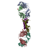
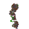
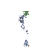
[English] 日本語
Yorodumi
- PDB-3mx0: Crystal Structure of EphA2 ectodomain in complex with ephrin-A5 -

+
Open data


ID or keywords:

Loading...

-
Basic information

Entry
Database: PDB / ID: 3mx0
TitleCrystal Structure of EphA2 ectodomain in complex with ephrin-A5
Components
  • Ephrin type-A receptor 2
  • Ephrin-A5Ephrin A5
KeywordsTRANSFERASE RECEPTOR/SIGNALLING PROTEIN / ectodomain / receptor-ligand complex / receptor-receptor interaction / TRANSFERASE RECEPTOR-SIGNALLING PROTEIN complex
Function / homology
Function and homology information


neurotrophin TRKC receptor binding / neurotrophin TRKB receptor binding / negative regulation of substrate adhesion-dependent cell spreading / regulation of cell-cell adhesion / synaptic membrane adhesion / notochord cell development / notochord formation / lens fiber cell morphogenesis / blood vessel endothelial cell proliferation involved in sprouting angiogenesis / negative regulation of lymphangiogenesis ...neurotrophin TRKC receptor binding / neurotrophin TRKB receptor binding / negative regulation of substrate adhesion-dependent cell spreading / regulation of cell-cell adhesion / synaptic membrane adhesion / notochord cell development / notochord formation / lens fiber cell morphogenesis / blood vessel endothelial cell proliferation involved in sprouting angiogenesis / negative regulation of lymphangiogenesis / axial mesoderm formation / collateral sprouting / pericyte cell differentiation / cellular response to follicle-stimulating hormone stimulus / cAMP metabolic process / positive regulation of bicellular tight junction assembly / regulation of blood vessel endothelial cell migration / positive regulation of collateral sprouting / negative regulation of chemokine production / regulation of insulin secretion involved in cellular response to glucose stimulus / ephrin receptor activity / leading edge membrane / bone remodeling / post-anal tail morphogenesis / neurotrophin TRKA receptor binding / chemorepellent activity / response to growth factor / activation of GTPase activity / regulation of cell morphogenesis / regulation of lamellipodium assembly / retinal ganglion cell axon guidance / positive regulation of synapse assembly / tight junction / branching involved in mammary gland duct morphogenesis / EPH-Ephrin signaling / neural tube development / RND1 GTPase cycle / RND2 GTPase cycle / RND3 GTPase cycle / mammary gland epithelial cell proliferation / regulation of focal adhesion assembly / RHOV GTPase cycle / regulation of GTPase activity / EPHA-mediated growth cone collapse / transmembrane receptor protein tyrosine kinase activator activity / growth factor binding / regulation of cell adhesion mediated by integrin / lamellipodium membrane / RHOU GTPase cycle / RHOG GTPase cycle / EPH-ephrin mediated repulsion of cells / basement membrane / negative regulation of phosphatidylinositol 3-kinase/protein kinase B signal transduction / GABA-ergic synapse / ephrin receptor signaling pathway / RAC2 GTPase cycle / RAC3 GTPase cycle / vasculogenesis / regulation of angiogenesis / regulation of microtubule cytoskeleton organization / cellular response to forskolin / keratinocyte differentiation / RAC1 GTPase cycle / transmembrane receptor protein tyrosine kinase activity / ephrin receptor binding / cell chemotaxis / negative regulation of angiogenesis / osteoclast differentiation / regulation of ERK1 and ERK2 cascade / phosphatidylinositol 3-kinase/protein kinase B signal transduction / skeletal system development / molecular function activator activity / cell motility / caveola / axon guidance / protein localization to plasma membrane / regulation of actin cytoskeleton organization / positive regulation of protein localization to plasma membrane / adherens junction / receptor protein-tyrosine kinase / neuron differentiation / ruffle membrane / osteoblast differentiation / cell surface receptor protein tyrosine kinase signaling pathway / intrinsic apoptotic signaling pathway in response to DNA damage / cell migration / positive regulation of peptidyl-tyrosine phosphorylation / virus receptor activity / lamellipodium / nervous system development / receptor complex / cell adhesion / positive regulation of cell migration / defense response to Gram-positive bacterium / cadherin binding / inflammatory response / positive regulation of protein phosphorylation / external side of plasma membrane / phosphorylation / focal adhesion
Similarity search - Function
Ephrin-A ectodomain / Ephrin receptor-binding domain / Ephrin, conserved site / Ephrin / Ephrin / Ephrin receptor-binding (ephrin RBD) domain signature. / Ephrin receptor-binding (ephrin RBD) domain profile. / Ephrin type-A receptor 2, ligand binding domain / Ephrin receptor type-A /type-B / Ephrin receptor ligand binding domain ...Ephrin-A ectodomain / Ephrin receptor-binding domain / Ephrin, conserved site / Ephrin / Ephrin / Ephrin receptor-binding (ephrin RBD) domain signature. / Ephrin receptor-binding (ephrin RBD) domain profile. / Ephrin type-A receptor 2, ligand binding domain / Ephrin receptor type-A /type-B / Ephrin receptor ligand binding domain / Tyrosine-protein kinase, receptor class V, conserved site / Ephrin receptor, transmembrane domain / Ephrin receptor ligand binding domain / Ephrin type-A receptor 2 transmembrane domain / Receptor tyrosine kinase class V signature 1. / Receptor tyrosine kinase class V signature 2. / Eph receptor ligand-binding domain profile. / Ephrin receptor ligand binding domain / Putative ephrin-receptor like / SAM domain (Sterile alpha motif) / SAM domain profile. / Sterile alpha motif. / Sterile alpha motif domain / Cupredoxins - blue copper proteins / Sterile alpha motif/pointed domain superfamily / Growth factor receptor cysteine-rich domain superfamily / Fibronectin type III domain / Cupredoxin / Fibronectin type 3 domain / Fibronectin type-III domain profile. / Galactose-binding-like domain superfamily / Fibronectin type III / Fibronectin type III superfamily / Tyrosine-protein kinase, catalytic domain / Tyrosine kinase, catalytic domain / Tyrosine protein kinases specific active-site signature. / Tyrosine-protein kinase, active site / Protein tyrosine and serine/threonine kinase / Serine-threonine/tyrosine-protein kinase, catalytic domain / Immunoglobulins / Protein kinase, ATP binding site / Protein kinases ATP-binding region signature. / Immunoglobulin-like fold / Protein kinase domain profile. / Protein kinase domain / Protein kinase-like domain superfamily / Immunoglobulin-like / Sandwich / Mainly Beta
Similarity search - Domain/homology
triacetyl-beta-chitotriose / Ephrin type-A receptor 2 / Ephrin-A5
Similarity search - Component
Biological speciesHomo sapiens (human)
MethodX-RAY DIFFRACTION / SYNCHROTRON / MOLECULAR REPLACEMENT / molecular replacement / Resolution: 3.506 Å
AuthorsHimanen, J.P. / Yermekbayeva, L. / Janes, P.W. / Walker, J.R. / Xu, K. / Atapattu, L. / Rajashankar, K.R. / Mensinga, A. / Lackmann, M. / Nikolov, D.B. / Dhe-Paganon, S.
CitationJournal: Proc.Natl.Acad.Sci.USA / Year: 2010
Title: Architecture of Eph receptor clusters.
Authors: Himanen, J.P. / Yermekbayeva, L. / Janes, P.W. / Walker, J.R. / Xu, K. / Atapattu, L. / Rajashankar, K.R. / Mensinga, A. / Lackmann, M. / Nikolov, D.B. / Dhe-Paganon, S.
History
DepositionMay 6, 2010Deposition site: RCSB / Processing site: RCSB
Revision 1.0Jun 30, 2010Provider: repository / Type: Initial release
Revision 1.1Jul 13, 2011Group: Version format compliance
Revision 1.2Nov 8, 2017Group: Refinement description / Category: software
Revision 2.0Jul 29, 2020Group: Atomic model / Data collection ...Atomic model / Data collection / Derived calculations / Structure summary
Category: atom_site / chem_comp ...atom_site / chem_comp / entity / entity_name_com / pdbx_branch_scheme / pdbx_chem_comp_identifier / pdbx_entity_branch / pdbx_entity_branch_descriptor / pdbx_entity_branch_link / pdbx_entity_branch_list / pdbx_entity_nonpoly / pdbx_molecule_features / pdbx_nonpoly_scheme / pdbx_struct_assembly_gen / struct_asym / struct_conn / struct_site / struct_site_gen
Item: _atom_site.B_iso_or_equiv / _atom_site.Cartn_x ..._atom_site.B_iso_or_equiv / _atom_site.Cartn_x / _atom_site.Cartn_y / _atom_site.Cartn_z / _atom_site.auth_asym_id / _atom_site.auth_seq_id / _atom_site.label_asym_id / _atom_site.label_entity_id / _chem_comp.name / _chem_comp.type / _pdbx_entity_nonpoly.entity_id / _pdbx_entity_nonpoly.name / _pdbx_struct_assembly_gen.asym_id_list / _struct_conn.pdbx_dist_value / _struct_conn.pdbx_leaving_atom_flag / _struct_conn.pdbx_role / _struct_conn.ptnr1_auth_asym_id / _struct_conn.ptnr1_auth_comp_id / _struct_conn.ptnr1_auth_seq_id / _struct_conn.ptnr1_label_asym_id / _struct_conn.ptnr1_label_atom_id / _struct_conn.ptnr1_label_comp_id / _struct_conn.ptnr1_label_seq_id / _struct_conn.ptnr2_auth_asym_id / _struct_conn.ptnr2_auth_seq_id / _struct_conn.ptnr2_label_asym_id
Description: Carbohydrate remediation / Provider: repository / Type: Remediation

-
Structure visualization

Structure viewerMolecule:
MolmilJmol/JSmol

Downloads & links

-
Assembly

Deposited unit
A: Ephrin type-A receptor 2
B: Ephrin-A5
C: Ephrin type-A receptor 2
D: Ephrin-A5
hetero molecules


Theoretical massNumber of molelcules
Total (without water)124,3318
Polymers122,6344
Non-polymers1,6984
Water0
1
A: Ephrin type-A receptor 2
B: Ephrin-A5
hetero molecules


Theoretical massNumber of molelcules
Total (without water)62,1664
Polymers61,3172
Non-polymers8492
Water0
TypeNameSymmetry operationNumber
identity operation1_555x,y,z1
Buried area2700 Å2
ΔGint2 kcal/mol
Surface area28950 Å2
MethodPISA
2
C: Ephrin type-A receptor 2
D: Ephrin-A5
hetero molecules


Theoretical massNumber of molelcules
Total (without water)62,1664
Polymers61,3172
Non-polymers8492
Water0
TypeNameSymmetry operationNumber
identity operation1_555x,y,z1
Buried area2900 Å2
ΔGint4 kcal/mol
Surface area28430 Å2
MethodPISA
Unit cell
Length a, b, c (Å)57.859, 89.049, 198.150
Angle α, β, γ (deg.)90.00, 96.22, 90.00
Int Tables number4
Space group name H-MP1211

-
Components

#1: Protein Ephrin type-A receptor 2 / Tyrosine-protein kinase receptor ECK / Epithelial cell kinase


Mass: 44996.477 Da / Num. of mol.: 2 / Fragment: Ectodomain (UNP Residues 27-435)
Source method: isolated from a genetically manipulated source
Source: (gene. exp.) Homo sapiens (human) / Gene: EPHA2, ECK / Plasmid: pDT101 / Cell line (production host): HEK-293 / Production host: Homo sapiens (human)
References: UniProt: P29317, receptor protein-tyrosine kinase
#2: Protein Ephrin-A5 / Ephrin A5 / EPH-related receptor tyrosine kinase ligand 7 / LERK-7 / AL-1


Mass: 16320.332 Da / Num. of mol.: 2 / Fragment: Ectodomain (UNP Residues 28-165)
Source method: isolated from a genetically manipulated source
Source: (gene. exp.) Homo sapiens (human) / Gene: EFNA5, EPLG7, LERK7 / Cell line (production host): HEK-293 / Production host: Homo sapiens (human) / References: UniProt: P52803
#3: Polysaccharide 2-acetamido-2-deoxy-beta-D-glucopyranose-(1-4)-2-acetamido-2-deoxy-beta-D-glucopyranose-(1-4)-2- ...2-acetamido-2-deoxy-beta-D-glucopyranose-(1-4)-2-acetamido-2-deoxy-beta-D-glucopyranose-(1-4)-2-acetamido-2-deoxy-beta-D-glucopyranose / triacetyl-beta-chitotriose


Type: oligosaccharide, Oligosaccharide / Class: Inhibitor / Mass: 627.594 Da / Num. of mol.: 2
Source method: isolated from a genetically manipulated source
Details: oligosaccharide / References: triacetyl-beta-chitotriose
DescriptorTypeProgram
DGlcpNAcb1-4DGlcpNAcb1-4DGlcpNAcb1-Glycam Condensed SequenceGMML 1.0
WURCS=2.0/1,3,2/[a2122h-1b_1-5_2*NCC/3=O]/1-1-1/a4-b1_b4-c1WURCSPDB2Glycan 1.1.0
[]{[(4+1)][b-D-GlcpNAc]{[(4+1)][b-D-GlcpNAc]{[(4+1)][b-D-GlcpNAc]{}}}}LINUCSPDB-CARE
#4: Sugar ChemComp-NAG / 2-acetamido-2-deoxy-beta-D-glucopyranose / N-Acetylglucosamine


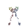
Type: D-saccharide, beta linking / Mass: 221.208 Da / Num. of mol.: 2
Source method: isolated from a genetically manipulated source
Formula: C8H15NO6
IdentifierTypeProgram
DGlcpNAcbCONDENSED IUPAC CARBOHYDRATE SYMBOLGMML 1.0
N-acetyl-b-D-glucopyranosamineCOMMON NAMEGMML 1.0
b-D-GlcpNAcIUPAC CARBOHYDRATE SYMBOLPDB-CARE 1.0
GlcNAcSNFG CARBOHYDRATE SYMBOLGMML 1.0

-
Experimental details

-
Experiment

ExperimentMethod: X-RAY DIFFRACTION / Number of used crystals: 1

-
Sample preparation

CrystalDensity Matthews: 4.14 Å3/Da / Density % sol: 70.28 %
Crystal growTemperature: 298 K / Method: vapor diffusion, hanging drop / pH: 5.8
Details: 20% PEG 3350, 200 mM Ammonium Citrate, 100 mM Na Citrate, pH 5.8, VAPOR DIFFUSION, HANGING DROP, temperature 298K

-
Data collection

DiffractionMean temperature: 100 K
Diffraction sourceSource: SYNCHROTRON / Site: APS / Beamline: 24-ID-C / Wavelength: 0.98792 Å
DetectorType: ADSC QUANTUM 315 / Detector: CCD
RadiationMonochromator: SI 111 CHANNEL / Protocol: SINGLE WAVELENGTH / Monochromatic (M) / Laue (L): M / Scattering type: x-ray
Radiation wavelengthWavelength: 0.98792 Å / Relative weight: 1
ReflectionResolution: 3.5→50 Å / Num. obs: 23143 / % possible obs: 91.4 % / Redundancy: 5.4 % / Rmerge(I) obs: 0.104 / Net I/σ(I): 8.8
Reflection shellResolution: 3.5→3.63 Å / Redundancy: 5.2 % / Rmerge(I) obs: 0.219 / Mean I/σ(I) obs: 6.2 / Rsym value: 0.22 / % possible all: 71.1

-
Phasing

PhasingMethod: molecular replacement

-
Processing

Software
NameVersionClassificationNB
DENZOdata reduction
SCALEPACKdata scaling
PHENIX1.6.1_357refinement
PDB_EXTRACT3.1data extraction
HKL-2000data collection
PHENIXphasing
RefinementMethod to determine structure: MOLECULAR REPLACEMENT / Resolution: 3.506→45.028 Å / Occupancy max: 1 / Occupancy min: 1 / SU ML: 0.38 / σ(F): 0.16 / Phase error: 33.16 / Stereochemistry target values: ML
RfactorNum. reflection% reflection
Rfree0.2951 1123 5.02 %
Rwork0.2338 --
obs0.2369 22383 88.12 %
all-23129 -
Solvent computationShrinkage radii: 0.9 Å / VDW probe radii: 1.11 Å / Solvent model: FLAT BULK SOLVENT MODEL / Bsol: 91.597 Å2 / ksol: 0.301 e/Å3
Displacement parameters
Baniso -1Baniso -2Baniso -3
1--20.6855 Å20 Å22.415 Å2
2--78.283 Å20 Å2
3----57.5975 Å2
Refinement stepCycle: LAST / Resolution: 3.506→45.028 Å
ProteinNucleic acidLigandSolventTotal
Num. atoms8462 0 112 0 8574
Refine LS restraints
Refine-IDTypeDev idealNumber
X-RAY DIFFRACTIONf_bond_d0.018852
X-RAY DIFFRACTIONf_angle_d1.47212024
X-RAY DIFFRACTIONf_dihedral_angle_d20.553202
X-RAY DIFFRACTIONf_chiral_restr0.0931286
X-RAY DIFFRACTIONf_plane_restr0.0061574
LS refinement shell
Resolution (Å)Rfactor RfreeNum. reflection RfreeRfactor RworkNum. reflection RworkRefine-ID% reflection obs (%)
3.506-3.6650.38191060.28831995X-RAY DIFFRACTION66
3.665-3.85820.31591150.2542248X-RAY DIFFRACTION75
3.8582-4.09980.27761330.2242482X-RAY DIFFRACTION83
4.0998-4.4160.28931440.21092694X-RAY DIFFRACTION90
4.416-4.860.26451510.18482858X-RAY DIFFRACTION95
4.86-5.56210.27281520.19332917X-RAY DIFFRACTION97
5.5621-7.00340.28041590.22212989X-RAY DIFFRACTION99
7.0034-45.0320.30761630.24593077X-RAY DIFFRACTION99

+
About Yorodumi

-
News

-
Feb 9, 2022. New format data for meta-information of EMDB entries

New format data for meta-information of EMDB entries

  • Version 3 of the EMDB header file is now the official format.
  • The previous official version 1.9 will be removed from the archive.

Related info.:EMDB header

External links:wwPDB to switch to version 3 of the EMDB data model

-
Aug 12, 2020. Covid-19 info

Covid-19 info

URL: https://pdbj.org/emnavi/covid19.php

New page: Covid-19 featured information page in EM Navigator.

Related info.:Covid-19 info / Mar 5, 2020. Novel coronavirus structure data

+
Mar 5, 2020. Novel coronavirus structure data

Novel coronavirus structure data

Related info.:Yorodumi Speices / Aug 12, 2020. Covid-19 info

External links:COVID-19 featured content - PDBj / Molecule of the Month (242):Coronavirus Proteases

+
Jan 31, 2019. EMDB accession codes are about to change! (news from PDBe EMDB page)

EMDB accession codes are about to change! (news from PDBe EMDB page)

  • The allocation of 4 digits for EMDB accession codes will soon come to an end. Whilst these codes will remain in use, new EMDB accession codes will include an additional digit and will expand incrementally as the available range of codes is exhausted. The current 4-digit format prefixed with “EMD-” (i.e. EMD-XXXX) will advance to a 5-digit format (i.e. EMD-XXXXX), and so on. It is currently estimated that the 4-digit codes will be depleted around Spring 2019, at which point the 5-digit format will come into force.
  • The EM Navigator/Yorodumi systems omit the EMD- prefix.

Related info.:Q: What is EMD? / ID/Accession-code notation in Yorodumi/EM Navigator

External links:EMDB Accession Codes are Changing Soon! / Contact to PDBj

+
Jul 12, 2017. Major update of PDB

Major update of PDB

  • wwPDB released updated PDB data conforming to the new PDBx/mmCIF dictionary.
  • This is a major update changing the version number from 4 to 5, and with Remediation, in which all the entries are updated.
  • In this update, many items about electron microscopy experimental information are reorganized (e.g. em_software).
  • Now, EM Navigator and Yorodumi are based on the updated data.

External links:wwPDB Remediation / Enriched Model Files Conforming to OneDep Data Standards Now Available in the PDB FTP Archive

-
Yorodumi

Thousand views of thousand structures

  • Yorodumi is a browser for structure data from EMDB, PDB, SASBDB, etc.
  • This page is also the successor to EM Navigator detail page, and also detail information page/front-end page for Omokage search.
  • The word "yorodu" (or yorozu) is an old Japanese word meaning "ten thousand". "mi" (miru) is to see.

Related info.:EMDB / PDB / SASBDB / Comparison of 3 databanks / Yorodumi Search / Aug 31, 2016. New EM Navigator & Yorodumi / Yorodumi Papers / Jmol/JSmol / Function and homology information / Changes in new EM Navigator and Yorodumi

Read more